. 2021 Aug 1;321(2):L429-L439.
doi: 10.1152/ajplung.00018.2021. Epub 2021 May 19.

Toxic effects of cell-free hemoglobin on the microvascular endothelium: implications for pulmonary and nonpulmonary organ dysfunction

Affiliations

Toxic effects of cell-free hemoglobin on the microvascular endothelium: implications for pulmonary and nonpulmonary organ dysfunction

Jamie E Meegan et al. Am J Physiol Lung Cell Mol Physiol. .

Abstract

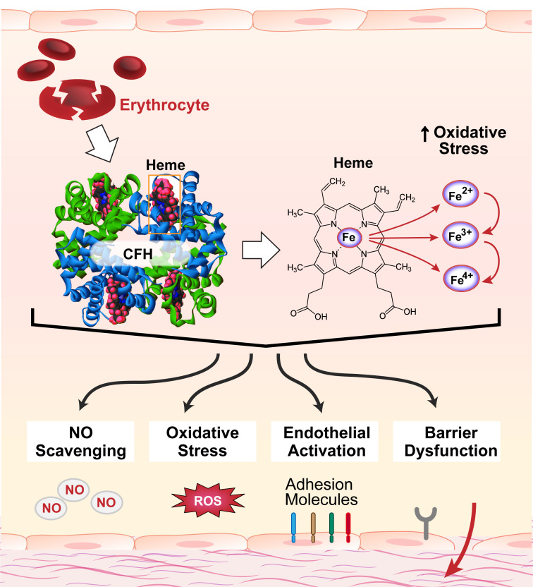
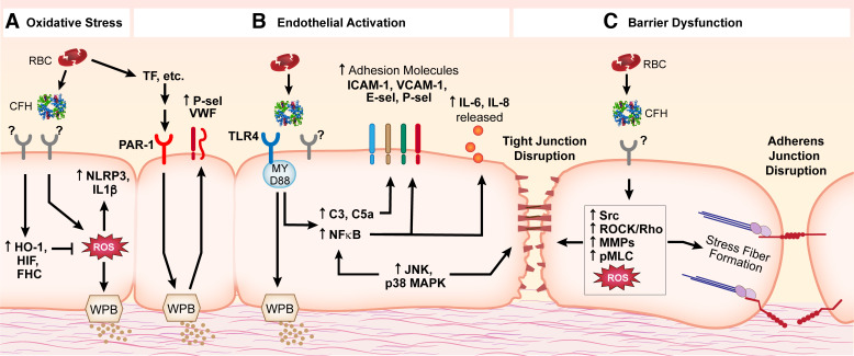
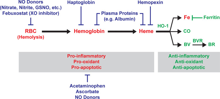
Levels of circulating cell-free hemoglobin are elevated during hemolytic and inflammatory diseases and contribute to organ dysfunction and severity of illness. Though several studies have investigated the contribution of hemoglobin to tissue injury, the precise signaling mechanisms of hemoglobin-mediated endothelial dysfunction in the lung and other organs are not yet completely understood. The purpose of this review is to highlight the knowledge gained thus far and the need for further investigation regarding hemoglobin-mediated endothelial inflammation and injury to develop novel therapeutic strategies targeting the damaging effects of cell-free hemoglobin.

Keywords: endothelium; heme; hemoglobin; inflammation; lung injury; microvascular endothelial dysfunction.


Conflict of interest statement

No conflicts of interest, financial or otherwise, are declared by the authors.

Figures

Figure 1.
Figure 1.
Circulating cell-free hemoglobin (CFH) and its released components contribute to endothelial injury via NO scavenging, oxidative stress, endothelial activation, and endothelial barrier dysfunction. CFH is released from damaged red blood cells during several hemolytic and inflammatory pathologies. Elevated circulating levels of CFH are associated with higher risk of organ dysfunction and mortality. Hemoglobin is composed of two α-globin and two β-globin subunits, each consisting of an iron-containing heme group. The iron can redox cycle from its oxygen-carrying ferrous (2+) form to ferric (3+, methemoglobin) or the highly reactive ferryl (4+) form, which can oxidize lipids and other substrates, increasing oxidative stress. Moreover, there is potential for the release of heme and/or iron from circulating CFH, adding complexity to the contributions of CFH to endothelial injury. Mechanisms demonstrated to be involved in CFH-mediated endothelial injury include NO scavenging, oxidative stress, endothelial activation, and endothelial barrier dysfunction. NO, nitric oxide; ROS, reactive oxygen species.
Figure 2.
Figure 2.
Potential signaling mechanisms involved in CFH-mediated endothelial injury. Though the entirety of the precise signaling mechanisms contributing to endothelial injury is yet to be elucidated, multiple pathways have been implicated. A: CFH causes increased ROS that lead to activation of NLRP3 signaling and activation of IL-1β, as well as degranulation of WPBs. Protective cellular mechanisms activated by CFH include HO-1, HIF, and FHC to counter oxidative stress. B: endothelial activation in response to CFH signals through the TLR4/MyD88 pathway leading to activation of MAPKs, NF-κB and complement signaling, upregulation of adhesion molecules, and release of pro-inflammatory cytokines; PAR-1 activation by CFH-mediated upregulation of the TF signaling cascade leads to increased coagulation signaling (including increased VWF and P-selectin). C: endothelial barrier dysfunction induced by CFH or its components is characterized by disruption of cell-cell junctions (adherens junctions and tight junctions) and reorganization of the actin cytoskeleton (stress fiber formation); signaling mechanisms that may be involved include upregulation of MAPKs, Src, Rho, MMPs, and pMLC signaling. Many of these signaling pathways are complex and overlapping. Of note, some studies show that TLR4/MAPK/NLRP3 signaling was not required in CFH-mediated endothelial injury; the precise signaling mechanisms involved most likely depend on context and require further investigation. CFH, circulating cell-free hemoglobin; C3, complement component 3; C5a, complement component 5a; E-sel, E-selectin; FHC, ferritin heavy chain; HIF, hypoxia-inducible factor; HO-1, heme oxygenase-1; ICAM-1, intercellular adhesion molecule 1; IL-1β, interleukin 1 beta; IL-6, interleukin 6; IL-8, interleukin 8; JNK, c-Jun N-terminal kinases; MAPK, mitogen-activated protein kinase; MMPs, matrix metalloproteinases; MyD88, myeloid differentiation primary response 88; NF-κB, nuclear factor kappa-light-chain-enhancer of activated B cells; NLRP3, NOD-, LRR- and pyrin domain-containing protein 3; PAR-1, protease-activated receptor-1; pMLC, phosphorylated myosin light chain; P-sel, P-selectin; ROCK, Rho-associated protein kinase; ROS, reactive oxygen species; TF, tissue factor; TLR4, Toll-like receptor 4; VCAM-1, vascular cell adhesion protein 1; VWF, von Willebrand factor; WPB, Weibel-Palade bodies.
Figure 3.
Figure 3.
Potential therapeutic strategies that target CFH-mediated endothelial injury. [Color key: Red = Injurious, Green = Cytoprotective, Blue = Potential therapies] Hemolysis (lysis of red blood cells) in the circulation releases CFH and subsequent free heme to affect endothelial function via oxidative stress and inflammation. Intracellular anti-inflammatory, anti-oxidant, and anti-apoptotic pathways driven by upregulation of HO-1 break down heme into labile iron (which leads to production of cytoprotective ferritin), CO, and biliverdin (which is converted to bilirubin by biliverdin reductase). Potential therapies against CFH-mediated endothelial injury are aimed at targeting hemolysis (NO donors such as nitrate, nitrite, GSNO, or Febuxostat), CFH (haptoglobin, plasma proteins), heme (hemopexin, plasma proteins), or oxidative stress/inflammation (acetaminophen, ascorbate, NO donors). BR, bilirubin; BV, biliverdin; BVR, biliverdin reductase; CFH, cell-free hemoglobin; CO, carbon monoxide; Fe, iron; GSNO, S-nitrosoglutathione; HO-1, heme oxygenase-1; RBC, red blood cells; XO, xanthine oxidase; NO, nitric oxide.

Similar articles

Cited by

References

    1. Hsu LL, Champion HC, Campbell-Lee SA, Bivalacqua TJ, Manci EA, Diwan BA, Schimel DM, Cochard AE, Wang X, Schechter AN, Noguchi CT, Gladwin MT. Hemolysis in sickle cell mice causes pulmonary hypertension due to global impairment in nitric oxide bioavailability. Blood 109: 3088–3098, 2007. [Erratum in Blood 111: 1772, 2008]. doi:10.1182/blood-2006-08-039438. - DOI - PMC - PubMed
    1. Detterich JA, Kato RM, Rabai M, Meiselman HJ, Coates TD, Wood JC. Chronic transfusion therapy improves but does not normalize systemic and pulmonary vasculopathy in sickle cell disease. Blood 126: 703–710, 2015. doi:10.1182/blood-2014-12-614370. - DOI - PMC - PubMed
    1. Atichartakarn V, Chuncharunee S, Archararit N, Udomsubpayakul U, Aryurachai K. Intravascular hemolysis, vascular endothelial cell activation and thrombophilia in splenectomized patients with hemoglobin E/beta-thalassemia disease. Acta Haematol 132: 100–107, 2014. doi:10.1159/000355719. - DOI - PubMed
    1. Reiter CD, Wang X, Tanus-Santos JE, Hogg N, Cannon RO 3rd, Schechter AN, Gladwin MT. Cell-free hemoglobin limits nitric oxide bioavailability in sickle-cell disease. Nat Med 8: 1383–1389, 2002. doi:10.1038/nm1202-799. - DOI - PubMed
    1. Donadee C, Raat NJ, Kanias T, Tejero J, Lee JS, Kelley EE, Zhao X, Liu C, Reynolds H, Azarov I, Frizzell S, Meyer EM, Donnenberg AD, Qu L, Triulzi D, Kim-Shapiro DB, Gladwin MT. Nitric oxide scavenging by red blood cell microparticles and cell-free hemoglobin as a mechanism for the red cell storage lesion. Circulation 124: 465–476, 2011. doi:10.1161/CIRCULATIONAHA.110.008698. - DOI - PMC - PubMed

Publication types

LinkOut - more resources