. 2021 Jul 1;321(1):L102-L115.
doi: 10.1152/ajplung.00618.2020. Epub 2021 May 19.

The proprotein convertase furin inhibits IL-13-induced inflammation in airway smooth muscle by regulating integrin-associated signaling complexes

Affiliations

The proprotein convertase furin inhibits IL-13-induced inflammation in airway smooth muscle by regulating integrin-associated signaling complexes

Yidi Wu et al. Am J Physiol Lung Cell Mol Physiol. .

Abstract

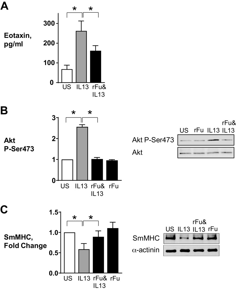
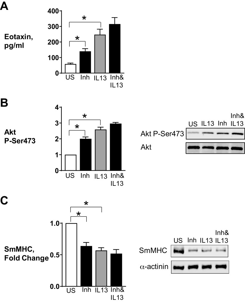
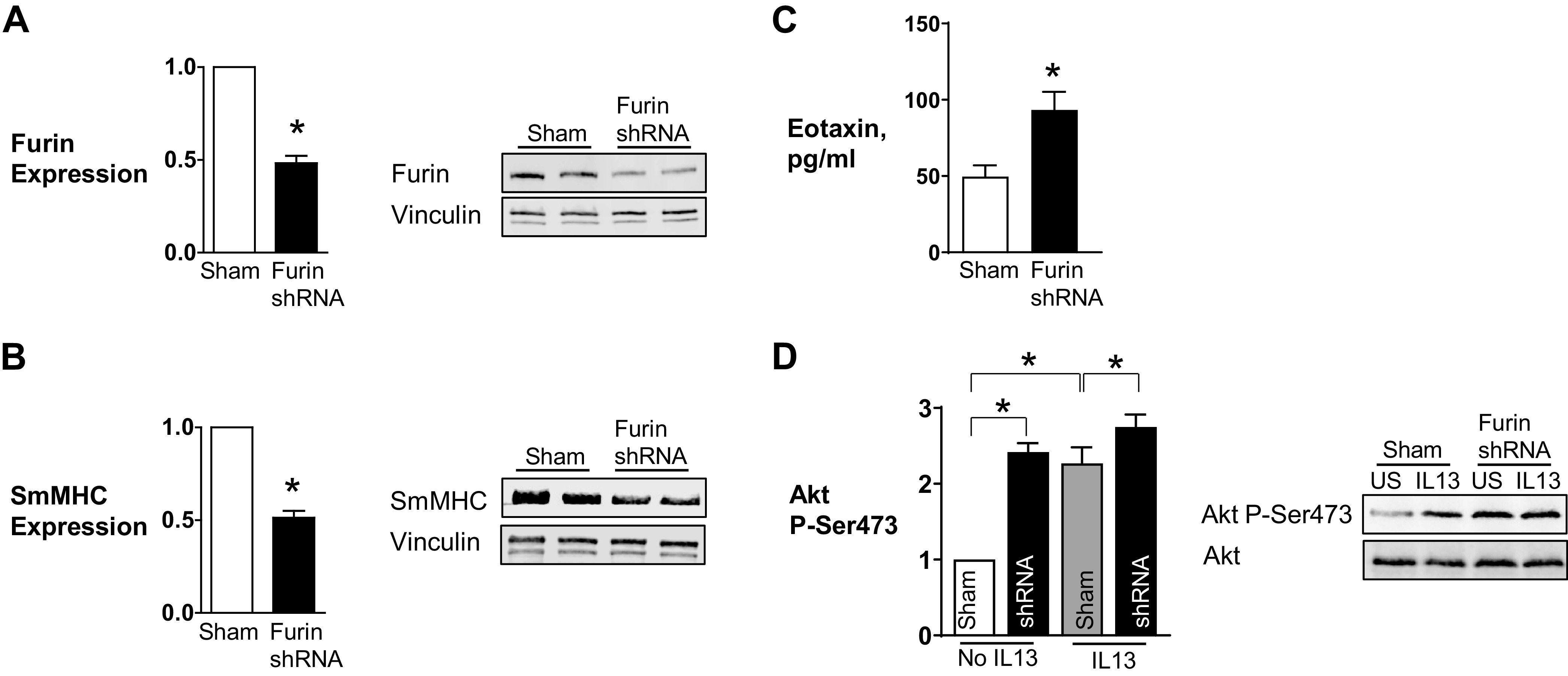
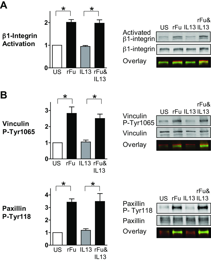
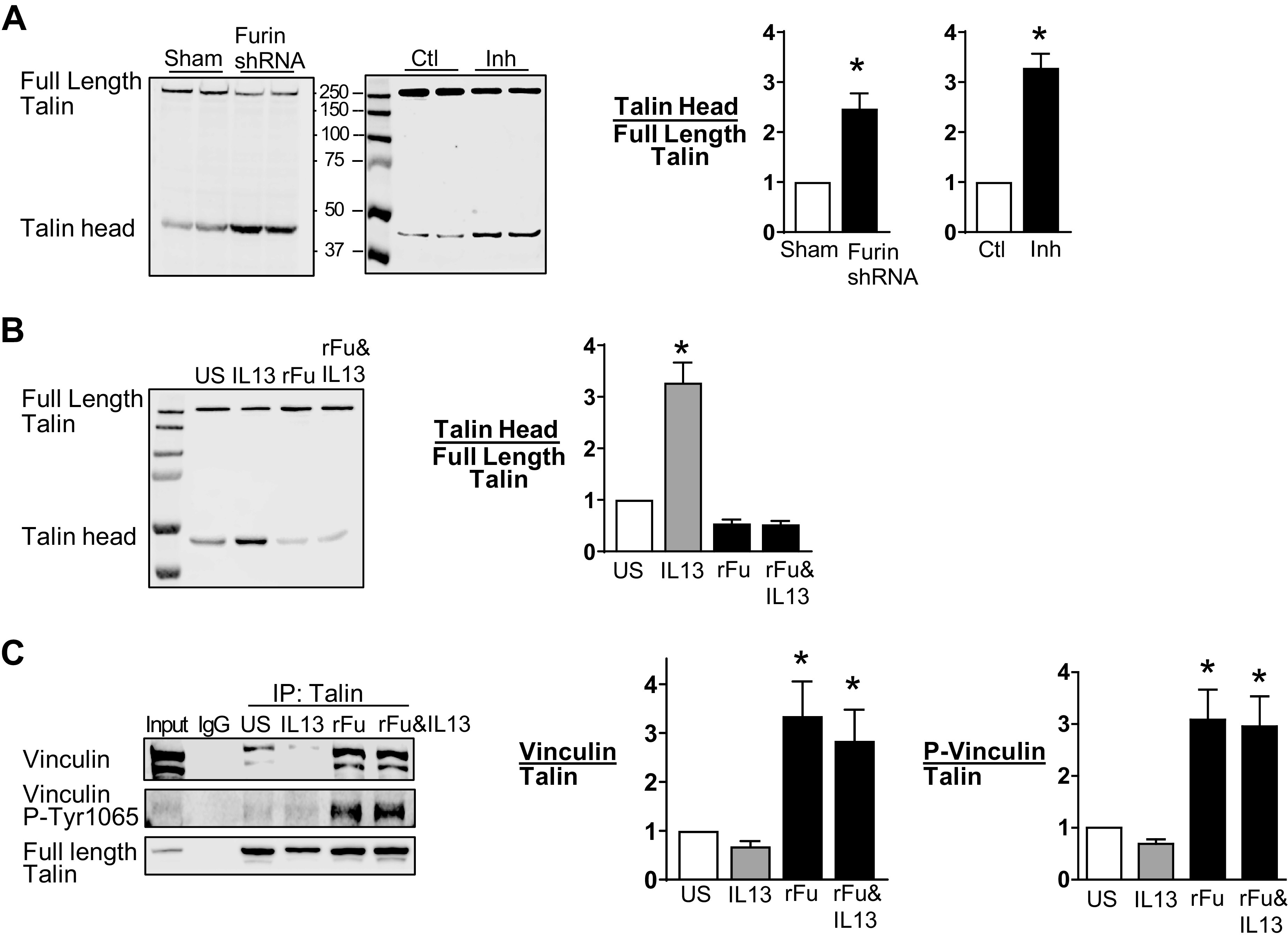
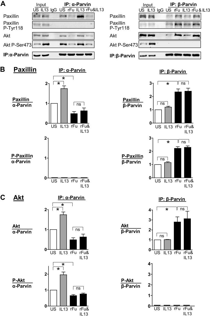
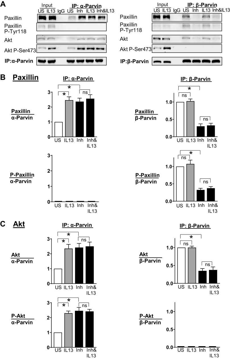
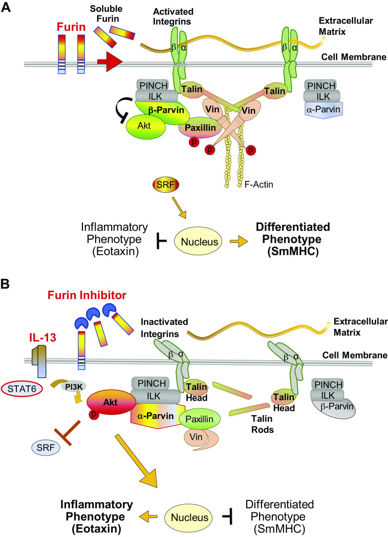
Furin is a proprotein convertase that regulates the activation and the inactivation of multiple proteins including matrix metalloproteinases, integrins, and cytokines. It is a serine endoprotease that localizes to the plasma membrane and can be secreted into the extracellular space. The role of furin in regulating inflammation in isolated canine airway smooth muscle tissues was investigated. The treatment of airway tissues with recombinant furin (rFurin) inhibited the activation of Akt and eotaxin secretion induced by IL-13, and it prevented the IL-13-induced suppression of smooth muscle myosin heavy chain expression. rFurin promoted a differentiated phenotype by activating β1-integrin proteins and stimulating the activation of the adhesome proteins vinculin and paxillin by talin. Activated paxillin induced the binding of Akt to β-parvin IPP [integrin-linked kinase (ILK), PINCH, parvin] complexes, which inhibits Akt activation. Treatment of tissues with a furin inhibitor or the depletion of endogenous furin using shRNA resulted in Akt activation and inflammatory responses similar to those induced by IL-13. Furin inactivation or IL-13 caused talin cleavage and integrin inactivation, resulting in the inactivation of vinculin and paxillin. Paxillin inactivation resulted in the coupling of Akt to α-parvin IPP complexes, which catalyze Akt activation and an inflammatory response. The results demonstrate that furin inhibits inflammation in airway smooth muscle induced by IL-13 and that the anti-inflammatory effects of furin are mediated by activating integrin proteins and integrin-associated signaling complexes that regulate Akt-mediated pathways to the nucleus. Furin may have therapeutic potential for the treatment of inflammatory conditions of the lungs and airways.

Keywords: IL-13; airway inflammation; airway smooth muscle; integrin complexes; signal transduction.


Conflict of interest statement

No conflicts of interest, financial or otherwise, are declared by the authors.

Figures

Figure 1.
Figure 1.
Recombinant furin (rFurin) prevents inflammatory signaling pathways in airway smooth muscle tissues. Airway smooth muscle tissues were treated with 0.5 µg/mL furin overnight and then incubated with or without 50 ng/mL IL-13 overnight or for 30 min. A: eotaxin secretion was significantly increased by stimulation with IL-13 (n = 6, P = 0.0001). rFurin significantly inhibited IL-13-induced eotaxin secretion (n = 6, P = 0.0133). B: IL-13 significantly increases Akt phosphorylation (n = 9, P = 0.0001). Furin significantly inhibits Akt phosphorylation induced by IL-13 (n = 9, P = 0.0001). C: expression of smooth muscle myosin heavy chain (SmMHC) was significantly inhibited in tissues stimulated with IL-13 (n = 7, P = 0.0001). Furin prevents inhibition of SmMHC expression induced by IL-13 (n = 7, P = 0.2526). All statistical analyses were performed by using one-way ANOVA with repeated measures. Values are means ± SE. *Significant difference between groups. rFu, rFurin; US, unstimulated.
Figure 2.
Figure 2.
Inhibition of furin activity promotes inflammatory signaling pathways in airway smooth muscle tissues. Airway smooth muscle tissues were treated with 100 µM Furin Inhibitor II in the presence or absence of 50 ng/mL IL-13 for measurement of Akt phosphorylation, smooth muscle myosin heavy chain (SmMHC), or eotaxin secretion. A: eotaxin secretion was significantly increased by Furin Inhibitor II (P = 0.0487) or IL-13 (P = 0.0001) in airway smooth muscle (ASM) tissues (n = 6). Inhibition of furin activity did not significantly increase eotaxin secretion induced by IL-13 (n = 6, P = 0.116). B: treatment of tissues with Furin Inhibitor II or IL-13 results in a significant increase in Akt phosphorylation (n = 8, P = 0.0001). C: expression of SmMHC was significantly inhibited by Furin Inhibitor II or IL-13 (n = 6, P = 0.0001). Furin inhibition did not significantly depress the expression of SmMHC of ASM tissues after IL-13 stimulation (n = 6, P = 0.058). Statistical analysis was performed by using one-way ANOVA with repeated measures. Values are means ± SE. *Significant difference between groups. Inh, Furin Inhibitor II; US, unstimulated.
Figure 3.
Figure 3.
Depletion of furin from airway smooth muscle tissues increases Akt phosphorylation and decreases smooth muscle myosin heavy chain (SmMHC) expression. A, B: representative immunoblots and mean expression levels of endogenous furin protein in extracts from tissues treated with furin shRNA or sham treated. ShRNA significantly reduced expression of furin (n = 10, P = 0.0001) (A) and SmMHC (n = 9, P = 0.0001) (B). C: the depletion of furin caused a significant increase in eotaxin secretion (n = 4, P = 0.0272). D: representative immunoblots of Akt from extracts of sham-treated or furin-depleted muscle tissues stimulated with IL-13 or unstimulated (US). Furin depletion resulted in a significant increase in Akt phosphorylation at Ser473 (n = 7, P = 0.0001). Furin inhibition also significantly potentiated IL-13-induced Akt phosphorylation (n = 7, P = 0.019). Data were analyzed by using a paired Student’s t test (A–C) or one-way ANOVA (D). Values are means ± SE. *Significant difference between groups.
Figure 4.
Figure 4.
Furin regulates the activation of β1-integrins and integrin-associated adhesion proteins in airway smooth muscle tissues. A: the effects of furin on the activation on β1-integrins were evaluated using an antibody (HUTS-4) that selectively reacts with activated β1-integrin. Representative immunoblots from extracts of recombinant furin (rFurin)-treated or -untreated muscle tissues stimulated with IL-13 or unstimulated (US). The incubation of tissues with rFurin caused the activation of β1-integrins (n = 9, P = 0.0001). rFurin also increased integrin activation in the presence of IL-13 (n = 9, P = 0.0001). B: representative immunoblots from extracts of rFurin-treated or -untreated muscle tissues stimulated with IL-13 or unstimulated (US). rFurin significantly increased vinculin and paxillin phosphorylation in airway smooth muscle tissues (vinculin: n = 5, P = 0.0004; paxillin: n = 6, P = 0.0001). rFurin also increased vinculin and paxillin phosphorylation in the presence of IL-13 (vinculin: n = 5, P = 0.0025; paxillin: n = 6, P = 0.0002). Data were analyzed by using one-way ANOVA. All values are means ± SE. *Significant difference between groups. rF, rFurin.
Figure 5.
Figure 5.
Furin Inhibitor II inhibits the activation of β1-integrins and integrin-associated adhesion proteins by acetylcholine (Ach) in airway smooth muscle tissues. A: representative immunoblots from extracts of inhibitor treated or untreated muscle tissues stimulated with ACh or unstimulated (US). ACh stimulation resulted in a marked increase in the activation of β1-integrin (n = 8, P = 0.0001) and incubation with Furin Inhibitor II inhibited the increase of activated β1-integrin induced by ACh (n = 8, P = 0.0001). B: representative immunoblots from extracts of untreated or inhibitor-treated muscle tissues stimulated with ACh or unstimulated (US). The increases in vinculin Tyr1065 phosphorylation and paxillin Tyr118 phosphorylation in response to 10−5 M ACh were significantly inhibited in tissues treated with Furin Inhibitor II (vinculin: n = 7, P = 0.0001; paxillin: n = 8, P = 0.0001). Data were analyzed by using one-way ANOVA. All values are means ± SE. *Significant difference between groups. Inh, Furin Inhibitor II.
Figure 6.
Figure 6.
Furin prevents IL-13-induced talin cleavage in airway smooth muscle tissues. A: representative immunoblots from extracts of tissues treated with furin shRNA or sham treated and treated with or without Furin Inhibitor II (Inh). Full-length talin and the talin head domain were analyzed by immunoblot with an N-terminal antibody to talin. Furin depletion significantly increased the ratio of the talin head domain to full-length talin (n = 6, P = 0.005). Furin inhibition also significantly increased the ratio of the talin head domain to full-length talin (n = 15, P = 0.0001). B: representative immunoblot of extracts from tissues treated with or without IL-13 in the presence or absence of recombinant furin (rFurin). IL-13 significantly increased the ratio of the talin head to full-length talin (n = 12, P = 0.0001). rFurin prevented the increase in talin cleavage induced by IL-13 (n = 12, P = 0.2972). C: talin was immunoprecipitated from tissue extracts, and immunocomplexes were blotted for vinculin and Tyr1065 phosphorylated vinculin. Vinculin and phosphorylated vinculin were significantly higher in tissues treated with rFurin or rFurin and IL-13 compared with untreated tissues (vinculin: P = 0.0026 for rFurin, P = 0.0184 for rFurin and IL-13; phosphorylated vinculin: P = 0.0014 for rFurin, P = 0.0026 for rFurin and IL-13) (n = 7). Data were analyzed by using a paired Student’s t test (A) or one-way ANOVA (B and C). All values are means ± SE. *Significant difference between groups. ctl, control; rFu, rFurin; US, unstimulated.
Figure 7.
Figure 7.
Furin promotes the association of Akt with β-parvin IPP [integrin-linked kinase (ILK), PINCH, parvin] complexes, which prevents IL-13-induced Akt activation. α-Parvin or β-parvin was immunoprecipitated from extracts of tissues treated with or without IL-13, recombinant furin (rFurin), or IL-13 and rFurin. A: α-parvin and β-parvin immunocomplexes were immunoblotted for Akt, pS473-Akt, paxillin, pY118-paxillin, and α-parvin or β-parvin. B: rFurin stimulated a significant increase in the association of phosphorylated paxillin with β-parvin IPP complexes (n = 4, P = 0.0001) and a significant decrease in the association of unphosphorylated paxillin with α-parvin complexes (n = 5, P = 0.0251). IL-13 stimulates the association of unphosphorylated paxillin with α-parvin IPP complexes (n = 5, P = 0.0018) and not with β-parvin complexes (n = 4, P = 0.6808). Furin prevents the interaction of unphosphorylated paxillin with α-parvin complexes in response to IL-13 (n = 5, P = 0.7880) and stimulates the interaction of phosphorylated paxillin with β-parvin complexes (n = 4, P = 0.0001). C: IL-13 stimulates the activation of Akt by α-parvin IPP complexes (n = 5, P = 0.0001). Furin alone does not stimulate the activation of Akt by α-parvin IPP complexes (n = 5, P = 0.0423), but furin prevents the activation of Akt by α-parvin IPP complexes caused by stimulation with IL-13 (n = 4, P = 0.7941). Statistical analysis was performed by using one-way ANOVA with repeated measures. Values are means ± SE. *Significant difference between groups. ns, no significant difference; rFu, rFurin; US, unstimulated.
Figure 8.
Figure 8.
Furin inhibition promotes the activation of Akt by α-parvin IPP [integrin-linked kinase (ILK), PINCH, parvin] complexes in airway smooth muscle tissues. α-Parvin or β-parvin was immunoprecipitated from extracts of tissues treated with or without IL-13, Furin Inhibitor II, or IL-13 and Furin Inhibitor II. A: α-parvin and β-parvin immunocomplexes were immunoblotted for Akt, pS473-Akt, paxillin, pY118-paxillin, and α-parvin or β-parvin. B: IL-13 and Furin Inhibitor II each stimulates a significant increase in the association of unphosphorylated paxillin with α-parvin IPP complexes (IL-13: n = 5, P = 0.0001; Furin Inhibitor II: n = 5, P = 0.0017). Furin Inhibitor II stimulates a significant decrease in the association of paxillin with β-parvin IPP complexes (n = 4, P = 0.0001). C: IL-13 and Furin Inhibitor II each induces Akt activation by stimulating the association of Akt with α-parvin IPP complexes (IL-13: n = 5, P = 0.0009; Furin Inhibitor II: n = 5, P = 0.0006). Furin Inhibitor II stimulates a significant decrease in the association of Akt with β-parvin IPP complexes (n = 4, P = 0.0001). Statistical analysis was performed by using one-way ANOVA with repeated measures. Values are means ± SE. *Significant difference between groups. Inh, Furin Inhibitor II; ns, no significant difference; US, unstimulated.
Figure 9.
Figure 9.
Proposed mechanism for the regulation of inflammation by furin in airway smooth muscle. A: furin activates integrin-mediated signaling pathways regulating Akt activity and phenotype expression in airway smooth muscle. Furin promotes the cross linking of β integrin proteins by talin, resulting in integrin activation and the high-affinity binding of integrin proteins to the extracellular matrix. Vinculin is activated by its binding to full-length talin, facilitating phosphorylation of its binding partner paxillin. Phosphorylated paxillin binds to β-parvin IPP (ILK, PINCH, parvin) complexes, which promotes the preferential binding of Akt to β-parvin instead of integrin-linked kinase (ILK). This prevents the activation of Akt by ILK and suppresses Akt-mediated synthetic pathways that induce an inflammatory phenotype. This promotes the localization of serum response factor (SRF) to the nucleus and the expression of a differentiated phenotype. α-Parvin IPP complexes are inactive because they are not recruited by phosphorylated paxillin. B: effect of furin inhibition or IL-13 stimulation on integrin-mediated signaling pathways regulating Akt activity and phenotype expression in airway smooth muscle. Stimulation with IL-13 or treatment with furin inhibitor leads to talin cleavage, which prevents the cross linking of β integrins by talin, resulting in integrin inactivation. This prevents the high-affinity binding of integrins to the extracellular matrix. Vinculin remains in an inactive complex with paxillin because vinculin cannot be activated by cleaved talin, and paxillin phosphorylation is therefore prevented. Unphosphorylated paxillin binds to α-parvin IPP complexes, which promotes the binding of Akt directly to ILK. ILK induces Akt activation resulting in an inflammatory phenotype. SRF localization to the nucleus is inhibited by activated Akt, which suppresses the expression of a differentiated smooth muscle phenotype. β-Parvin IPP complexes remain inactive, as they are not recruited by unphosphorylated paxillin. smMHC, smooth muscle myosin heavy chain.

Similar articles

Cited by

References

    1. Desai LP, Wu Y, Tepper RS, Gunst SJ. Mechanical stimuli and IL-13 interact at integrin adhesion complexes to regulate expression of smooth muscle myosin heavy chain in airway smooth muscle tissue. Am J Physiol Lung Cell Mol Physiol 301: L275–L284, 2011. doi:10.1152/ajplung.00043.2011. - DOI - PMC - PubMed
    1. Hirst SJ, Hallsworth MP, Peng Q, Lee TH. Selective induction of eotaxin release by interleukin-13 or interleukin-4 in human airway smooth muscle cells is synergistic with interleukin-1beta and is mediated by the interleukin-4 receptor alpha-chain. Am J Respir Crit Care Med 165: 1161–1171, 2002. doi:10.1164/ajrccm.165.8.2107158. - DOI - PubMed
    1. Huang Y, Gunst SJ. Phenotype transitions induced by mechanical stimuli in airway smooth muscle are regulated by differential interactions of parvin isoforms with paxillin and Akt. Am J Physiol Lung Cell Mol Physiol 318: L1036–L1055, 2020. doi:10.1152/ajplung.00506.2019. - DOI - PMC - PubMed
    1. Lockett AD, Wu Y, Gunst SJ. Elastase alters contractility and promotes an inflammatory synthetic phenotype in airway smooth muscle tissues. Am J Physiol Lung Cell Mol Physiol 314: L626–L634, 2018. doi:10.1152/ajplung.00334.2017. - DOI - PMC - PubMed
    1. Moore PE, Church TL, Chism DD, Panettieri RA Jr, Shore SA. IL-13 and IL-4 cause eotaxin release in human airway smooth muscle cells: a role for ERK. Am J Physiol Lung Cell Mol Physiol 282: L847–L853, 2002. doi:10.1152/ajplung.00245.2001. - DOI - PubMed

Publication types

MeSH terms

LinkOut - more resources