. 2022 Jan;101(1):73-82.
doi: 10.1177/00220345211013722. Epub 2021 May 19.

Alveolar Bone Marrow Gli1+ Stem Cells Support Implant Osseointegration

Affiliations

Alveolar Bone Marrow Gli1+ Stem Cells Support Implant Osseointegration

Y Yi et al. J Dent Res. 2022 Jan.

Abstract

Osseointegration is the key issue for implant success. The in vivo properties of cell populations driving the osseointegration process have remained largely unknown. In the current study, using tissue clearing-based 3-dimensional imaging and transgenic mouse model-based lineage tracing methods, we identified Gli1+ cells within alveolar bone marrow and their progeny as the cell population participating in extraction socket healing and implant osseointegration. These Gli1+ cells are surrounding blood vessels and do not express lineage differentiation markers. After tooth extraction and delayed placement of a dental implant, Gli1+ cells were activated into proliferation, and their descendants contributed significantly to new bone formation. Ablation of Gli1+ cells severely compromised the healing and osseointegration processes. Blockage of canonical Wnt signaling resulted in impaired recruitment of Gli1+ cells and compromised bone healing surrounding implants. Collectively, these findings demonstrate that Gli1+ cells surrounding alveolar bone marrow vasculature are stem cells supporting dental implant osseointegration. Canonical Wnt signal plays critical roles in regulating Gli1+ stem cells.

Keywords: Wnt signaling pathway; alveolar bone; bone-implant interface; dental implantation; hedgehogs; periodontium.


Conflict of interest statement

Declaration of Conflicting Interests: The authors declared no potential conflicts of interest with respect to the research, authorship, and/or publication of this article.

Figures

Figure 1.
Figure 1.
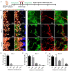
Gli1+ cells within alveolar bone marrow were surrounding blood vessels and largely negative for pericyte markers and osteogenic lineage markers. (A) Experimental timeline. (B) The alveolar bones were collected on day 5 after inducton. TdTomato+ cells were visualized in the alveolar bone marrow space (a). Sections at the mandibular first molar furcation of Gli1-creERT2;Ai14 mice were stained with antibodies against endothelium markers laminin (b) and GS-IB4 (c), pericyte markers NG2 (d) and CD146 (e), stem cell markers α–smooth muscle actin (αSMA) (f) and LepR (g), and lineage markers including periostin (h), Sp7 (i), and type I collagen (j). d′–f′, h′–j′ are enlarged images of boxed areas in d–f, h–j, respectively. g′ is the LepR antibody staining of gingiva on the same section from g, serving as a positive control. (C) Percentiles of double-positive cells (i.e., cells that express both tdTomato and specific markers) among tdTomato+ cells on sections (n = 4).
Figure 2.
Figure 2.
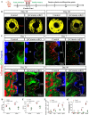
Gli1+ cells within alveolar bone marrow were activated after tooth extraction. They migrated along blood vessels and contributed to new bone formation at the extraction site. (A) Experimental timeline of tooth extraction and sample collection. (B) Gli1-creERT2;Ai14 mice extraction site, 1 d after mandibular molar extraction (a). Two-hour EdU incorporation assay showed dividing Gli1+ cells around vasculature 1 day after tooth extraction (b, c). Tamoxifen was injected 5 d prior to the surgery. (C) Percentages of tdTomato+/EdU+ cells among tdTomato+ cells within the alveolar bone marrow near socket before and after extraction (n = 4). ****P < 0.0001. (D) Distribution of Gli1+ cells and their derivatives on 7 d (a), 14 d (d), and 28 d (g) after tooth extraction. Immunostaining of laminin (b, e, h) and Sp7 (c, f, i) showed the association of tdTomato+ cells with blood vessels and osteoblasts, respectively. White arrows indicate blood vessels; arrows with star indicate Sp7+/tdTomato+ cells. (E) Percentiles of tdTomato+ cells within the extraction socket at different time points (n = 4). **P < 0.01. (F) Number of tdTomato+ cells within each field view at different distances from blood vessels at various time points (n = 4). (G) Percentiles of Sp7+/tdTomato+ cells among tdTomato+ cells within the extraction socket at different time points (n = 4). ****P < 0.0001. ns, no significant difference.
Figure 3.
Figure 3.
Gli1+ cells within alveolar bone marrow contributed to the implant osseointegration. (A) Experimental timeline of delayed implant placement and sample collection. (B) Tissue clearing–based 3-dimensional images of Gli1-creERT2;Ai14 mouse mandibles with implants showed contribution of Gli1+ cells toward osseointegration on day 1, 7, 14, 28, or 90 after implant placement. Tamoxifen was injected 5 d ahead of implant surgery. (C) Normalized tdTomato+ cell numbers at different time points were quantified relative to day 1 (n = 4). **P < 0.01. ***P < 0.001. ****P < 0.0001. ns, no significant difference. (D) Percentiles of tdTomato+ cells among all cells within the alveolar bone marrow at different time points (n = 4). *P < 0.05. ***P < 0.001. ns, no significant difference.
Figure 4.
Figure 4.
Migration of Gli1+ cells and their derivatives during osseointegration were closely associated with blood vessels. (A) Experimental timeline. (B) Tissue clearing–based 3-dimensional images of Cdh5-creERT2;Ai14 mouse mandibles with implants showed angiogenesis surrounding the titanium implants on day 1, 7, or 28 after implant placement. (C) Representative images of Gli1-creERT2;Ai14 mandibles with implants stained with GS-IB4 showed an association between peri-implant Gli1+ cells and vasculature. (DF) Percentages of tdTomato+ cells at different distances from GS-IB4+ blood vessels among all tdTomato+ cells on day 1 (D), 7 (E), or 28 (F) after implant placement (n = 4).
Figure 5.
Figure 5.
Canonical Wnt signaling pathway is required for Gli1+ cell-mediated implant osseointegration. (A) Experimental timeline. (B) Three-dimensional (3-D) reconstruction of micro–computed tomography of Gli1-creERT2;Ai14 (control) and Gli1-creERT2;β-cateninflox/flox;Ai14 (β-catenin inducible conditional knockout [icKO]) littermate mice 28 d and 90 d after implant placement. (C) Representative 3-D images showed Gli1+ cell contribution and new bone formation in control or β-catenin icKO group on day 1, 7, 28, or 90 after implant placement. (D) Quantitative bone volume fraction analysis of newly formed bone (n = 4). **P < 0.01. (E) Quantitative bone–implant contact (BIC) analysis (n = 4). **P < 0.01. ***P < 0.001. (F) Normalized tdTomato+ cell numbers in control and β-catenin icKO groups at different time points were quantified relative to day 1 (n = 4). *P < 0.05. **P < 0.01. ***P < 0.001. ns, no significant difference. (G) Percentiles of tdTomato+ cells in the alveolar bone of control and β-catenin icKO sample at different time points (n = 4). ***P < 0.001. ns, no significant difference.

Similar articles

Cited by

References

    1. Branemark PI, Adell R, Breine U, Hansson BO, Lindstrom J, Ohlsson A. 1969. Intra-osseous anchorage of dental prostheses. I. Experimental studies. Scand J Plast Reconstr Surg. 3(2):81–100. - PubMed
    1. Buser D, Sennerby L, De Bruyn H. 2017. Modern implant dentistry based on osseointegration: 50 years of progress, current trends and open questions. Periodontol; 2000. 73(1):7–21. - PubMed
    1. Calvo-Guirado JL, Ortiz-Ruiz AJ, Negri B, Lopez-Mari L, Rodriguez-Barba C, Schlottig F. 2010. Histological and histomorphometric evaluation of immediate implant placement on a dog model with a new implant surface treatment. Clin Oral Implants Res. 21(3):308–315. - PubMed
    1. Canellas JVDS, Medeiros PJD, Figueredo CMDS, Fischer RG, Ritto FG. 2019. Which is the best choice after tooth extraction, immediate implant placement or delayed placement with alveolar ridge preservation? A systematic review and meta-analysis. J Craniomaxillofac Surg. 47(11):1793–1802. - PubMed
    1. Chappuis V, Engel O, Reyes M, Shahim K, Nolte LP, Buser D. 2013. Ridge alterations post-extraction in the esthetic zone: a 3D analysis with CBCT. J Dent Res. 92(12 Suppl):195S–201S. - PMC - PubMed

Publication types

LinkOut - more resources