. 2021 Jul 5;220(7):e202101057.
doi: 10.1083/jcb.202101057. Epub 2021 May 19.

Per1/Per2-Igf2 axis-mediated circadian regulation of myogenic differentiation

Affiliations

Per1/Per2-Igf2 axis-mediated circadian regulation of myogenic differentiation

Nobuko Katoku-Kikyo et al. J Cell Biol. .

Abstract

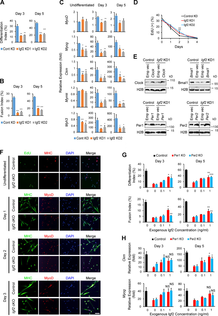
Circadian rhythms regulate cell proliferation and differentiation, but circadian control of tissue regeneration remains elusive at the molecular level. Here, we show that proper myoblast differentiation and muscle regeneration are regulated by the circadian master regulators Per1 and Per2. Depletion of Per1 or Per2 suppressed myoblast differentiation in vitro and muscle regeneration in vivo, demonstrating their nonredundant functions. Both Per1 and Per2 were required for the activation of Igf2, an autocrine promoter of myoblast differentiation, accompanied by Per-dependent recruitment of RNA polymerase II, dynamic histone modifications at the Igf2 promoter and enhancer, and the promoter-enhancer interaction. This circadian epigenetic priming created a preferred time window for initiating myoblast differentiation. Consistently, muscle regeneration was faster if initiated at night, when Per1, Per2, and Igf2 were highly expressed compared with morning. This study reveals the circadian timing as a significant factor for effective muscle cell differentiation and regeneration.


Figures

Figure 1.
Figure 1.
Regeneration of TA muscle in Per1−/−, Per2−/−, and Per1−/−:Per2−/− mice. (A) H&E staining of day 4.5 TA muscle sections. TA muscle was injured with barium chloride at ZT14 on day 0, and EdU was injected i.p. 96 h later for F and G. The muscle was harvested 12 h later at day 4.5. Bar, 100 µm. (B) Size distribution of H&E-stained myofibers containing centrally located nuclei on day 4.5. The minimal Feret’s diameter in each myofiber was measured. n = 8 mice in each group, including 4 males and 4 females, in B and C. (C) Average of the minimal Feret’s diameters of myofibers with centrally located nuclei on day 4.5. (D) Immunofluorescence staining of TA muscle with antibodies against eMHC and laminin (showing the border of each myofiber) on day 4.5. DNA was counterstained with DAPI. Bar, 100 µm. (E) Average of the minimal Feret’s diameters of eMHC+ areas on day 4.5. n = 4 mice. (F) Immunofluorescence staining of TA muscle sections with the MyoD antibody and an EdU kit. Bar, 25 µm. (G) Frequency of positive cells for EdU uptake and MyoD staining shown in F. n = 4 mice. (H) Sirius red staining of days 7 and 14 after injury and uninjured TA muscle. Bar, 200 µm. (I) The area percentage of Sirius red+ fibrosis in the groups shown in H. (J–L) Differentiation index (J), fusion index (K), and the frequency of EdU uptake (L) during the differentiation of primary myoblasts prepared from WT and Per KO mice. Data represent biological triplicates with n = 80–130 nuclei in each replicate. Data are presented as mean + SEM. *, P < 0.05; **, P < 0.01; and ***, P < 0.001 with Student’s t test compared with WT.
Figure S1.
Figure S1.
Regeneration of TA muscle and myoblast differentiation comparing Per1−/−, Per2−/−, and Per1−/−:Per2−/− mice. (A) Average of the minimal Feret’s diameters of myofibers with centrally located nuclei on day 14. TA muscle was injured with barium chloride at ZT14 on day 0 and harvested 14 d later. n = 8 mice with 4 males and 4 females in each group in A and B. (B) Average of the minimal Feret’s diameters of myofibers in uninjured mice. (C–F) EdU uptake and immunofluorescence staining of undifferentiated (C) and differentiating primary myoblasts on day 1 (D), day 2 (E), and day 3 (F) with antibodies against MHC and MyoD. DNA was counterstained with DAPI. Bars, 100 µm. *, P < 0.05 and ***, P < 0.001 with Student’s t test compared with WT mice. Data are presented as mean + SEM.
Figure S2.
Figure S2.
RNA-seq analysis of Per KO C2C12 cells. (A) Relative expression levels of Per1 and Per2 mRNAs in C2C12 cells after KD of each gene. The expression level with control scrambled shRNA was defined as 1.0 for each gene. ***, P < 0.001 with Student’s t test. Data are presented as mean + SEM of biological triplicates. (B) Indel frequency in Per1 KO and Per2 KO C2C12 cells analyzed with TIDE software (https://tide.nki.nl/). (C) Western blotting demonstrating down-regulation of Per1 and Per2 in KD and KO cells. Histone H2B was used as a loading control (Cont). (D) Venn diagrams displaying the number of genes whose expression levels were >200% or <50% of those of control KO cells. n = 1 in D–G. (E) The number of genes that were commonly up-regulated (> Cont × 2) or down-regulated (< Cont × 0.5) more than twofold in Per1 KO and Per2 KO cells compared with control KO cells. (F) Scatterplots comparing control, Per1 KO, and Per2 KO C2C12 cells. (G) Gene ontology (GO) terms relevant to muscle differentiation that were enriched in the genes commonly down-regulated in Per1 KO and Per2 KO cells compared with control KO cells. FC, fold change.
Figure 2.
Figure 2.
Differentiation of C2C12 cells after depletion of Per1 and Per2. (A) Immunofluorescence staining of C2C12 cells with MHC antibody during differentiation with 5% HS. Per1 and Per2 were depleted with shRNA (KD) and CRISPR-Cas9 (KO). Nontargeting sequences were used as a control for each. Bar, 100 µm. (B and C) Differentiation index (B) and Fusion index (C) on days 3 and 5. (D) Temporal profile of the frequency of EdU+ nuclei in KO cells during differentiation. (E) Relative expression levels of five muscle genes determined by qPCR during differentiation. The value obtained with undifferentiated control cells treated with scrambled shRNA was defined as 1.0 for each gene. (F) Heat map comparing the transcriptome of KO cells. Day 0 refers to undifferentiated cells in F and G. n = 1 in F and G. (G) Principal component analysis of KO cells. Data are presented as mean + or ± SEM of biological triplicates unless indicated otherwise. Each replicate includes n = 1,000–1,500 nuclei in B–D. *, P < 0.05; **, P < 0.01; and ***, P < 0.001 with Student’s t test compared with control.
Figure S3.
Figure S3.
RNA-seq analysis of Per KO cells and differentiation of Igf2 KD cells. (A) List of genes belonging to the gene ontology terms shown in Fig. S2 G. Four genes commonly down-regulated in Per1 KO and Per2 KO cells in an undifferentiated state and during differentiation are highlighted in yellow. n = 1 in A and B. (B) Relative expression level of Igf2 mRNA determined by qPCR in C2C12 cells after depletion of Per1 and Per2. The value obtained with control scrambled shRNA on day 0 (before differentiation) was defined as 1.0. (C) Relative expression level of Igf2 mRNA after KD with two shRNA clones. The expression level with control shRNA was defined as 1.0. (D) MHC staining of differentiating C2C12 cells after Igf2 KD with two shRNAs. Bar, 200 µm. **, P < 0.01 and ***, P < 0.001 with Student’s t test compared with control cells (Cont). Data are presented as mean + SEM of biological triplicates. FC, fold change.
Figure S4.
Figure S4.
Per1/Per2–Igf2 axis and myoblast differentiation. (A and B) Differentiation index (A) and fusion index (B) during differentiation of Igf2 KD cells. (C) Relative expression levels of five muscle genes during differentiation of Igf2 KD cells. The value obtained with day 0 control (Cont) KD cells was defined as 1.0. (D) Temporal profile of the frequency of EdU+ nuclei in Igf2 KD cells during differentiation. (E) Western blotting of Clock, Bmal1, Per1, and Per2 after retrovirus-mediated transduction of these genes in C2C12 cells. Empty vector (Emp Vec) was used as a control. Cells transduced with Igf2 shRNA clone 1 (Igf2 KD1) and those with control shRNA were compared. Histone H2B was used as a loading control. (F) EdU uptake and immunofluorescence staining of undifferentiated and differentiating primary myoblasts with antibodies against MHC and MyoD. DNA was counterstained with DAPI. Bar, 100 µm. (G) Differentiation index (top) and fusion index (bottom) of control and Per KO C2C12 cells cultured with exogenous Igf2 at different concentrations for 3 and 5 d. Igf2 was not added to the control cells. (H) Relative expression levels of Ckm (top) and Myog (bottom) of control and Per KO C2C12 cells treated with exogenous Igf2 for 3 and 5 d. The expression level of the control cells before differentiation was defined as 1.0. Data are presented as mean + or ± SEM of biological triplicates. Each replicate includes n = 1,000–1,500 nuclei in A and B. *, P < 0.05; **, P < 0.01; and ***, P < 0.001 with Student’s t test compared with control. NS, P ≥ 0.05. Asterisks were only added to the values with 1 ng/ml Igf2 in G and H.
Figure 3.
Figure 3.
Disrupted differentiation of C2C12 cells by Igf2 KD. (A) Relative expression levels of Cav3, Csrp3, and Myoz1 in Igf2 KD cells. (B) Relative expression levels of Igf2 and muscle genes during differentiation of C2C12 cells. The cells were transduced with empty vector (Emp Vec), Per1, Per2, CBE (Clock, Bmal1, and empty vector), CBP1 (Clock, Bmal1, and Per1), and CBP2 (Clock, Bmal1, and Per2) before differentiation. Igf2 KD cells and control cells were compared. (C and D) Differentiation index (C) and fusion index (D) of the cells used in B. Data are presented as mean + SEM of biological triplicates. Each replicate includes n = 1,000–1,500 nuclei in C and D. *, P < 0.05; **, P < 0.01; and ***, P < 0.001 with Student’s t test compared with control KD cells in A and with empty vector cells in B–D.
Figure 4.
Figure 4.
Impaired muscle regeneration in satellite cell–specific Igf2 cKO mice. (A) Experimental scheme for satellite cell–specific Igf2 cKO by injection of TMX and muscle regeneration from barium chloride (BaCl2)–induced injury. (B) Relative expression level of Igf2 in isolated satellite cells comparing control (Cont) and Igf2 cKO cells. (C) H&E staining of TA muscle sections prepared from uninjured and regeneration day 7 mice. TA muscle was injured with BaCl2 at ZT14 on day 0. Bar, 100 µm. (D) Size distribution of H&E-stained myofibers containing centrally located nuclei at the indicated time points. The minimal Feret’s diameter in each myofiber was measured. n = 3 for each genotype. (E) Average of the minimal Feret’s diameters of myofibers with centrally located nuclei. (F) Immunofluorescence staining of TA muscle with antibodies against eMHC and laminin on day 4. DNA was counterstained with DAPI. Bar, 25 µm. (G) Average of the minimal Feret’s diameters of eMHC+ areas on day 4. n = 4 for each genotype. (H) Immunofluorescence staining of TA muscle sections with MyoD antibody and the EdU kit. Mice were injected with EdU on day 2 after injury, and TA muscle was harvested on day 3 for staining. Bar, 25 µm. (I) Frequency of positive cells for EdU uptake and MyoD staining shown in H. n = 3 for each genotype. (J and K) Differentiation index (J) and fusion index (K) of primary myoblasts prepared from control and Igf2 cKO mice, representing biological triplicates with n = 80–110 nuclei in each replicate. Data are presented as mean + SEM. *, P < 0.05; **, P < 0.01; and ***, P < 0.001 with Student’s t test compared with control.
Figure 5.
Figure 5.
Circadian oscillation of the expression and epigenetics of the Igf2 gene. (A) Western blotting with control and Per KO C2C12 cells harvested every 4 h after circadian synchronization. Histone H2B was used as a loading control. (B) Relative expression levels of Igf2, Per1, and Per2 in TA muscle measured by qPCR. The value of WT mice at ZT2 was defined as 1.0. n = 3 mice with technical triplicates for each. (C) Igf2 concentration in the C2C12 cell supernatant measured with ELISA. The culture medium was not replaced for 48 h before measurement. (D) Igf2 concentration in the C2C12 cell supernatant after circadian synchronization. Cells were treated with dexamethasone between −1 h and 0 h for synchronization. The culture medium was replaced with fresh growth medium at 0 h and was not changed until the harvest at the indicated time point. The concentration indicates the accumulated Igf2 in the medium. (E) The change of the Igf2 concentration in D was highlighted by displaying the change of the concentration between two time points. (F) ChIP-seq analyses of the Igf2 enhancer within the Nctc1 gene downloaded from the GEO database. See Materials and methods for the accession number of each dataset. R1–R3 indicate the regions amplified by PCR in G and in Fig. S5 A. (G) ChIP-PCR analyses of the indicated proteins in control and Per KO C2C12 cells at region R2. Relative abundance compared with control IgG is shown. Peak values of control cells that are higher than those of Per1 KO and Per2 KO cells are highlighted with asterisks. (H) ChIP-seq analyses of the Igf2 promoters downloaded from the GEO database. See Materials and methods for the accession number of each dataset. R4–R6 indicate the regions amplified by PCR in I and in Fig. S5 D. Variants 1, 2, and 3 correspond to NM_010514, NM_001122736, and NM_001122737, respectively. Pr1–Pr3 indicate promoter 1 to promoter 3. Pol II, RNA polymerase II. (I) ChIP-PCR analyses of indicated proteins in control and Per KO C2C12 cells at the R5 region. (J) Nascent transcript analysis with a nuclear run-on assay comparing control and Per KO cells. Synchronized C2C12 cells were labeled with EU for 4 h before harvesting every 4 h, and EU+ RNA was isolated with a kit, followed by RT-PCR of the indicated genes. The values were normalized against Clock mRNA, whose expression was not influenced by circadian rhythms. The value with control cells at 24 h after synchronization was defined as 1.0. Data are presented as mean ± SEM of biological triplicates. *, P < 0.05; **, P < 0.01; and ***, P < 0.001 with Student’s t test compared with control. #, 24 h rhythmicity with Cosinor (P < 0.05); ##, P < 0.01; and ###, P < 0.001.
Figure S5.
Figure S5.
Per1/Per2–Igf2 axis and circadian timing–dependent myogenesis and muscle regeneration. (A) ChIP-PCR analyses of indicated proteins in control and Per KO C2C12 cells at regions R1 and R3 shown in Fig. 5 F. Relative abundance compared with control IgG is shown. Peak time points when the control cell values were higher than those of Per1 KO and Per2 KO cells are highlighted with asterisks. (B) Locations of the PCR primers specific to two variants and common to all three variants of Igf2. (C) qPCR results of the Igf2 variant mRNAs in control (Cont) and Per KO cells. The PCR products obtained with the common primers largely represented the expression levels of variant 3 because the levels of variants 1 and 2 were by far lower than the level of variant 3. (D) ChIP-PCR analyses of the indicated proteins in control and Per KO C2C12 cells at regions R4 and R6 in Fig. 5 H. Relative abundance compared with control IgG is shown. Peak time points when the control cell values were higher than those of Per1 KO and Per2 KO cells are highlighted with asterisks. Pol II, RNA polymerase II. (E–G) Analyses of differentiation index (E), fusion index (F), and relative expression levels of differentiation-specific genes (G) with C2C12 cells that were induced to differentiate at the indicated post-synchronization time points. Control and Igf2 KD cells prepared with two shRNA clones were compared. (H) Average Feret’s diameters of myofibers with centrally located nuclei on day 14. TA muscle was injured with barium chloride at ZT2 or ZT14 on day 0 and harvested 14 d later. Mean + SEM of 8 mice, including 4 males and 4 females in each group, is shown. 2 and 14 at the end of each genotype indicate the injury time at ZT2 and ZT14, respectively. Data are presented as mean + or ± SEM of biological triplicates. Each replicate includes n = 1,000–1,500 nuclei in E and F. *, P < 0.05; **, P < 0.01; and ***, P < 0.001 with Student’s t test compared with control values in A and D, control common primers in B, and different time points in control cells in E–G. #, 24 h rhythmicity with Cosinor (P < 0.05); ##, P < 0.01; and ###, P < 0.001.
Figure 6.
Figure 6.
Circadian regulation of the Igf2 gene and C2C12 cell differentiation. (A) Locations of the primers used in the 3C experiments and BamHI sites in relation to the Igf2 promoter 3 and enhancer. The primer shown in red was used in combination with one of the primers shown in black in 3C, and the results were plotted in B and C. (B and C) Relative cross-linking frequency obtained with 3C comparing different time points (B) and Per KO cells and control (C). The value obtained with the Clock gene primers was defined as 1.0. (D) Schedule of circadian synchronization and initiation of differentiation. After incubation with dexamethasone between −1 h and 0 h, the culture medium was replaced with growth medium containing 10% FBS at 0 h. The culture medium was replaced with differentiation medium (DM) containing 5% HS at different time points every 4 h (arrows). Differentiation was continued for 48 h from each starting point before fixation or harvest for various analyses. (E) Immunofluorescence staining of C2C12 cells with MHC antibody and Hoechst 48 h after starting differentiation at the indicated time points shown in D. Bar, 200 µm. (F–H) Analyses of differentiation index (F), fusion index (G), and relative expression of differentiation-specific genes (H) with C2C12 cells that were induced to differentiate at the indicated post-synchronization time points. *, P < 0.05; **, P < 0.01; and ***, P < 0.001 with Student’s t test comparing two time points in B, control and Per KO cells in C, and different time points in control cells in F–H. Graphs show mean ± SEM of biological triplicates. Each replicate includes n = 1,000–1,500 nuclei in F and G. #, 24-h rhythmicity with Cosinor (P < 0.05); ##, P < 0.01; and ###, P < 0.001.
Figure 7.
Figure 7.
Differential regeneration efficiency of TA muscle depending on the circadian timing of the injury. (A) Size distribution of H&E-stained myofibers containing centrally located nuclei on day 4.5. TA muscle was injured with barium chloride at ZT2 or ZT14 on day 0 and harvested on day 4.5. The minimal Feret’s diameter of each myofiber was calculated. n = 8 mice with 4 males and 4 females in each group in A and B. (B) Average of the minimal Feret’s diameters of myofibers with centrally located nuclei on day 4.5. 2 and 14 at the end of each genotype indicate the injury time at ZT2 and ZT14, respectively. (C) Immunofluorescence staining of WT TA muscle injured at ZT2 and ZT14 with antibodies against eMHC and laminin on day 4.5. DNA was counterstained with DAPI. Bar, 100 µm. (D) Average of the minimal Feret’s diameters of the eMHC+ areas on day 4.5. n = 4 mice. (E) Immunofluorescence staining of WT TA muscle sections with the MyoD antibody and an EdU kit. TA muscle was injured with barium chloride at ZT2 or ZT14 on day 0, and EdU was injected i.p. 96 or 120 h later. The muscle was harvested 12 h later at day 4.5 or 5.5. Bar, 25 µm. (F) Frequency of positive cells for EdU uptake and MyoD staining in TA muscle sections shown in E. n = 4 mice. (G) Sirius red staining of WT TA muscle on day 14 after injury. Bar, 200 µm. (H) The area percentage of fibrosis indicated by positive Sirius red staining on days 7 and 14. Data are presented as mean + SEM. *, P < 0.05; **, P < 0.01; and ***, P < 0.001 with Student’s t test. The values at ZT14 in Fig. 1 were reused in these figures.

References

    1. Akashi, M., Okamoto A., Tsuchiya Y., Todo T., Nishida E., and Node K.. 2014. A positive role for PERIOD in mammalian circadian gene expression. Cell Rep. 7:1056–1064. 10.1016/j.celrep.2014.03.072 - DOI - PubMed
    1. Allard, J.B., and Duan C.. 2018. IGF-binding proteins: why do they exist and why are there so many? Front. Endocrinol. (Lausanne). 9:117. 10.3389/fendo.2018.00117 - DOI - PMC - PubMed
    1. Alzhanov, D., and Rotwein P.. 2016. Characterizing a distal muscle enhancer in the mouse Igf2 locus. Physiol. Genomics. 48:167–172. 10.1152/physiolgenomics.00095.2015 - DOI - PMC - PubMed
    1. Alzhanov, D.T., McInerney S.F., and Rotwein P.. 2010. Long range interactions regulate Igf2 gene transcription during skeletal muscle differentiation. J. Biol. Chem. 285:38969–38977. 10.1074/jbc.M110.160986 - DOI - PMC - PubMed
    1. Andrews, J.L., Zhang X., McCarthy J.J., McDearmon E.L., Hornberger T.A., Russell B., Campbell K.S., Arbogast S., Reid M.B., Walker J.R., et al. . 2010. CLOCK and BMAL1 regulate MyoD and are necessary for maintenance of skeletal muscle phenotype and function. Proc. Natl. Acad. Sci. USA. 107:19090–19095. 10.1073/pnas.1014523107 - DOI - PMC - PubMed

Publication types

MeSH terms