. 2021 Jul 6;13(7):evab108.
doi: 10.1093/gbe/evab108.

Coxiella burnetii and Related Tick Endosymbionts Evolved from Pathogenic Ancestors

Affiliations

Coxiella burnetii and Related Tick Endosymbionts Evolved from Pathogenic Ancestors

Amanda E Brenner et al. Genome Biol Evol. .

Abstract

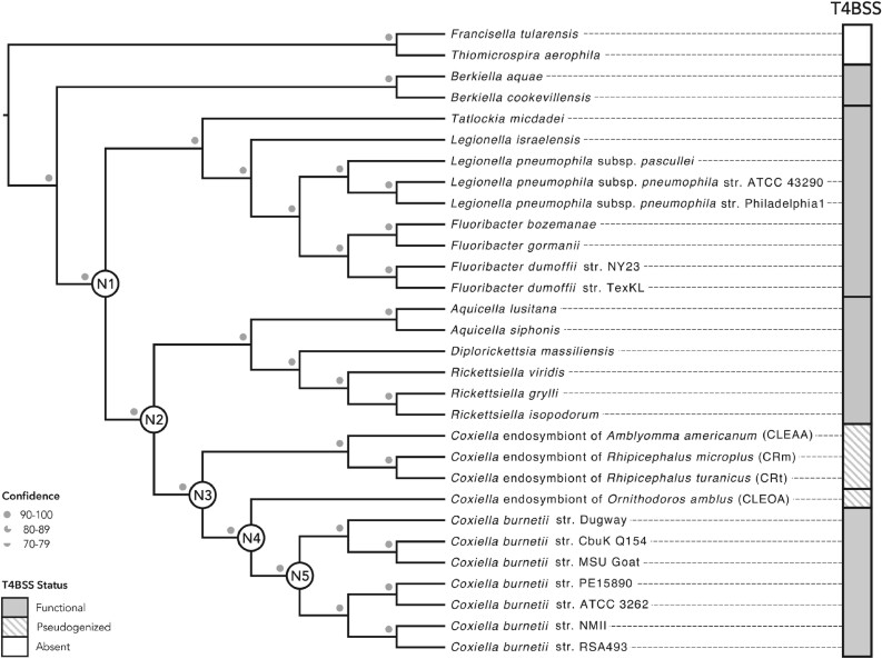
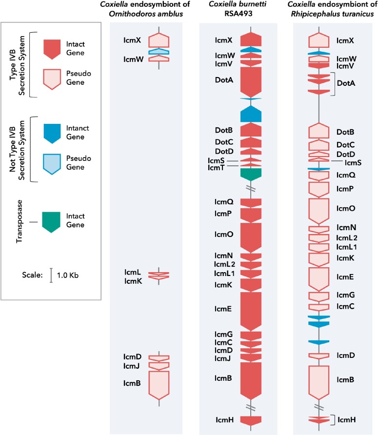
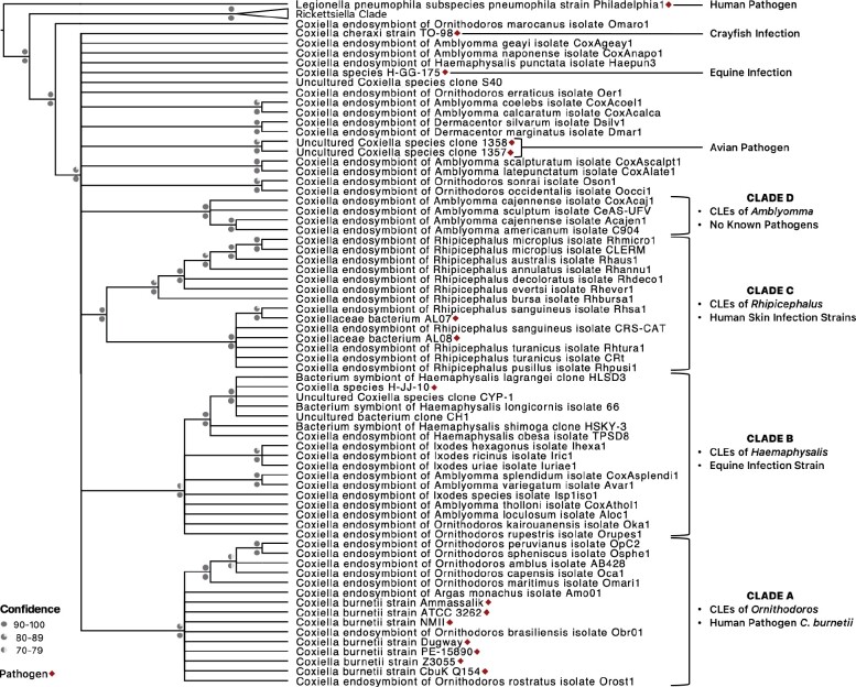
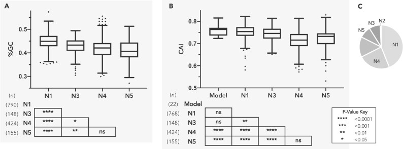
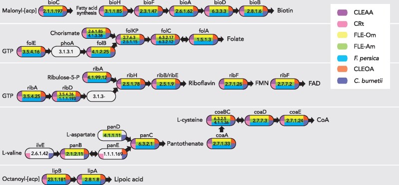
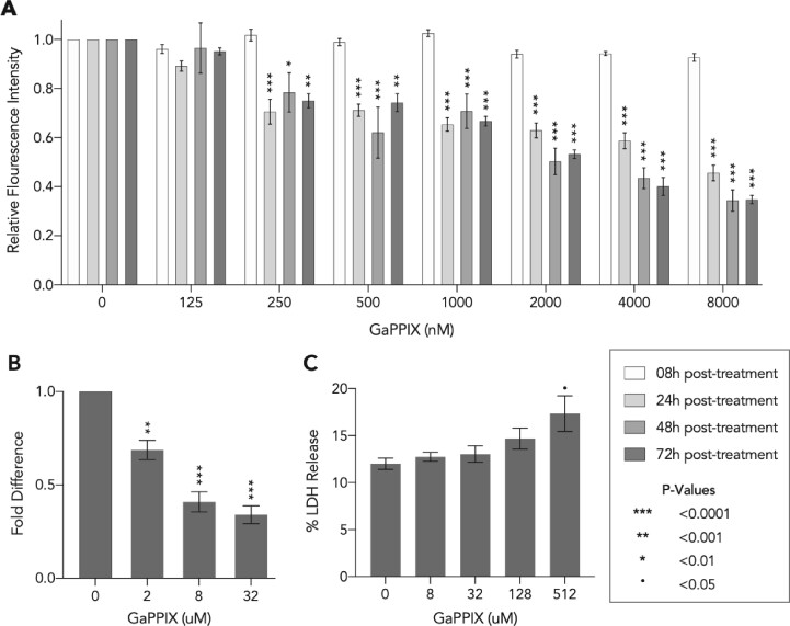
Both symbiotic and pathogenic bacteria in the family Coxiellaceae cause morbidity and mortality in humans and animals. For instance, Coxiella-like endosymbionts (CLEs) improve the reproductive success of ticks-a major disease vector, while Coxiella burnetii causes human Q fever, and uncharacterized coxiellae infect both animals and humans. To better understand the evolution of pathogenesis and symbiosis in this group of intracellular bacteria, we sequenced the genome of a CLE present in the soft tick Ornithodoros amblus (CLEOA) and compared it to the genomes of other bacteria in the order Legionellales. Our analyses confirmed that CLEOA is more closely related to C. burnetii, the human pathogen, than to CLEs in hard ticks, and showed that most clades of CLEs contain both endosymbionts and pathogens, indicating that several CLE lineages have evolved independently from pathogenic Coxiella. We also determined that the last common ancestorof CLEOA and C. burnetii was equipped to infect macrophages and that even though horizontal gene transfer (HGT) contributed significantly to the evolution of C. burnetii, most acquisition events occurred primarily in ancestors predating the CLEOA-C. burnetii divergence. These discoveries clarify the evolution of C. burnetii, which previously was assumed to have emerged when an avirulent tick endosymbiont recently gained virulence factors via HGT. Finally, we identified several metabolic pathways, including heme biosynthesis, that are likely critical to the intracellular growth of the human pathogen but not the tick symbiont, and show that the use of heme analog is a promising approach to controlling C. burnetii infections.

Keywords: Coxiella; endosymbiont; heme; pathogen; tick.


Figures

Fig. 1.
Fig. 1.
CLEOA is the closet relative of Coxiella burnetii. Maximum likelihood and Bayesian trees built using 117 single-copy protein-coding genes were combined to generate the shown phylogenomic tree. Bootstrap support and posterior probabilities agreed at all branchpoints and are depicted as a single confidence value. The Dot/Icm Type IVB secretion system (T4BSS), which is critical to pathogenesis, is found in all members of the order Legionellales, but has been pseudogenized in CLEs. Nodes N1–N5 mark major branching points in the evolution of C. burnetii.
Fig. 2.
Fig. 2.
CLEs contain nonfunctional T4BSS. Comparison of T4BSS genes in CLEOA, Coxiella burnetii RSA493 and CRt indicate that the secretion system has been rendered nonfunctional in tick endosymbionts. Filled blocks represent intact genes. Outlined blocks represent pseudogenized genes.
Fig. 3.
Fig. 3.
CLE clades contain both tick endosymbionts and pathogens. A 16S rDNA-based phylogenetic tree is shown. Bootstrap support and posterior probabilities are labeled above and below branchpoints, respectively. Nodes with ≤70% bootstrap support were collapsed to polytomies. Taxa colors represent the continent from which the host was derived. Established pathogens are marked with asterisks. Clades A–D were originally defined by Duron et al. (2015a).
Fig. 4.
Fig. 4.
HGT was a major contributor to gene accumulation in C. burnetii’s ancestors. Nodes N1–N5 (as labeled in fig. 1) represent major branchpoints in the evolution of C. burnetii. (A and B) depict %GC and Codon Adaptation Index (CAI) distributions, respectively, for genes originating at each node. Boxes illustrate each distribution’s interquartile range while the black line dividing the box represents the median. Whiskers represent minimum and maximum values, excluding outliers (black diamonds) which were determined using the Tukey method. P-values shown in tables are for pairwise T-tests (pooled SD, BKY adjusted). Genes gained at N2 were excluded due to small sample size (n =13). (C) Coxiella burnetii genome composition based on nodes of gene origin: N1: 43.9%, N2: 0.7%, N3: 8.2%, N4: 22.6%, N5: 8.6%. The unlabeled portion represents potentially spurious genes (n =224) as well as genes with undefined nodes of origin (n =44).
Fig. 5.
Fig. 5.
CLEs and FLEs encode B vitamin and cofactor biosynthetic pathways. CLEOA, CRt, CLEAA, FLE-Om (Francisella endosymbiont of Ornithodoros moubata; LVCE00000000), FLE-Am (Francisella endosymbiont of Amblyomma maculatum; LNCT00000000), and Francisella persica (FLE in Argas arboreus; NZ_CP013022) contain intact pathways for the synthesis of B vitamins and cofactors. Enzymes catalyzing each step are labeled with gene names and EC numbers. Blocks with no color indicate that a functional copy of a gene was not detected in that genome. Depictions of metabolic pathways modified from Gerhart et al. (2018).
Fig. 6.
Fig. 6.
A heme analog reduces Coxiella burnetii growth. (A) Bacteria growing in ACCM-2 were exposed to concentrations of GaPPIX shown in x-axis and were quantified using PicoGreen at 8 h, 24 h, 48 h, and 72 h post-treatment. Data shown are mean fluorescence intensity (± SE) compared to the vehicle control (0 nM). Statistical significance was analyzed using two-way repeated measures ANOVA followed by Dunnett's test (n = 5). (B) At 72 h post-treatment, bacterial growth within THP-1 cells was quantified using qPCR. Data shown as mean fold difference (±SE) compared to control. (C) At 24 h post-GaPPIX treatment of THP-1 cells, lactate dehydrogenase (LDH) activity was determined by measuring the level of resorufin formation using an LDH cytotoxicity assay. The cytotoxicity was reported as the percentage LDH released compared to the maximum LDH activity. Data shown as mean percentage LDH released (±SEM). For both (B and C), statistical significance was analyzed using one-way ANOVA followed by Dunnett's test (n = 3).

Similar articles

Cited by

References

    1. Albertsen M, et al. 2013. Genome sequences of rare, uncultured bacteria obtained by differential coverage binning of multiple metagenomes. Nat Biotechnol. 31(6):533–538. - PubMed
    1. Almeida AP, et al. 2012. Coxiella symbiont in the tick Ornithodoros rostratus (Acari: argasidae). Ticks Tick Borne Dis. 3(4):203–206. - PubMed
    1. Alneberg J, et al. 2014. Binning metagenomic contigs by coverage and composition. Nat Methods. 11(11):1144–1146. - PubMed
    1. Angelakis E, et al. 2016. Candidatus Coxiella massiliensis infection. Emerg Infect Dis. 22(2):285–288. - PMC - PubMed
    1. Arivett BA, et al. 2015. Antimicrobial activity of gallium protoporphyrin IX against Acinetobacter baumannii strains displaying different antibiotic resistance phenotypes. Antimicrob Agents Chemother. 59(12):7657–7665. - PMC - PubMed

Publication types

LinkOut - more resources