

Eosinophilic Vacuolated Tumor of the Kidney: A Review of Evolving Concepts in This Novel Subtype With Additional Insights From a Case With MTOR Mutation and Concomitant Chromosome 1 Loss
- PMID: 34009776
- PMCID: PMC8205969
- DOI: 10.1097/PAP.0000000000000299
Eosinophilic Vacuolated Tumor of the Kidney: A Review of Evolving Concepts in This Novel Subtype With Additional Insights From a Case With MTOR Mutation and Concomitant Chromosome 1 Loss
Abstract
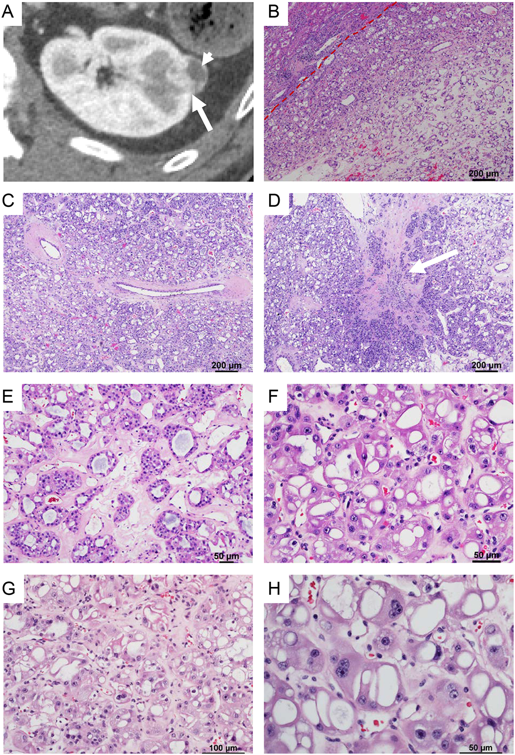
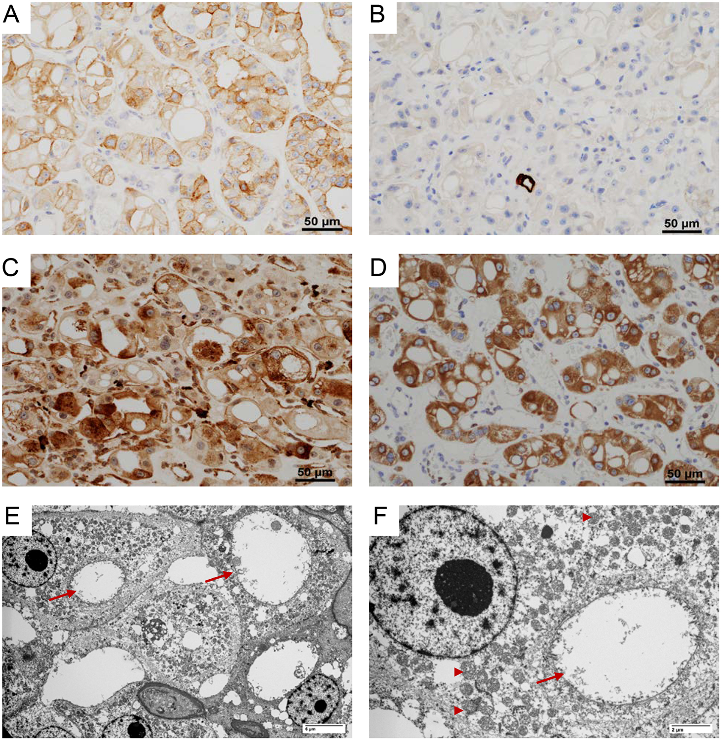
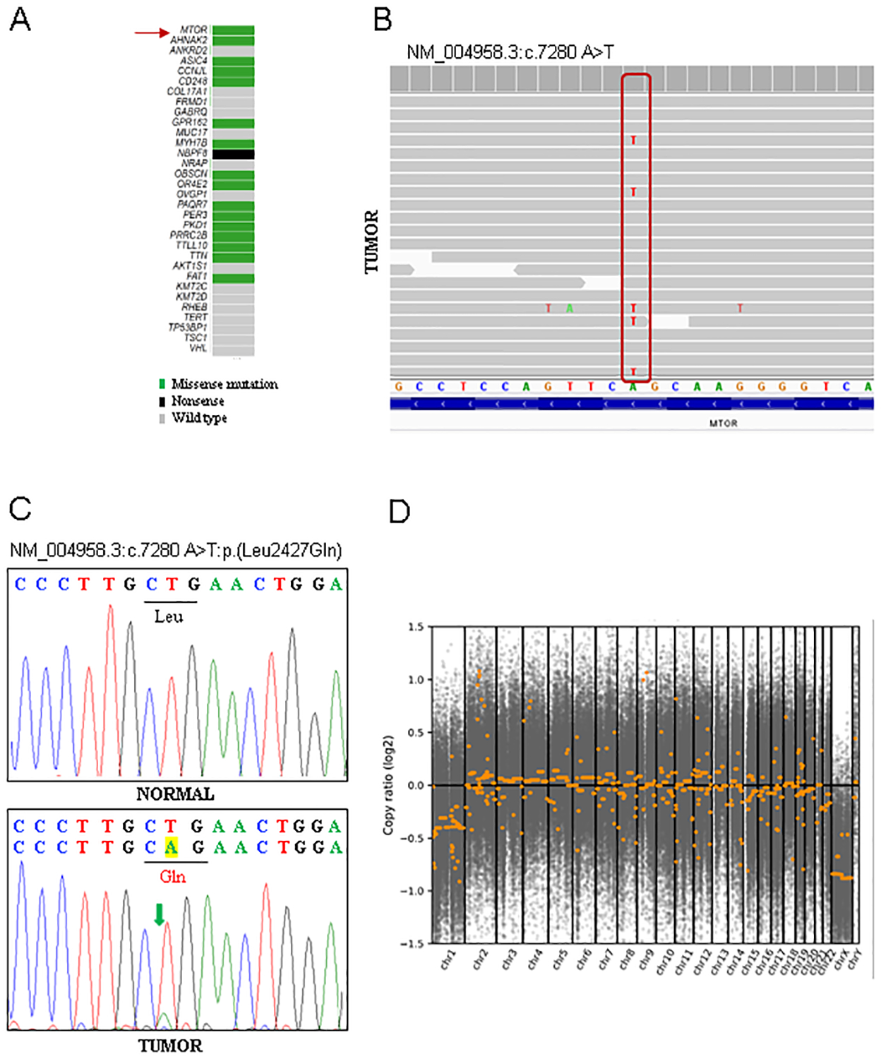
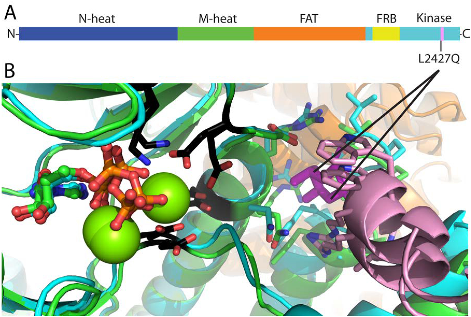
Recent advances in molecular genetics have expanded our knowledge of renal tumors and enabled a better classification. These studies have revealed that renal tumors with predominantly "eosinophilic/oncocytic" cytoplasm include several novel biological subtypes beyond the traditionally well-recognized renal oncocytoma and an eosinophilic variant of chromophobe renal cell carcinoma. Herein, we present a comprehensive review of the eosinophilic vacuolated tumor (EVT) building upon a case report including radiology, histopathology, electron microscopy, and next-generation sequencing. EVTs are characterized by mTORC1 activation. We speculate that loss of chromosome 1 in EVT with MTOR mutation may be driven in part by an advantage conferred by loss of the remaining MTOR wild-type allele. mTORC1 is best known for its role in promoting protein translation and it is interesting that dilated cisterns of rough endoplasmic reticulum (ER) likely account for the cytoplasmic vacuoles seen by light microscopy. We present an integrated view of EVT as well as cues that can assist in the differential diagnosis.
Copyright © 2021 Wolters Kluwer Health, Inc. All rights reserved.
Conflict of interest statement
The authors have no conflicts of interest to disclose.
Figures
References
-
- McKenney JK, Przybycin CG, Trpkov K, Magi-Galluzzi C. Eosinophilic solid and cystic renal cell carcinomas have metastatic potential. Histopathology. 2018;72(6):1066–1067. - PubMed
-
- Trpkov K, Hes O, Bonert M, et al. Eosinophilic, Solid, and Cystic Renal Cell Carcinoma: Clinicopathologic Study of 16 Unique, Sporadic Neoplasms Occurring in Women. Am J Surg Pathol. 2016;40(1):60–71. - PubMed
-
- Trpkov K, Bonert M, Gao Y, et al. High-grade oncocytic tumour (HOT) of kidney in a patient with tuberous sclerosis complex. Histopathology. 2019;75(3):440–442. - PubMed
-
- He H, Trpkov K, Martinek P, et al. “High-grade oncocytic renal tumor”: morphologic, immunohistochemical, and molecular genetic study of 14 cases. Virchows Arch. 2018;473(6):725–738. - PubMed
Publication types
MeSH terms
Substances
Grants and funding
LinkOut - more resources
Full Text Sources
Other Literature Sources
Medical
Miscellaneous
