Case Reports
. 2021 Jun 3;108(6):1115-1125.
doi: 10.1016/j.ajhg.2021.04.019. Epub 2021 May 18.

A human importin-β-related disorder: Syndromic thoracic aortic aneurysm caused by bi-allelic loss-of-function variants in IPO8

Affiliations
Case Reports

A human importin-β-related disorder: Syndromic thoracic aortic aneurysm caused by bi-allelic loss-of-function variants in IPO8

Ilse Van Gucht et al. Am J Hum Genet. .

Abstract

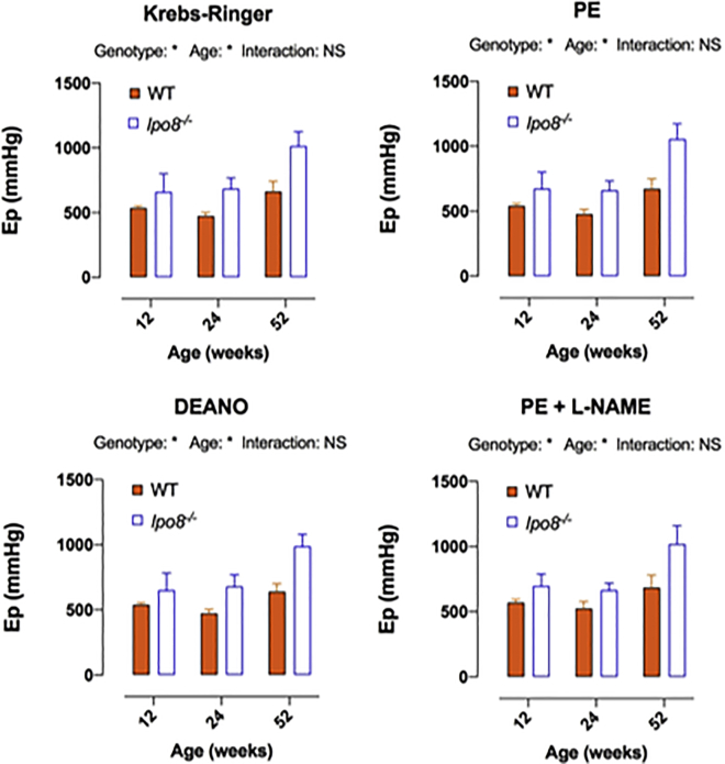
Importin 8, encoded by IPO8, is a ubiquitously expressed member of the importin-β protein family that translocates cargo molecules such as proteins, RNAs, and ribonucleoprotein complexes into the nucleus in a RanGTP-dependent manner. Current knowledge of the cargoes of importin 8 is limited, but TGF-β signaling components such as SMAD1-4 have been suggested to be among them. Here, we report that bi-allelic loss-of-function variants in IPO8 cause a syndromic form of thoracic aortic aneurysm (TAA) with clinical overlap with Loeys-Dietz and Shprintzen-Goldberg syndromes. Seven individuals from six unrelated families showed a consistent phenotype with early-onset TAA, motor developmental delay, connective tissue findings, and craniofacial dysmorphic features. A C57BL/6N Ipo8 knockout mouse model recapitulates TAA development from 8-12 weeks onward in both sexes but most prominently shows ascending aorta dilatation with a propensity for dissection in males. Compliance assays suggest augmented passive stiffness of the ascending aorta in male Ipo8-/- mice throughout life. Immunohistological investigation of mutant aortic walls reveals elastic fiber disorganization and fragmentation along with a signature of increased TGF-β signaling, as evidenced by nuclear pSmad2 accumulation. RT-qPCR assays of the aortic wall in male Ipo8-/- mice demonstrate decreased Smad6/7 and increased Mmp2 and Ccn2 (Ctgf) expression, reinforcing a role for dysregulation of the TGF-β signaling pathway in TAA development. Because importin 8 is the most downstream TGF-β-related effector implicated in TAA pathogenesis so far, it offers opportunities for future mechanistic studies and represents a candidate drug target for TAA.

Keywords: Loeys-Dietz syndrome; Shprintzen-Goldberg syndrome; TGF-beta; importin 8; knockout mouse model; thoracic aortic aneurysm.


Conflict of interest statement

The Department of Molecular and Human Genetics at Baylor College of Medicine receives revenue from clinical genetic testing conducted at Baylor Genetics Laboratories. A.B. is an employee of GeneDx, Inc.

Figures

Figure 1
Figure 1
Familial screening and clinical characterization of individuals with bi-allelic IPO8 variants (A) Pedigrees of the families with their respective pathogenic variants. Squares represent males, while circles represent females, filled symbols denote affected individuals, a double line connecting spouses symbolizes consanguinity, and m/m1/m2 denote the presence of the respective IPO8 variants and a + sign represents the wild-type IPO8 allele. Variants are annotated against GenBank: NM_006390.3. (B) Clinical phenotyping. Proband 1-II:3 showing prominent forehead, hypertelorism, mild ptosis left eye, retrognathia, pectus excavatum, umbilical hernia, joint hypermobility with thumb abduction, and camptodactyly of the second toe. CT angiography of proband 2-II:1 demonstrating dilatation of the common carotid arteries along with marked tortuosity of the common carotid and internal carotid artery, mild tortuosity of the vertebral arteries, and enlargement of the anterior and middle cerebral arteries bilaterally. Proband 3-II:3 presenting with frontal bossing with bitemporal flattening, retrognathia, downturned corners of the mouth, and flat feet. Proband 5-II:2 showing prominent forehead, significant hypertelorism with flat nasal bridge, mild ptosis of left eye, and retrognathia. Proband 6-II:1 demonstrating dolichocephaly, retrognathia, malar flattening, downslanting palpebral fissures, and hypertelorism. Magnetic resonance angiography (MRA) revealing tortuous intracranial and extracranial arterial vessels, most prominently involving the superior cervical internal carotid arteries with dilation of the left internal carotid artery at the carotid bifurcation. CT scan (pre-surgical) showing os odontoideum with cervical spinal canal stenosis (arrows).
Figure 2
Figure 2
Progressive TAA development in Ipo8−/− mice (A) Log of weight-corrected aortic root diameters in male and female mice combined (N = 17/group). (B) Log of weight-corrected ascending aortic diameters in male and female mice combined (N = 17/group). (C) Log of weight-corrected ascending aortic diameters in male mice only (10 Ipo8−/− versus 9 WT). The error bars show the standard error of the mean (SEM). p values, which represent the interaction term between genotype and age, were calculated via mixed model analysis. WT, wild-type.
Figure 3
Figure 3
Trend toward increased ascending aortic passive stiffness in Ipo8−/− mice at a distention pressure of 120–160 mmHg Age- and genotype-dependency of the Peterson modulus (Ep) of ascending aortic segments of male Ipo8−/− and wild-type mice under control (Krebs-Ringer), maximally relaxed (DEANO), and contracted (PE or PE + L-NAME) conditions at 12 (5 Ipo8−/− versus 4 WT), 24 (4 Ipo8−/− versus 4 WT), and 52 (4 Ipo8−/− versus 2 WT) weeks of age. The error bars show the SEM. Two-way ANOVA p values are shown (p < 0.05). Sidak post hoc testing did not reveal statistically significant genotype-based differences in Ep. PE, phenylephrine; DEANO, diethylamine NONOate; L-NAME, N(Ω)-nitro-L-arginine methyl ester; Ep, Peterson modulus; WT, wild-type; NS, non-significant.
Figure 4
Figure 4
Elastic fiber deterioration and nuclear pSmad2 accumulation in the ascending aorta of Ipo8−/− mice (A) Histological and immunohistochemistry images demonstrating marked elastin disorganization and fragmentation as well as prominent nuclear pSmad2 accumulation in Ipo8−/− mice. Scale bar represents 50 μm. (B) Elastic fiber integrity scores and nuclear pSmad2 grades of the ascending aorta of all ages combined (12 [3 Ipo8−/− versus 3 WT], 24 [3 Ipo8−/− versus 3 WT], and 52 weeks [3 Ipo8−/− versus 2 WT]). Elastin grades can range from 1 to 4: grade 1 sections present with continuous and well-organized elastic bundles and grade 4 sections display vastly disorganized fibers, marked fiber fragmentation, and a thickened aortic wall. For pSmad2, grades 1, 2, 3, and 4 denote sections in which respectively <25%, 25%–50%, 50%–75%, and 75%–100% of nuclei stained positive. Averaged age-combined scores of blinded observations of three independent researchers are shown. The error bars depict the SEM. p values were calculated via two-way ANOVA statistics (p < 0.05, ∗∗∗p < 0.001). WT, wild-type.
Figure 5
Figure 5
mRNA expression analysis of TGF-β-related genes reveals decreased Smad6 and Smad7 levels as well as increased Mmp2 and Ccn2 (Ctgf) levels in the ascending aorta of Ipo8−/− mice Ascending aortic samples of 16-week-old Ipo8−/− and WT males were used (N = 12/group). The error bars depict the SEM. p values were calculated via mixed model statistics (p < 0.05, ∗∗p < 0.01). WT, wild-type; NS, non-significant.

References

    1. Faggion Vinholo T., Brownstein A.J., Ziganshin B.A., Zafar M.A., Kuivaniemi H., Body S.C., Bale A.E., Elefteriades J.A. Genes Associated with Thoracic Aortic Aneurysm and Dissection: 2019 Update and Clinical Implications. Aorta (Stamford) 2019;7:99–107. - PMC - PubMed
    1. Verstraeten A., Luyckx I., Loeys B. Aetiology and management of hereditary aortopathy. Nat. Rev. Cardiol. 2017;14:197–208. - PubMed
    1. Dietz H.C., Cutting G.R., Pyeritz R.E., Maslen C.L., Sakai L.Y., Corson G.M., Puffenberger E.G., Hamosh A., Nanthakumar E.J., Curristin S.M. Marfan syndrome caused by a recurrent de novo missense mutation in the fibrillin gene. Nature. 1991;352:337–339. - PubMed
    1. Loeys B.L., Chen J., Neptune E.R., Judge D.P., Podowski M., Holm T., Meyers J., Leitch C.C., Katsanis N., Sharifi N. A syndrome of altered cardiovascular, craniofacial, neurocognitive and skeletal development caused by mutations in TGFBR1 or TGFBR2. Nat. Genet. 2005;37:275–281. - PubMed
    1. Cannaerts E., Kempers M., Maugeri A., Marcelis C., Gardeitchik T., Richer J., Micha D., Beauchesne L., Timmermans J., Vermeersch P. Novel pathogenic SMAD2 variants in five families with arterial aneurysm and dissection: further delineation of the phenotype. J. Med. Genet. 2019;56:220–227. - PubMed

Publication types

MeSH terms