. 2021 May 18;35(7):109147.
doi: 10.1016/j.celrep.2021.109147.

GluA2 overexpression in oligodendrocyte progenitors promotes postinjury oligodendrocyte regeneration

Affiliations

GluA2 overexpression in oligodendrocyte progenitors promotes postinjury oligodendrocyte regeneration

Rabia R Khawaja et al. Cell Rep. .

Abstract

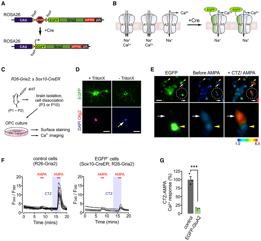
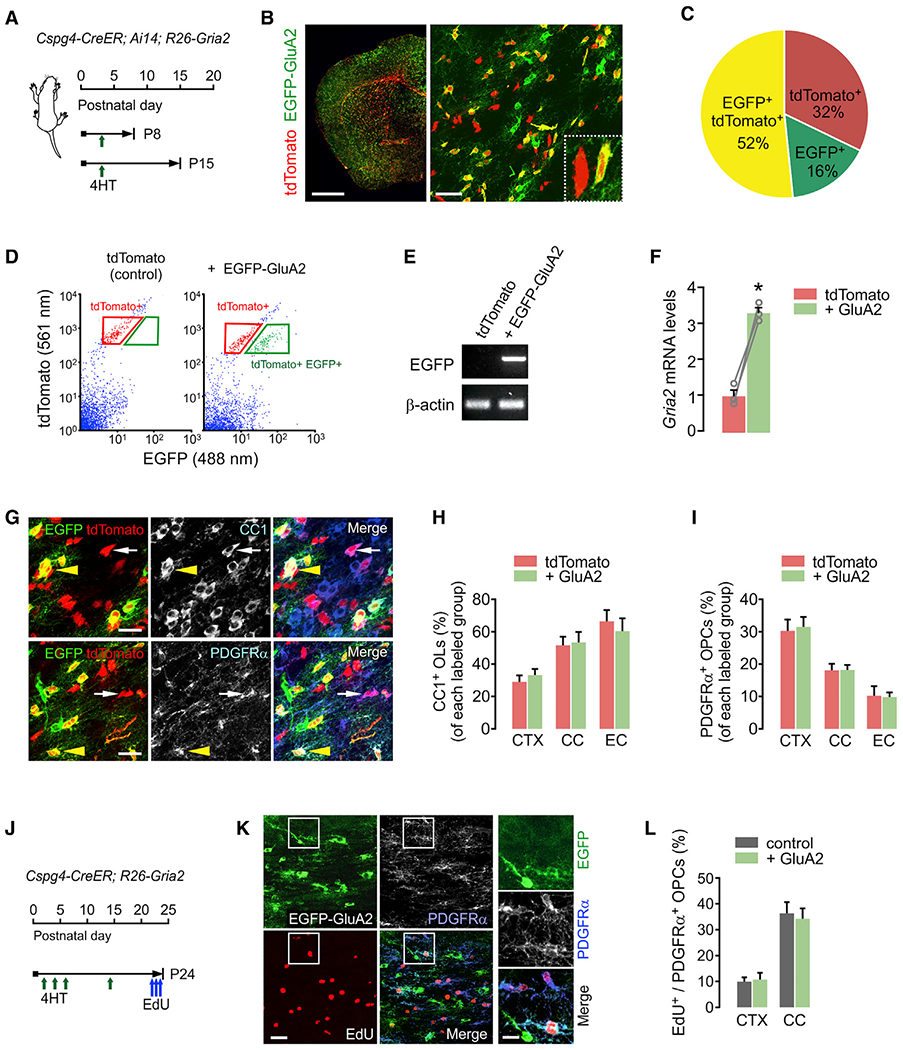
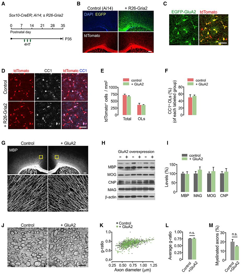
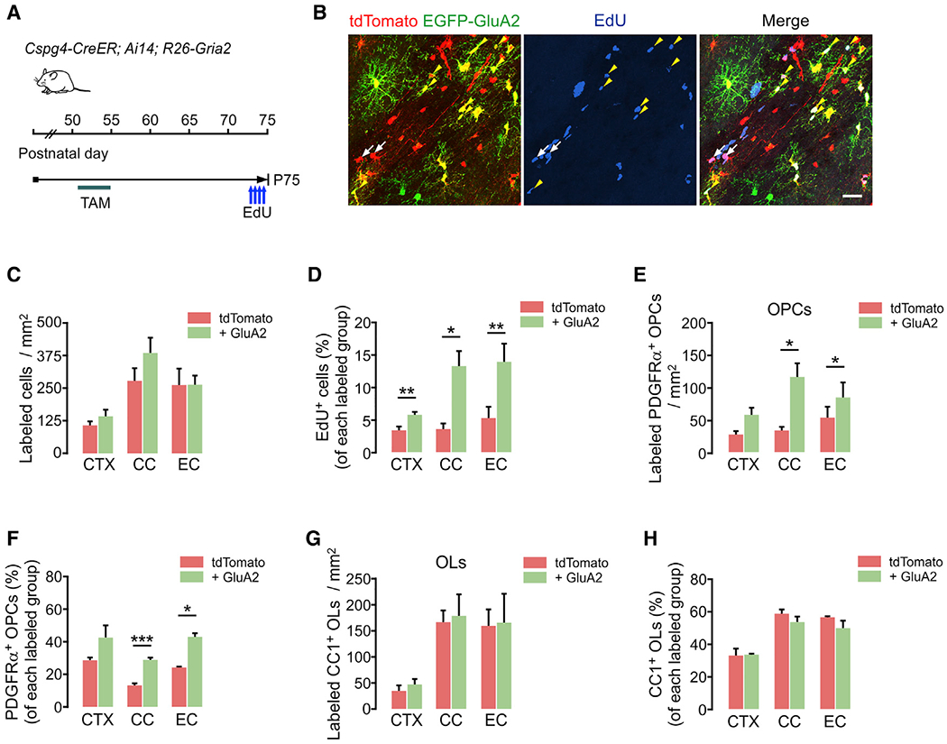
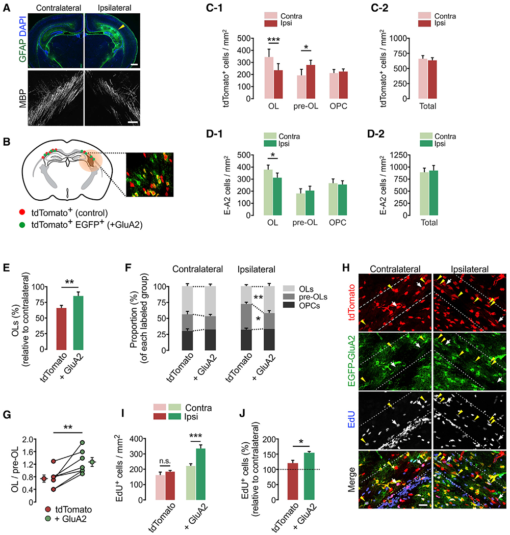
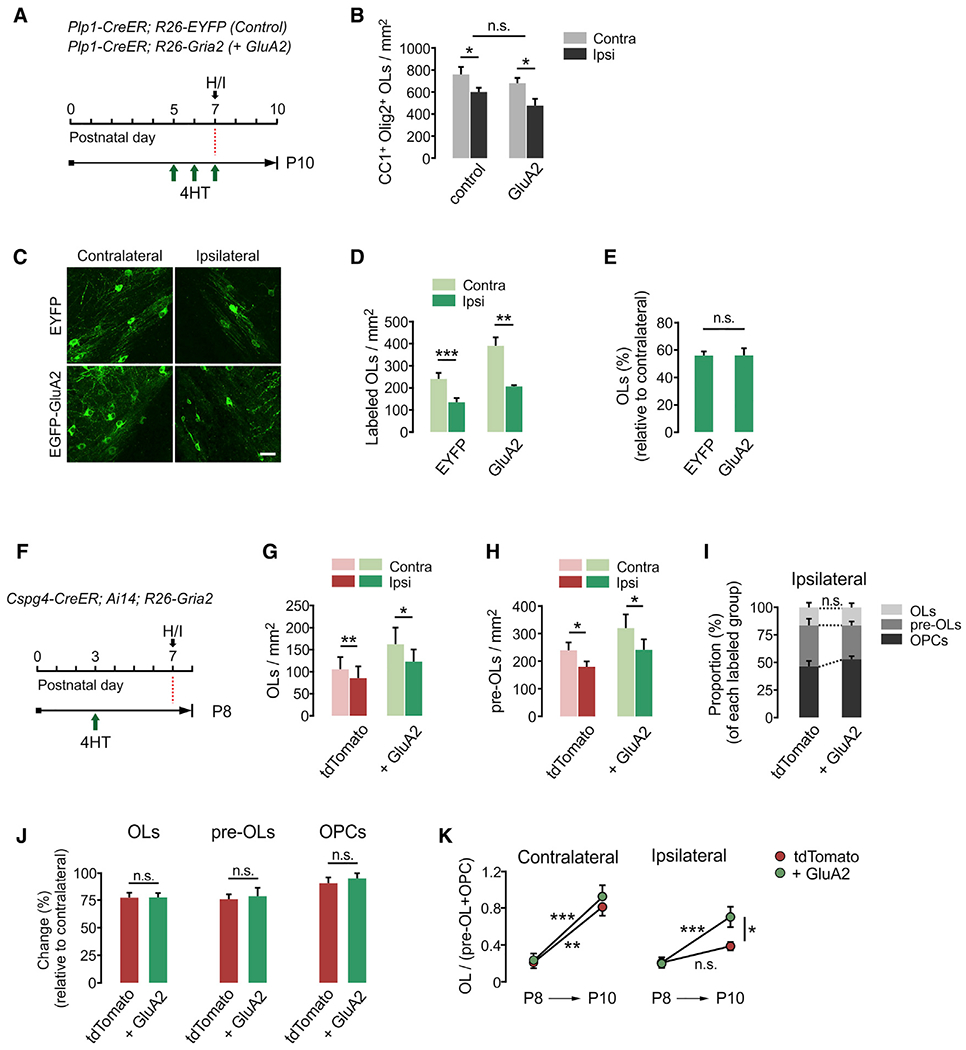
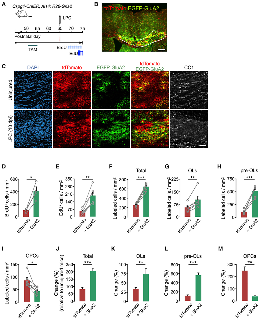
Oligodendrocyte precursor cells (OPCs) are essential for developmental myelination and oligodendrocyte regeneration after CNS injury. These progenitors express calcium-permeable AMPA receptors (AMPARs) and form direct synapses with neurons throughout the CNS, but the roles of this signaling are unclear. To enable selective alteration of the properties of AMPARs in oligodendroglia, we generate mice that allow cell-specific overexpression of EGFP-GluA2 in vivo. In healthy conditions, OPC-specific GluA2 overexpression significantly increase their proliferation in an age-dependent manner but did not alter their rate of differentiation into oligodendrocytes. In contrast, after demyelinating brain injury in neonates or adults, higher GluA2 levels promote both OPC proliferation and oligodendrocyte regeneration, but do not prevent injury-induced initial cell loss. These findings indicate that AMPAR GluA2 content regulates the proliferative and regenerative behavior of adult OPCs, serving as a putative target for better myelin repair.

Keywords: AMPA receptor; GluA2; OPC; calcium; hypoxic-ischemia; injury; oligodendrocyte; remyelination.


Conflict of interest statement

Declaration of interests The authors declare no competing interests.

Figures

Figure 1.
Figure 1.. Cell-specific GluA2 overexpression disrupts AMPA-dependent calcium response in OPCs
(A) Diagram of CAG-loxP-STOP-loxP-EGFP-Gria2 transgene placed on the Rosa26 locus. (B) Expected outcomes after insertion of overexpressed EGFP-GluA2 into the cell membrane. Presence of calcium-impermeable of GluA2 in AMPAR complexes may block calcium entry through AMPARs, regardless of subunit composition. A1–A4 are GluA1–GluA4, respectively. (C) Illustration of experiments for EGFP-GluA2 validation. After 4HT injections (0.2 mg per subcutaneous [s.c.] injection, at P1 and P2), R26-Gria2 ± Sox10-CreER mice were killed at P3 (calcium imaging) or P10 (immunopanning and surface detection of EGFP). (D) Immunodetection of EGFP expressed on the cell surface. OPCs were isolated via immunopanning from 4HT-administered Sox10-CreER;R26-Gria2 mice and stained for EGFP and Olig2 with or without Triton X-100 for permeabilization. After secondary antibody probing, cells were permeabilized for DAPI staining. White arrow highlights no Olig2 detection. Scale bars: 25 μm. (E) Upper panel: images of cultured EGFP+ OPCs (left) and their calcium responses before (center) and after CTZ (100 μM) + AMPA (20 μM) (right). Lower panel: enlarged views of 2 adjacent cells (EGFP+ and EGFP OPCs) in the circled area of the upper panel. Yellow arrowheads and white arrows indicate EGFP+ and EGFP cells, respectively. Scale bar: 50μm. (F) Traces of the calcium responses of individual cells as ratios of fluorescence collected after excitation at 340 and 380 nm. Red bars and shaded regions represent durations of AMPA (1 min) and CTZ (2.5 min) applications, respectively. Calcium responses of OPCs collected from an R26-Gria2 (left) and EGFP+ OPCs from a Sox10-CreER; R26-Gria2 mouse (right). (G) EGFP-GluA2 attenuates CTZ + AMPA-elicited calcium responses in OPCs. Peak responses averaged over all cells for each coverslip. n = 3 coverslips per group. Data are represented as means ± SEMs. Unpaired Student’s t test. ***p < 0.001. See also Figures S1 and S2.
Figure 2.
Figure 2.. GluA2 overexpression does not change OPC proliferation or differentiation into OLs in early life
(A) Timelines for 4HT injection and mouse sampling. Cspg4-CreER; Ai14; R26-Gria2 triple Tg pups received a single 4HT injection (0.2 mg, s.c.) at P3 and were sampled at either P8 or P15. (B) Fluorescence (left) and confocal (right) images of tdTomato or EGFP-expressing cells in the brain (left: P15; right: P8). Right panel inset: 2 cells expressing tdTomato alone (control) or both tdTomato and EGFP-GluA2 (E-A2 cell), in the external capsule (EC). Scale bars: 1 mm (left), 50 μm (right). (C) Pie chart for cells expressing tdTomato, EGFP, or both tdTomato and EGFP in the CC at P8 (P3+5). n = 4 mice. (D) Scatterplot of flow cytometry and gating strategy for isolation of tdTomato+ EGFP and E-A2 cells. The tdTomato+ cells from an age-matched Cspg4-CreER; Ai14 littermate (P3+12) were used as the gating control (left). (E) RT-PCR of EGFP with the isolated cells. The absence or presence of EGFP mRNA confirmed the effective separation of control and E-A2 cells. (F) Relative mRNA levels of Gria2 in tdTomato+ and E-A2 (+ GluA2) cells isolated from the same brain. n = 3 mice. Paired Student’s t test. *p < 0.05. (G) Confocal images of tdTomato+ CC1+ OLs (top panel) and tdTomato+ PDGFRα+ OPCs (bottom panel) with or without EGFP-GluA2 expression. White arrows and yellow arrowheads indicate tdTomato+ EGFP (control) and E-A2 cells, respectively. Scale bars: 25 μm. (H and I) Percentages of OLs (H) and OPCs (I) among tdTomato+ EGFP cells (tdTomato) and E-A2 cells (+ GluA2) in the CTX, CC, and EC. n = 5 mice. Paired Student’s t test. (H) Control versus + GluA2 group, CTX: p = 0.227; CC: p = 0.498; EC: p = 0.102. (I) CTX: p = 0.776; CC: p = 0.957; EC: p = 0.895. (J) Timeline for injections of 4HT and EdU, and mouse sampling. Cspg4-CreER; R26-Gria2 mice received 4HT at P2, P4, P6, and P14, and 3 injections of EdU between P22 and P23, before sampling at P24. (K) Confocal images of EdU+ PDGFRα+ OPCs in a Cspg4-CreER; R26-Gria2 mouse (P24). Right panels: enlargement of the left panel insets. Scale bars: 25 μm (left) and 10 μm (right). (L) Percentage of EdU+ cells among EGFP (control) and EGFP+ OPCs (+ GluA2). Paired Student’s t test (control versus + GluA2 group, CTX: p = 0.305, n = 4 mice; CC: p = 0.613, n = 5 mice). Data are represented as means ± SEMs.
Figure 3.
Figure 3.. No overt changes in myelination after GluA2 overexpression
(A) Timeline for 4HT injections and mouse sampling. Sox10-CreER; Ai14 (control) and Sox10-CreER; Ai14; R26-Gria2 (+ GluA2) mice received 4HT at P9, P11, and P13 (0.2 mg per s.c. injection) and killed at P35. (B) Fluorescence images showing widespread tdTomato expression with or without EGFP-GluA2. Scale bar: 200 μm. (C) Confocal images of tdTomato and EGFP in the CTX of a Sox10-CreER; Ai14; R26-Gria2 mouse. Scale bar: 50 μm. (D) Confocal images of cortical tdTomato+ CC1+ OLs. Scale bar: 50 μm. (E) Densities of all tdTomato+ cells and tdTomato+ CC1+ OLs in the CTX (control versus + GluA2 group, total tdTomato+ cells: p = 0.246; tdTomato+CC1+ OLs: p = 0.972). (F) Percentage of CC1+ OLs among tdTomato+ cells. n = 5 (Sox10-CreER; Ai14, control) and 4 (Sox10-CreER; Ai14; R26-Gria2, + GluA2) mice for (E and F). (G) Fluorescence and confocal images of MBP in R26-Gria2 (control) and Sox10-CreER; R26-Gria2 (+ GluA2) mice. Upper panels: boxes correspond to confocal images in lower panels (optical thickness = 7 μm). Scale bars: 200 μm (up) and 50 μm (bottom). (H and I) Western blot analysis of myelin proteins from cortical lysates of R26-Gria2 and Sox10-CreER; R26-Gria2 mice (H) and its quantification (I). Control versus + GluA2 group, MBP: p = 0.877; MAG: p = 0.337; MOG: p = 0.611; CNP: p = 0.636. n = 3 mice per group. (J) Representative electron micrographs of callosal axons. Scale bar: 2 μm. (K) Scatterplots of g-ratios. More than 150 myelinated axons per mouse were analyzed. (L and M) Average g-ratio (L, control versus + GluA2 group, p = 0.880) and percentage of myelinated axons (M, control versus + GluA2 group. p = 0.12). For quantification of the percentage of myelinated axons, 700–900 axons were analyzed per mouse. n = 3 mice per group. Data are represented as means ± SEMs. Unpaired Student’s t test for (E), (F), (I), (L), and (M). See also Figure S1.
Figure 4.
Figure 4.. GluA2 overexpression increases OPC proliferation in the adult brain
(A) Timeline for tamoxifen and EdU injections and sampling of Cspg4-CreER; Ai14; R26-Gria2 mice. Tamoxifen was injected (40 mg/kg per i.p. injection) 10 times between P51 and P55, and mice were sampled at P75. EdU was injected 4 times over 2 days before sampling. (B) Confocal images of EdU-incorporated tdTomato+ or E-A2 cells in EC. White arrows and yellow arrowheads indicate EdU+ tdTomato+ EGFP (control) and EdU+ E-2A (+ GluA2) cells, respectively. Scale bar: 25 μm. (C) Densities of tdTomato+ (control) and E-2A (+ GluA2) cells (control versus + GluA2 group, CTX: p = 0.11; CC: p = 0.26; EC: p = 0.99). (D) Percentages of EdU+ cells among tdTomato+ (control) and E-A2 cells (+ GluA2) (control versus + GluA2 group, CTX: p = 0.0084; CC: p = 0.021; EC: p = 0.0091). (E) Densities of tdTomato+ PDGFRα+ OPCs with or without EGFP-GluA2 expression (control versus + GluA2 group, CTX: p = 0.069; CC: p = 0.048; EC: p = 0.047). (F) Percentages of OPCs among tdTomato+ (control) and E-A2 cells (+ GluA2) (control versus + GluA2 group, CTX: p = 0.18; CC: p = 0.0009; EC: p = 0.017). (G) Densities of tdTomato+ CC1+ OLs with or without EGFP-GluA2 expression (control versus + GluA2 group, CTX: p = 0.41; CC: p = 0.84; EC: p = 0.93). (H) Percentages of OLs among tdTomato+ (control) and E-A2 cells (+ GluA2) (control versus + GluA2 group, CTX: p = 0.89; CC: p = 0.25; EC: p = 0.23). Data are represented as means ± SEMs, n = 4 mice (C–H). Paired Student’s t test for (C–H). *p < 0.05, **p < 0.01, and ***p < 0.001.
Figure 5.
Figure 5.. GluA2 overexpression in OPCs promotes post-injury OL development and cell proliferation in the young brain
(A) Fluorescence brain images showing astrogliosis and loss of MBP on the injured side, ipsilateral to ligated artery, at 3 dpi. Scale bars: 500 μm (top) and 50 μm (bottom). (B) Schematic diagram of mosaic analysis of tdTomato+ control (red dots) and E-A2 cells (+ GluA2, green dots) in the injured EC (shaded) in Cspg4-CreER; Ai14; R26-Gria2 mice. Inset: confocal image of tdTomato and EGFP at P10. (C-1) Differential changes in density of tdTomato+ OL lineage cells according to maturation stage after H/I injury (contralateral versus ipsilateral side, OL: p = 0.0004, n = 11 mice; pre-OL: p = 0.0172, n = 8 mice; OPCs: p = 0.434, n = 9 mice). (C-2) Total tdTomato+ cells in the EC at 3 dpi (P10). Contralateral versus ipsilateral side, p = 0.461; n = 11 mice. (D-1) H/I injury-induced changes in densities of E-A2 cells according to maturation stage. Contralateral versus ipsilateral side, OLs: p = 0.031, n = 11 mice; pre-OLs: p = 0.436, n = 8 mice; OPCs: p = 0.561, n = 9 mice. (D-2) Total E-A2 cells in the EC at 3 dpi (P10). Contralateral versus ipsilateral side, p = 0.552, n = 11 mice. (E) Loss of tdTomato+ control and E-A2 (+ GluA2) OLs on the ipsilateral (injured) relative to the contralateral side (control versus + GluA2 group, p = 0.009, n = 11 mice). (F) Proportion of OLs, pre-OLs and OPCs among labeled cells (control versus + GluA2 group, ipsilateral OLs: p = 0.0097; ipsilateral pre-OLs: p = 0.019, n = 8 mice). (G) Ratios of (OL to pre-OL) on ipsilateral side of each mouse. Separate circles with error bars represent means ± SEM for each group (control versus + GluA2 group, p = 0.006, n = 7 mice). (H) Confocal images of EdU+ cells in H/I-injured Cspg4-CreER; Ai14; R26-EGFP-Gria2 mice at P10. White arrows and yellow arrowheads indicate EdU+ tdTomato+ EGFP (control) and EdU+ E-A2 (+ GluA2) cells, respectively. Scale bar: 25 μm. (I) Densities of EdU+ cells. Contralateral versus ipsilateral side, control: p = 0.181; + GluA2: p = 0.0005, n = 5 mice. (J) Percentages of EdU+ cells in the ipsilateral EC relative to the contralateral EC (control versus + GluA2 group, p = 0.041, n = 5 mice). Data are represented as means ± SEMs. Paired Student’s t test for (E), (G), (I), (J). *p < 0.05, **p < 0.01, and ***p < 0.001. See also Figures S3 and S4.
Figure 6.
Figure 6.. Beneficial effects of GluA2 overexpression become evident through post-injury OL development
(A) Timeline for 4HT injection, H/I injury, and mouse sampling. Plp1-CreER; R26-EYFP (control) and Plp1-CreER; R26-Gria2 (GluA2) mice received daily 4HT injections (0.2 mg per s.c. injection) between P5 and P7, before injury induction. (B) Densities of CC1+ Olig2+ OLs in EC at 3 dpi. Contralateral versus ipsilateral side, control: p = 0.019; GluA2: p = 0.016. Paired Student’s t test. n = 4 (Plp1-CreER; R26-EYFP) and 5 (Plp1-CreER; R26-Gria2) mice. (C) Confocal images of OLs expressing EYFP or EGFP-GluA2 at 3 dpi. Scale bar: 25 μm. (D) Densities of EYFP (or EGFP-GluA2)-labeled CC1+ OLs in EC. Contralateral versus ipsilateral side, EYFP: p = 0.004; GluA2: p = 0.005. Paired Student’s t test. (E) Percentage losses of EYFP (or EGFP-GluA2)-labeled OLs in the ipsilateral EC relative to the contralateral (EYFP versus GluA2, p = 0.987). Unpaired Student’s t test. n = 5 (Plp1-CreER; R26-EYFP) and 6 (Plp1-CreER; R26-Gria2) mice for (D) and (E). (F) Timeline for 4HT injection, H/I injury, and sampling of Cspg4-CreER; Ai14; R26-Gria2 mice. Mice were sampled at 1 dpi (P8) to examine early cell loss after injury. (G and H) Densities of fluorescent protein-labeled CC1+ OLs (G) (contralateral versus ipsilateral side, tdTomato: p = 0.006; + GluA2: p = 0.026) and CC1 PDGFRα pre-OLs (H) (contralateral versus ipsilateral side, tdTomato: p = 0.025; + GluA2: p = 0.0348) in EC at P8. (I) Proportion of OLs, pre-OLs, and OPCs among tdTomato+ EGFP and E-A2 cells on the ipsilateral side at P8 (tdTomato versus + GluA2, OLs: p = 0.969; pre-OLs: p = 0.115; OPCs: p = 0.099). (J) Changes in percentages of fluorescent protein-labeled OLs, pre-OLs, and OPCs in the ipsilateral EC relative to contralateral at P8 (tdTomato versus + GluA2, OLs: p = 0.975; pre-OLs: p = 0.932; OPCs: p = 0.139). (K) Ratios of (OL/Pre-OL + OPC) at P8 and P10. tdTomato versus + GluA2 on the contralateral side at P8: p > 0.05; at P10: p > 0.05; P8 versus P10 on the contralateral side, tdTomato: p < 0.01; + GluA2: p < 0.001; tdTomato versus + GluA2 on the ipsilateral side at P8: p > 0.05; P10:p< 0.05; P8 versus P10 on the ipsilateral side, tdTomato: p > 0.05; + GluA2: p < 0.001. Two-way ANOVA with post hoc comparison by Bonferroni test. n = 5 (P8) and 9 (P10) mice. Data are represented as means ± SEMs. *p < 0.05, **p < 0.01, and ***p < 0.001. Paired Student’s t test for (G)–(J). n = 5 mice for (G) and (H).
Figure 7.
Figure 7.. GluA2 overexpression in adult OPCs promotes cell proliferation and OL regeneration after acute demyelination
(A) Timeline for tamoxifen injections, LPC, BrdU, and EdU, and sampling of Cspg4-CreER; Ai-14; R26-Gria2 mice. Tamoxifen (40 mg/kg per intraperitoneal [i.p.] injection) was administered 10 times between P51 and P55, and mice were sampled at P75. Demyelination was induced by 1% LPC injection into the CC at P65. BrdU (between P68 and P74) and EdU (between P73 and P74) were also administered daily. (B) Fluorescence image of tdTomato+ and EGFP+ cells in the LPC-administered CC and surrounding regions. Scale bar: 500 μm. (C) Confocal images of tdTomato, EGFP-GluA2, and CC1+ cells in the CC of uninjured (top) or the LPC-injected (bottom) mice. Scale bar: 50 μm. (D) Densities of BrdU-incorporated tdTomato+ and E-A2 cells (tdTomato+ control versus E-A2, p = 0.011). (E) Densities of EdU-incorporated tdTomato+ and E-A2 cells (tdTomato+ control versus E-A2, p = 0.0025). (F–I) Densities of tdTomato+ and E-A2 cells analyzed according to cell stage. (F) Total tdTomato+ and E-A2 cells (tdTomato+ control versus E-A2, p = 0.0006). (G) CC1+ OLs (tdTomato+ control versus E-A2, p = 0.0088). (H) CC1 PDGFRα pre-OLs (tdTomato+ control versus E-A2, p = 0.0004). (I) PDGFRα+ OPCs (tdTomato+ control versus E-A2, p = 0.02). Paired Student’s t test (for D–I). n = 4 (D) and n = 6 (E—I) mice. (J–M) Percentage of changes in densities of tdTomato+ and E-A2 cells in LPC-injected (10 dpi) mice compared to uninjured mice (P51 + 24) at P75. (J) Total labeled cells (tdTomato+ control versus E-A2, p = 0.0005). (K) CC1+ OLs (tdTomato+ control versus E-A2, p = 0.0066). (L) CC1 PDGFRα pre-OLs (tdTomato+ control versus E-A2, p = 0.0004). (M) PDGFRα+ OPCs (tdTomato+ control versus E-A2, p = 0.0012). Unpaired Student’s t test. n = 4 (uninjured) and n = 6 (LPC-injected) mice. Data are represented as means ± SEMs. *p < 0.05, **p < 0.01, and ***p < 0.001.

References

    1. Alizadeh A, Dyck SM, and Karimi-Abdolrezaee S (2015). Myelin damage and repair in pathologic CNS: challenges and prospects. Front. Mol. Neurosci 8, 35. - PMC - PubMed
    1. Baraban M, Koudelka S, and Lyons DA (2018). Ca2+ activity signatures of myelin sheath formation and growth in vivo. Nat. Neurosci 21, 19–23. - PMC - PubMed
    1. Beattie MS, Ferguson AR, and Bresnahan JC (2010). AMPA-receptor trafficking and injury-induced cell death. Eur. J. Neurosci 32, 290–297. - PMC - PubMed
    1. Bergles DE, Roberts JD, Somogyi P, and Jahr CE (2000). Glutamatergic synapses on oligodendrocyte precursor cells in the hippocampus. Nature 405, 187–191. - PubMed
    1. Berret E, Barron T, Xu J, Debner E, Kim EJ, and Kim JH (2017). Oligodendroglial excitability mediated by glutamatergic inputs and Nav1.2 activation. Nat. Commun 8, 557. - PMC - PubMed

Publication types

Substances

LinkOut - more resources