

Ocular redness - I: Etiology, pathogenesis, and assessment of conjunctival hyperemia
- PMID: 34010701
- PMCID: PMC8328962
- DOI: 10.1016/j.jtos.2021.05.003
Ocular redness - I: Etiology, pathogenesis, and assessment of conjunctival hyperemia
Abstract
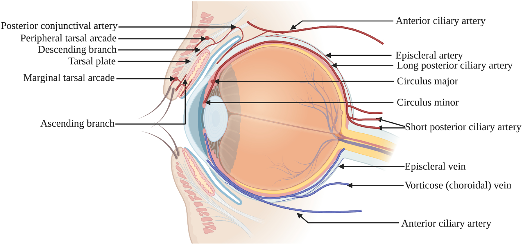
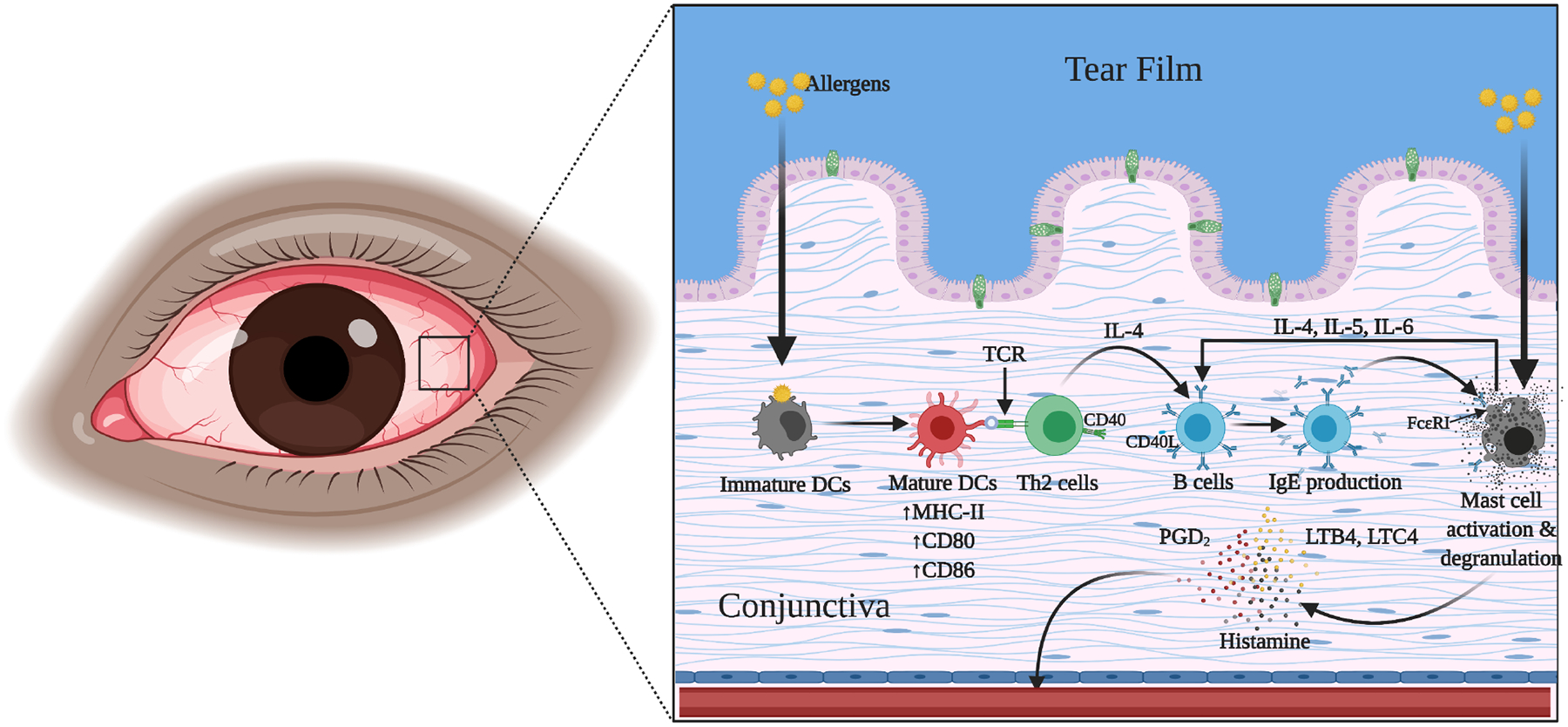
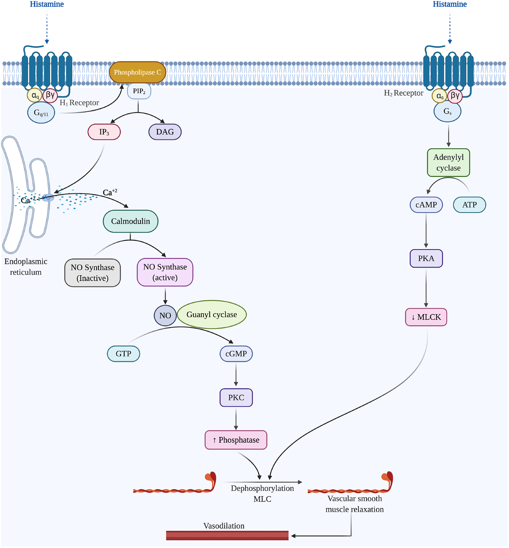
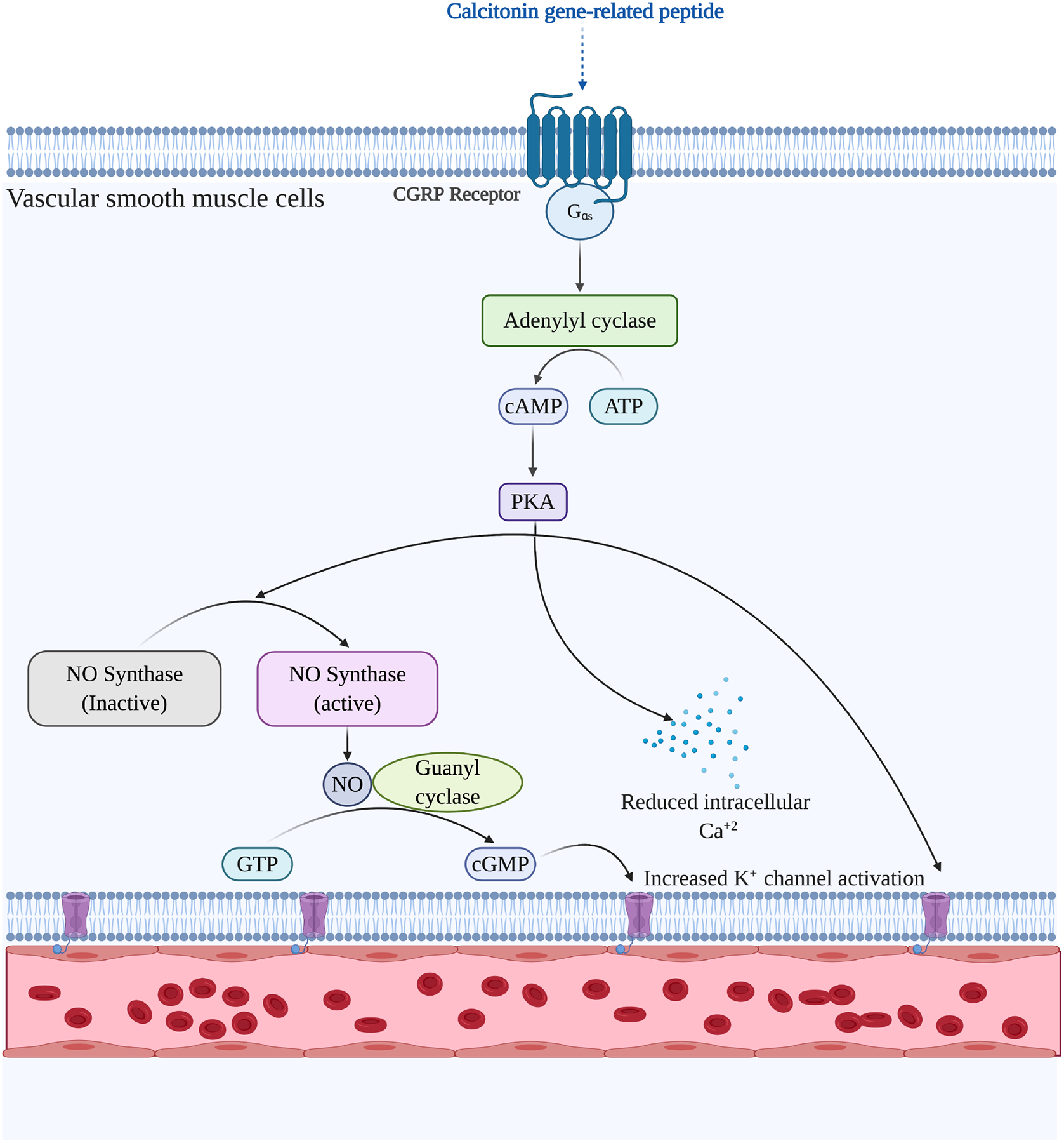
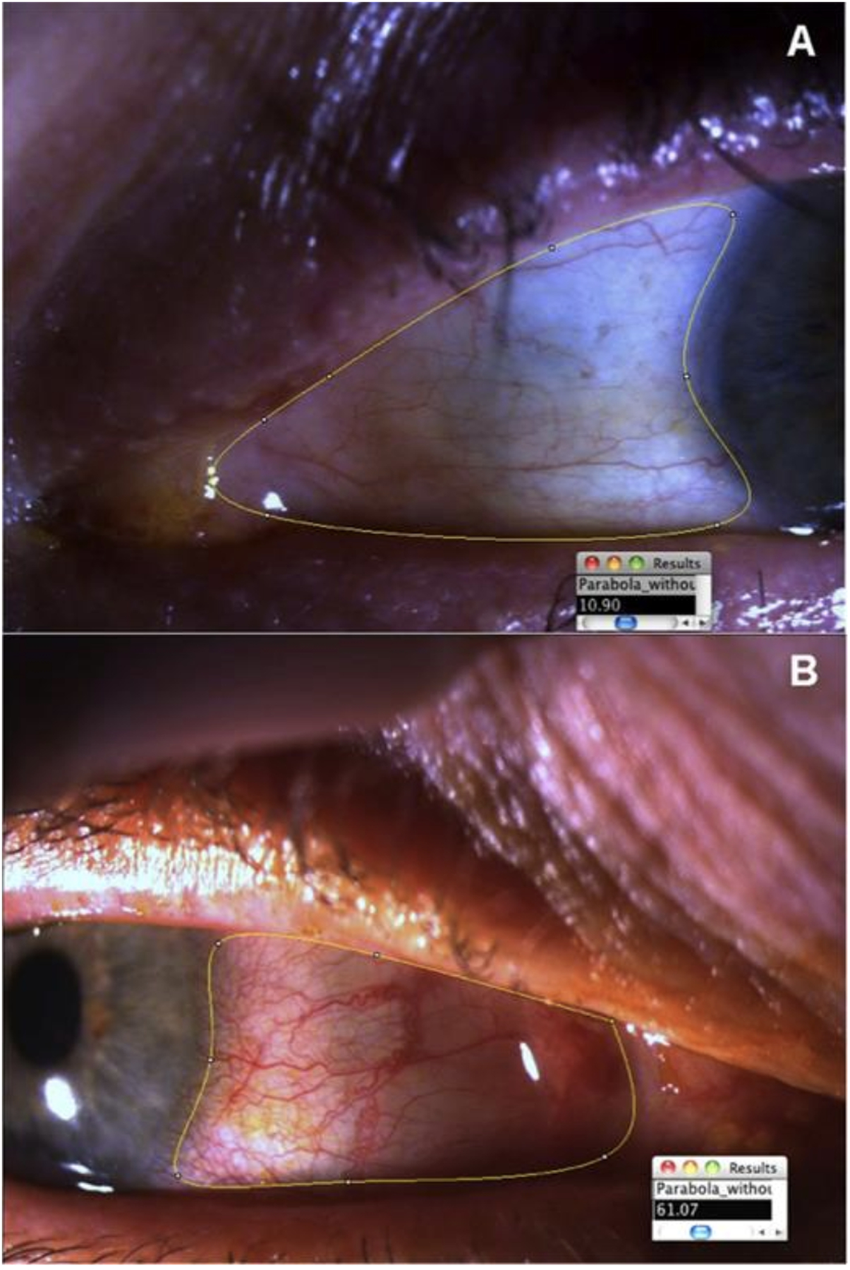
The translucent appearance of the conjunctiva allows for immediate visualization of changes in the circulation of the conjunctival microvasculature consisting of extensive branching of superficial and deep arterial systems and corresponding drainage pathways, and the translucent appearance of the conjunctiva allows for immediate visualization of changes in the circulation. Conjunctival hyperemia is caused by a pathological vasodilatory response of the microvasculature in response to inflammation due to a myriad of infectious and non-infectious etiologies. It is one of the most common contributors of ocular complaints that prompts visits to medical centers. Our understanding of these neurogenic and immune-mediated pathways has progressed over time and has played a critical role in developing targeted novel therapies. Due to a multitude of underlying etiologies, patients must be accurately diagnosed for efficacious management of conjunctival hyperemia. The diagnostic techniques used for the grading of conjunctival hyperemia have also evolved from descriptive and subjective grading scales to more reliable computer-based objective grading scales.
Keywords: Conjunctiva; Conjunctival vasculature; Conjunctivitis, ocular redness, conjunctival hyperemia; Microcirculation.
Copyright © 2021 Elsevier Inc. All rights reserved.
Conflict of interest statement
Figures
References
Publication types
MeSH terms
Grants and funding
LinkOut - more resources
Full Text Sources
Other Literature Sources
Medical
