. 2021 Aug 1;106(8):2170-2179.
doi: 10.3324/haematol.2021.279000.

Antibody-mediated procoagulant platelets in SARS-CoV-2-vaccination associated immune thrombotic thrombocytopenia

Affiliations

Antibody-mediated procoagulant platelets in SARS-CoV-2-vaccination associated immune thrombotic thrombocytopenia

Karina Althaus et al. Haematologica. .

Abstract

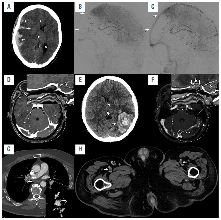
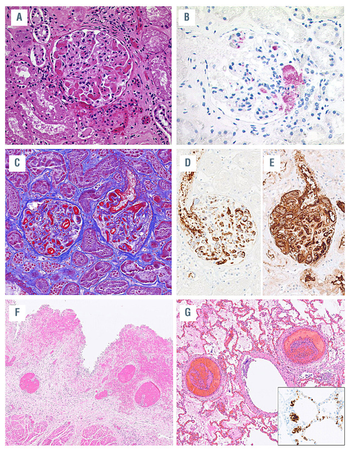
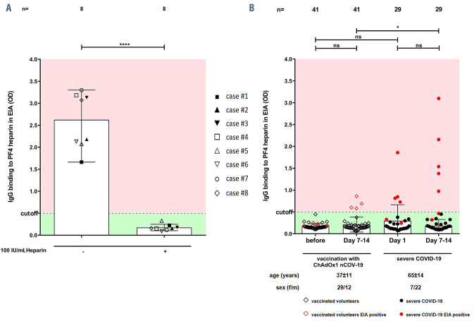
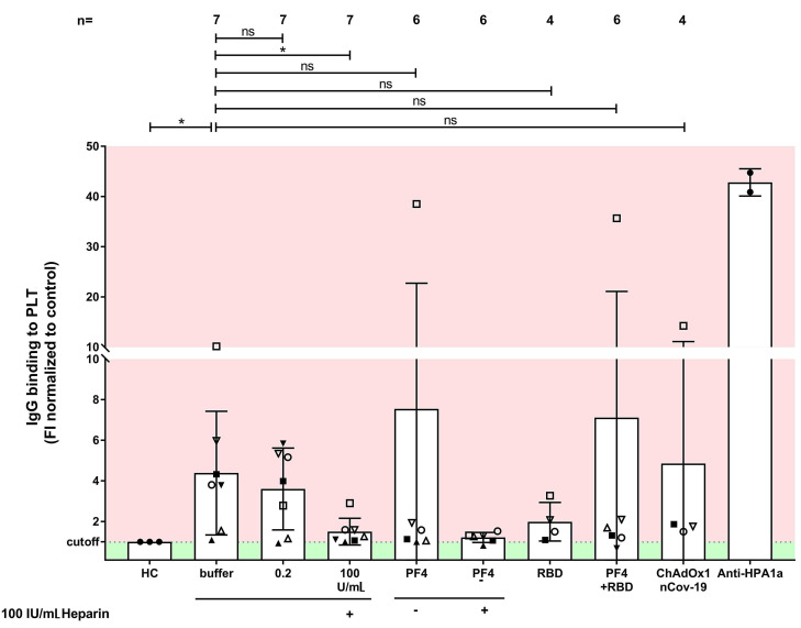
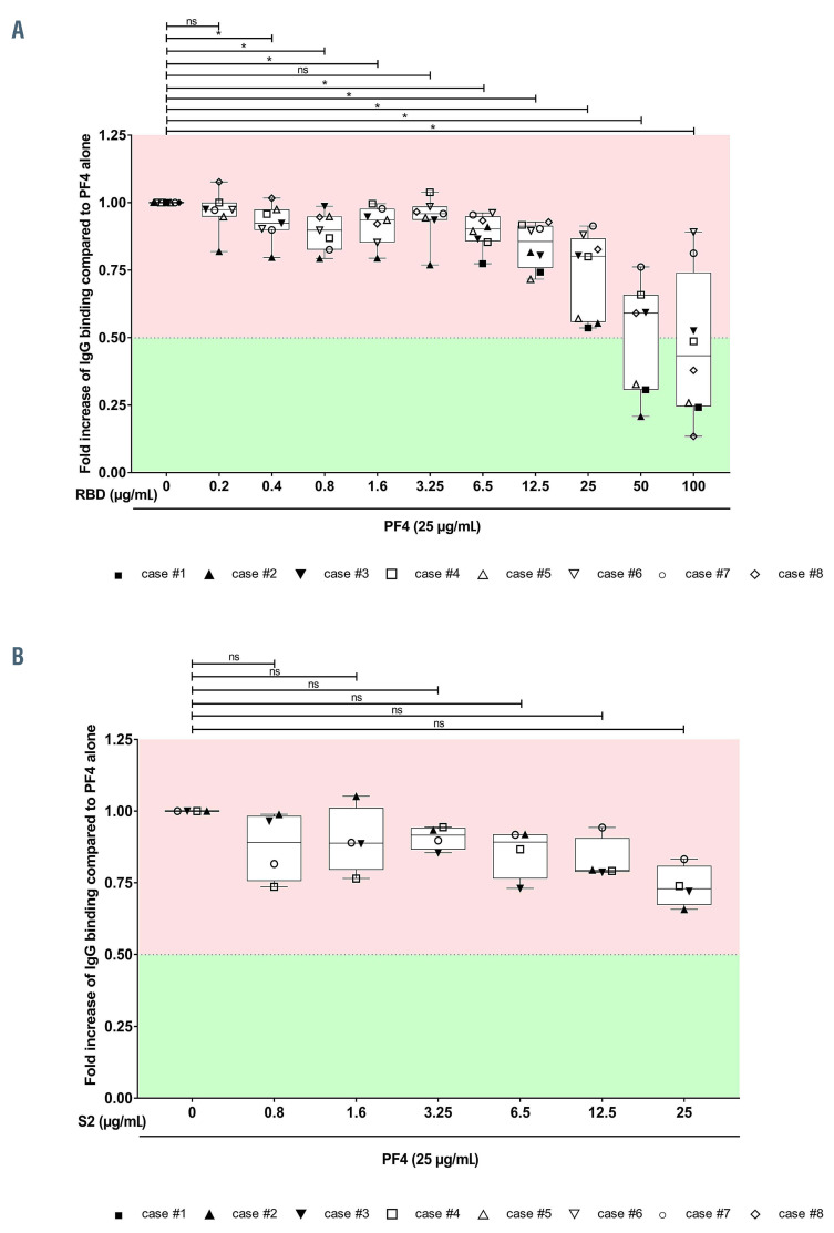
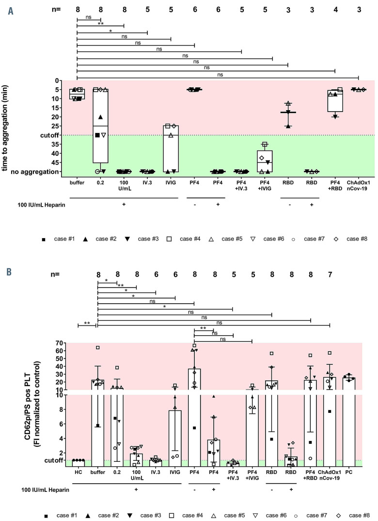
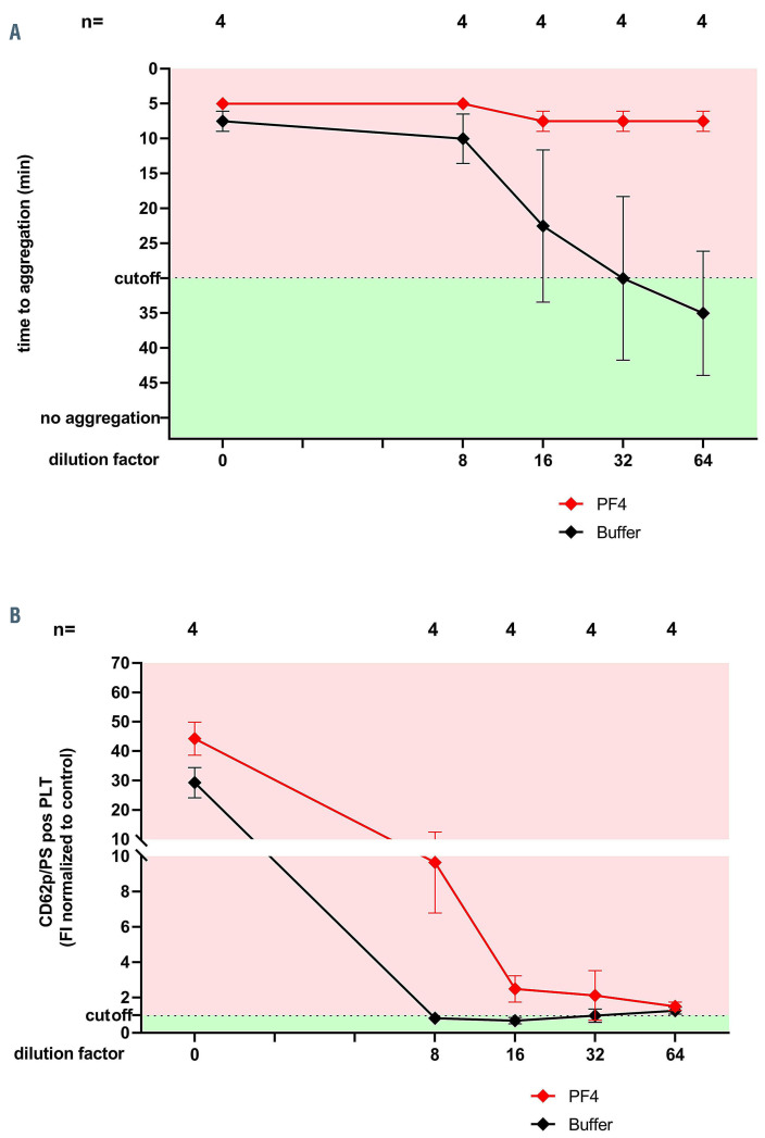
The COVID-19 pandemic has resulted in significant morbidity and mortality worldwide. To prevent severe infection, mass COVID-19 vaccination campaigns with several vaccine types are currently underway. We report pathological and immunological findings in 8 patients who developed vaccine-induced immune thrombotic thrombocytopenia (VITT) after administration of SARS-CoV-2 vaccine ChAdOx1 nCoV-19. We analyzed patient material using enzyme immune assays, flow cytometry and heparin-induced platelet aggregation assay and performed autopsies on two fatal cases. Eight patients (5 female, 3 male) with a median age of 41.5 years (range, 24 to 53) were referred to us with suspected thrombotic complications 6 to 20 days after ChAdOx1 nCoV-19 vaccination. All patients had thrombocytopenia at admission. Patients had a median platelet count of 46.5 x10⁹/L (range, 8 to 92). Three had a fatal outcome and 5 were successfully treated. Autopsies showed arterial and venous thromboses in various organs and the occlusion of glomerular capillaries by hyaline thrombi. Sera from VITT patients contain high titer antibodies against platelet factor 4 (PF4) (OD 2.59±0.64). PF4 antibodies in VITT patients induced significant increase in procoagulant markers (P-selectin and phosphatidylserine externalization) compared to healthy volunteers and healthy vaccinated volunteers. The generation of procoagulant platelets was PF4 and heparin dependent. We demonstrate the contribution of antibody-mediated platelet activation in the pathogenesis of VITT.


Figures

Figure 1.
Figure 1.
Imaging example of three illustrative cases. Imaging examples of case #1 (A to C), case #8 (D to F) and case #4 (G and H). In case #1, non-enhanced computed tomography imaging (A) showed a parenchymal and subdural hemorrhage (arrows in A), causing a midline shift (arrowheads in A). Digital subtraction angiography was performed (B) showing thrombosis of the right sigmoid and transverse sinus, superior sagittal sinus (arrows in B), and straight sinus. Angiography after mechanical recanalization (C) shows the recanalized cerebral sinuses (superior sagittal sinus marked with arrows). In case #8, cerebral imaging 7 days after vaccination was unremarkable (curved reconstruction of the left transverse and sigmoid sinus shown in the right upper corner of D and F). She worsened, which led to a repeated cerebral imaging, showing a large intraparenchymal hemorrhage in the left temporal lobe (arrow in E), causing midline shift (arrowhead in E), caused by a thrombosis of the transverse and sigmoid sinus (arrows in F), as well as of the adjacent tentorial veins. In case #4, a thrombus in the right pulmonary artery was observed (arrows in E; coronal reconstruction shown in the right lower corner of G). Further imaging also revealed thrombi in the femoral veins on both sides (arrows in H).
Figure 2.
Figure 2.
Histopathological findings in case #2 and case #3. (A) Case #2: occlusion of glomerular capillary loops by hyaline thrombi. Hematoxylin-Eosin (H&E) staining, original magnification 200x. (B) Deposition of platelets in glomerular vessels documented by CD42b staining, immunoperoxidase staining, magnification 200x. (C) Case #3: occlusion of glomerular capillary loops by hyaline thrombi with fibrin deposits highlighted in red, Masson’s trichrome stain, magnification 200x. (D) Immunostaining for CD61 and (E) fibrin demonstrate the massive intravascular deposits of fibrin and platelets, immunoperoxidase staining, magnification 200x. (F) Thrombotic occlusions of submucosal vessels in the urinary bladder with hemorrhage. H&E staining, magnification 40x. (F) Thrombotic occlusion of medium-sized pulmonary vessels. H&E stining, magnification 40x. (G) Insert shows platelet deposits in pulmonary capillaries stained for CD61, immunoperoxidase staining, magnification 400x.
Figure 3.
Figure 3.
Binding profile of sera from vaccine-induced immune thrombotic thrombocytopenia. (A) Results of the PF4/heparin immunoglobulin G (IgG)-enzyme immune assay (EIA) in patients with vaccine-induced immune thrombotic thrombocytopenia (VITT) with and without 100 IU/mL heparin. All VITT patients showed an enhanced binding which was significantly inhibited at high dose of heparin (100 IU/mL). (B) PF4-seroconversion after vaccination and severe SARS-CoV2 infection was followed up. IgG PF4/heparin antibody binding results in healthy volunteers before and after vaccination and COVID-19 patients in intensive care units showed four vaccinated volunteers displaying a positive EIA result after 7-14 days post-vaccination (red empty diamonds). OD: optical density.
Figure 4.
Figure 4.
Immunoglobulin G binding to platelets by flow cytometry in sera of vaccine-induced immune thrombotic thrombocytopenia patients. Immunoglobulin G (IgG) binding to healthy washed platelets (PLT) after incubation with sera from vaccine-induced immune thrombotic thrombocytopenia (VITT) patients was measured (assessed by flow cytometry and expressed as fold increase (FI) normalized to controls). VITT patients showed significantly higher binding at the baseline in comparison to healthy controls, which was inhibited by high dose heparin. ns: not significant; *P<0.05, **P<0.01, ***P<0.001 and ****P<0.0001.
Figure 5.
Figure 5.
Immunoglobulin G binding to SARS-CoV-2 Spike-RBD and S2 in the presence and absence of PF4. (A) Immunoglobulin G (IgG) binding to SARSCoV- 2 Spike-RBD was assessed by IgGenzyme immune assay (EIA) and expressed as fold increase to PF4 alone. (B) IgG binding to SARS-CoV-2 S2/PF4 complexes was assessed by EIA and expressed as fold increase to PF4 alone. ns: not significant; *P<0.05, **P<0.01, ***P<0.001 and ****P<0.0001.
Figure 6.
Figure 6.
Antibody-mediated platelet activation and generation of procoagulant platelets. Results of the platelet activation assay (HIPA) with modifications in the vaccine-induced immune thrombotic thrombocytopenia (VITT) patients. Each dot represents the median of four different donors. (A) All VITT patients presented platelet (PLT) activation with buffer alone, which was significantly increased by PF4 but inhibited with high dose of heparin. Procoagulant platelets (CD62P/phosphatidylserine [PS] positive) in different settings were analyzed via Annexin V-FITC and CD62p-APC antibody staining. (B) Where indicated, platelets were treated with PF4, 0.2 U/mL and 100 IU/mL heparin, RBD and ChAdOx1 nCoV-19A. Data are presented as mean ± standard deviation of the measured fold increase compared to control. ns: not significant; *P<0.05, **P<0.01, ***P<0.001 and ****P<0.0001. The number of sera tested is reported in each graph. Dotted lines represent the cutoffs determined testing sera from healthy donors. FI: fold increase.
Figure 7.
Figure 7.
Antibody-mediated platelet activation and generation of procoagulant platelets with diluted sera. (A) Results of heparininduced platelet aggregation assay (HIPA) at different titrations of sera from vaccine-induced immune thrombotic thrombocytopenia (VITT) patients. Note that diluted sera (from 1:64) activated platelets only in the presence e of PF4. (B) Effect of sera from VITT patients at different titrations on the development of procoagulant platelets. Note that diluted sera (from 1:8) activated platelets only in the presence e of PF4. Data are presented as mean ± standard deviation of the measured fold increase (FI) compared to control. ns: not significant; *P<0.05; **P<0.01, ***P<0.001 and ****P<0.0001. The number of sera tested is reported in each graph. Dotted lines represent the cutoffs determined testing sera from healthy donors.

Comment in

References

    1. Grasselli G, Greco M, Zanella A, et al. . Risk factors associated with mortality among patients with COVID-19 in intensive care units in Lombardy, Italy. JAMA Intern Med. 2020;180(10):1345-1355. - PMC - PubMed
    1. Castells MC, Phillips EJ. Maintaining safety with SARS-CoV-2 vaccines. N Engl J Med. 2021;384(7):643-649. - PMC - PubMed
    1. Althaus K, Marini I, Zlamal J, et al. . Antibody-induced procoagulant platelets in severe COVID-19 infection. Blood. 2021; 137(8):1061-1071. - PMC - PubMed
    1. Wolf ME, Luz B, Niehaus L, Bhogal P, Bazner H, Henkes H. Thrombocytopenia and intracranial venous sinus thrombosis after "COVID-19 vaccine AstraZeneca" Exposure. J Clin Med. 2021;10(8):1599. - PMC - PubMed
    1. Becker M, Strengert M, Junker D, et al. . Exploring beyond clinical routine SARSCoV- 2 serology using MultiCoV-Ab to evaluate endemic coronavirus cross-reactivity. Nat Commun. 2021;12(1):1152. - PMC - PubMed

Publication types