. 2021 May 19;12(1):292.
doi: 10.1186/s13287-021-02348-z.

Liver development is restored by blastocyst complementation of HHEX knockout in mice and pigs

Affiliations

Liver development is restored by blastocyst complementation of HHEX knockout in mice and pigs

M Ruiz-Estevez et al. Stem Cell Res Ther. .

Abstract

Background: There are over 17,000 patients in the US waiting to receive liver transplants, and these numbers are increasing dramatically. Significant effort is being made to obtain functional hepatocytes and liver tissue that can for therapeutic use in patients. Blastocyst complementation is a challenging, innovative technology that could fundamentally change the future of organ transplantation. It requires the knockout (KO) of genes essential for cell or organ development in early stage host embryos followed by injection of donor pluripotent stem cells (PSCs) into host blastocysts to generate chimeric offspring in which progeny of the donor cells populate the open niche to develop functional tissues and organs.

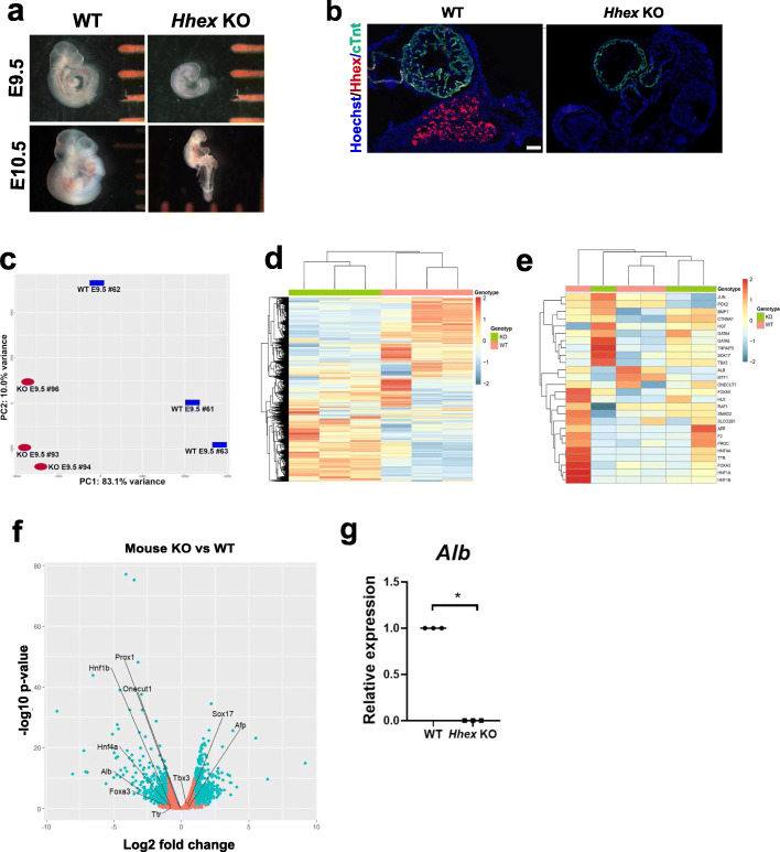
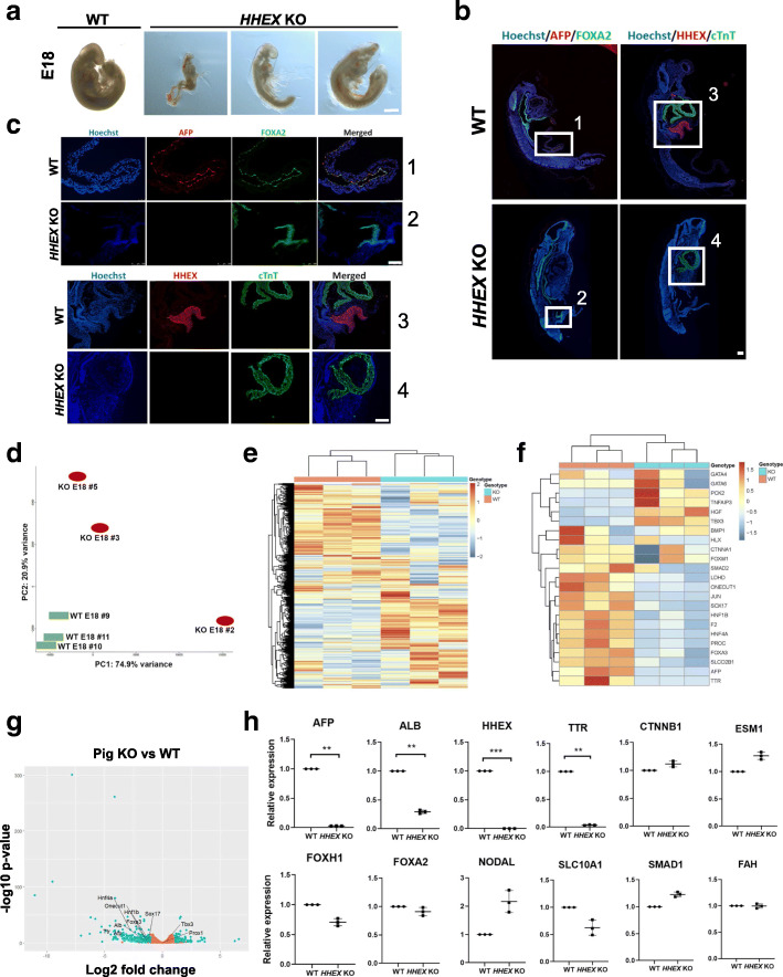
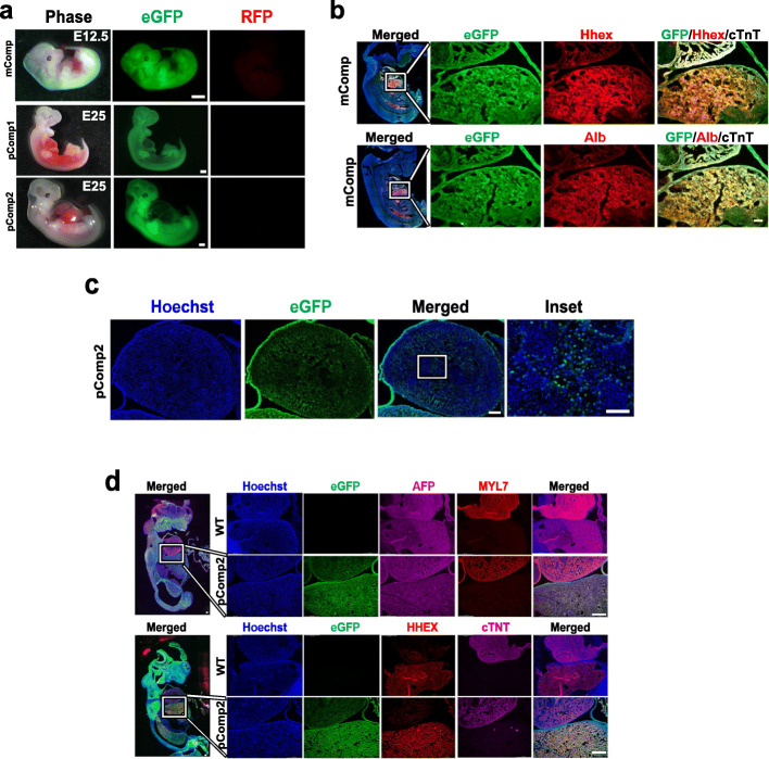
Methods: The HHEX gene is necessary for proper liver development. We engineered loss of HHEX gene expression in early mouse and pig embryos and performed intraspecies blastocyst complementation of HHEX KO embryos with eGFP-labeled PSCs in order to rescue the loss of liver development.

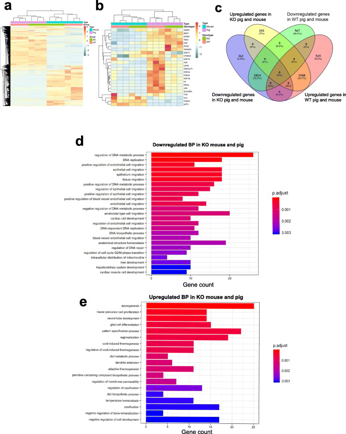
Results: Loss of HHEX gene expression resulted in embryonic lethality at day 10.5 in mice and produced characteristics of lethality at day 18 in pigs, with absence of liver tissue in both species. Analyses of mouse and pig HHEX KO fetuses confirmed significant loss of liver-specific gene and protein expression. Intraspecies blastocyst complementation restored liver formation and liver-specific proteins in both mouse and pig. Livers in complemented chimeric fetuses in both species were comprised of eGFP-labeled donor-derived cells and survived beyond the previously observed time of HHEX KO embryonic lethality.

Conclusions: This work demonstrates that loss of liver development in the HHEX KO can be rescued via blastocyst complementation in both mice and pigs. This complementation strategy is the first step towards generating interspecies chimeras for the goal of producing human liver cells, tissues, and potentially complete organs for clinical transplantation.

Keywords: Development; Embryo; Gene editing; Stem cells; Transplantation.


Conflict of interest statement

D.F.C. serves as C.S.O./Senior Vice President of Research and Development at Recombinetics, Inc.; W.C.L. served as C.S.O. of Regenevida a Division of Recombinetics, Inc. during the initial phase of this project; all the authors with Recombinetics, Inc. affiliation are employees and shareholders of the company; the other authors declare that they have no competing interests.

Figures

Fig. 1
Fig. 1
Characterization of the HHEX KO mouse embryos compared to WT. a Phenotype of the Hhex KO E9.5 and E10.5 mice. Separation between orange bars is 1 mm. b IHC images of E9.5 Hhex KO mouse with Hoechst (blue), anti-HHEX (red), and anti-cTnT (green) antibodies. Bars, 100 μm. cg Gene expression analyses (RNA-seq, cf; qRT-PC, g) in the Hhex KO E9.5 mice vs WT (N = 3 each group). c Principal component analysis. d Heatmap showing all the significantly up- and downregulated genes. e Heatmap showing liver-related genes whose expressions are significantly up- or downregulated. f Volcano plot showing significantly up- and downregulated genes (both in blue) and unaffected genes (orange), highlighting some liver-related genes. g Relative expression of transcripts. *p < 0.05
Fig. 2
Fig. 2
Characterization of the HHEX KO pig embryos compared to WT. a Phenotype of the HHEX KO E18 pigs. b, c IHC images of E18 HHEX KO pig embryos with Hoechst (blue), anti-AFP/HHEX (red), and anti-FOXA2/cTnT (green) antibodies. White insets in b correspond to numbered enlarged images in c. Bars: all are 100 μm, except for the bar in a, which is 500 μm. dh Gene expression analyses (RNA-seq, dg; qRT-PC, h) in the HHEX KO E18 pigs vs WT (N = 3 each group). d Principal component analysis. e Heatmap showing all the significantly up- and downregulated genes. f Heatmap showing liver-related genes whose expressions are significantly up- or downregulated. g Volcano plot showing significantly up- and downregulated genes (both in blue) and unaffected genes (orange), highlighting some liver-related genes. h Relative expression of transcripts. **p < 0.01; **p < 0.001
Fig. 3
Fig. 3
Mouse and pig RNA-seq expression data comparison. a, b Heatmaps combining mouse and pig data with all the significantly affected genes (a) and only those related to liver (b). c Venn diagram showing significantly down- and upregulated common genes between both species. d, e Bar plots reporting the top significantly down- (d) and upregulated (e) biological processes (BPs) related to the 362 down- and 393 upregulated common genes for both HHEX KO mouse and pig. The color code is proportional to significance. N = 6 per group
Fig. 4
Fig. 4
Mouse and pig complemented embryos. a Whole embryo images showing eGFP expression (RFP channel shown to indicate absence of signal and no autofluoresence). b, d IHC images of the mouse (b) and pig (d) complemented embryos showing the liver-heart area in the insets in the magnifications. c Heart area of one of the complemented pig embryos with the inset highlighting presence of both eGFP+ and eGFP cells within the tissue. eGFP, enhanced green fluorescent protein; RFP, red fluorescent protein; mComp, mouse complemented embryo; pComp1, pig complemented embryo 1; pComp2, pic complemented embryo 2; MYL7, myosin light chain 7; cTNT, cardiac troponin T (white signal in mouse); ALB, albumin. Bars, 1 mm in a, 100 μm b and d whole embryos, and in b magnifications; 500 μm in d magnifications and in c, except for its inset, which is 250 μm

References

    1. Si-Tayeb K, Noto FK, Nagaoka M, Li J, Battle MA, Duris C, North PE, Dalton S, Duncan SA. Highly efficient generation of human hepatocyte-like cells from induced pluripotent stem cells. Hepatology. 2010;51(1):297–305. doi: 10.1002/hep.23354. - DOI - PMC - PubMed
    1. Asgari S, Moslem M, Bagheri-Lankarani K, Pournasr B, Miryounesi M, Baharvand H. Differentiation and transplantation of human induced pluripotent stem cell-derived hepatocyte-like cells. Stem Cell Rev Reports. 2013;9(4):493–504. doi: 10.1007/s12015-011-9330-y. - DOI - PubMed
    1. Wu XB, Tao R. Hepatocyte differentiation of mesenchymal stem cells. Hepatobiliary Pancreat Dis Int. 2012;11(4):360–371. doi: 10.1016/S1499-3872(12)60193-3. - DOI - PubMed
    1. Pettinato G, Thompson MT, Fisher RA. Human embryoid bodies to hepatocyte-like clusters: preparing for translation. Liver Res. 2017;1(2):88–95. doi: 10.1016/j.livres.2017.08.004. - DOI
    1. Zhou Q, Li L, Li J. Stem cells with decellularized liver scaffolds in liver regeneration and their potential clinical applications. Liver Int. 2015;35(3):687–694. doi: 10.1111/liv.12581. - DOI - PubMed

Publication types