. 2021 May 19;23(1):56.
doi: 10.1186/s13058-021-01430-x.

Novel mechanism for OSM-promoted extracellular matrix remodeling in breast cancer: LOXL2 upregulation and subsequent ECM alignment

Affiliations

Novel mechanism for OSM-promoted extracellular matrix remodeling in breast cancer: LOXL2 upregulation and subsequent ECM alignment

Simion C Dinca et al. Breast Cancer Res. .

Abstract

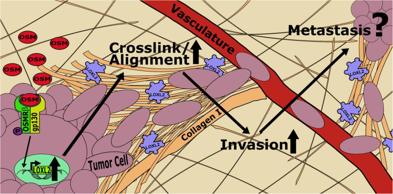
Background: Invasive ductal carcinoma (IDC) is a serious problem for patients as it metastasizes, decreasing 5-year patient survival from > 95 to ~ 27%. The breast tumor microenvironment (TME) is often saturated with proinflammatory cytokines, such as oncostatin M (OSM), which promote epithelial-to-mesenchymal transitions (EMT) in IDC and increased metastasis. The extracellular matrix (ECM) also plays an important role in promoting invasive and metastatic potential of IDC. Specifically, the reorganization and alignment of collagen fibers in stromal ECM leads to directed tumor cell motility, which promotes metastasis. Lysyl oxidase like-2 (LOXL2) catalyzes ECM remodeling by crosslinking of collagen I in the ECM. We propose a novel mechanism whereby OSM induces LOXL2 expression, mediating stromal ECM remodeling of the breast TME.

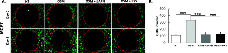
Methods: Bioinformatics was utilized to determine survival and gene correlation in patients. IDC cell lines were treated with OSM (also IL-6, LIF, and IL-1β) and analyzed for LOXL2 expression by qRT-PCR and immunolabelling techniques. Collagen I contraction assays, 3D invasion assays, and confocal microscopy were performed with and without LOXL2 inhibition to determine the impact of OSM-induced LOXL2 on the ECM.

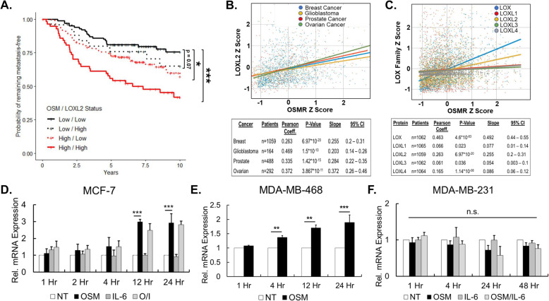
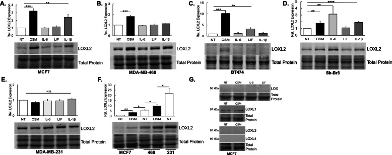
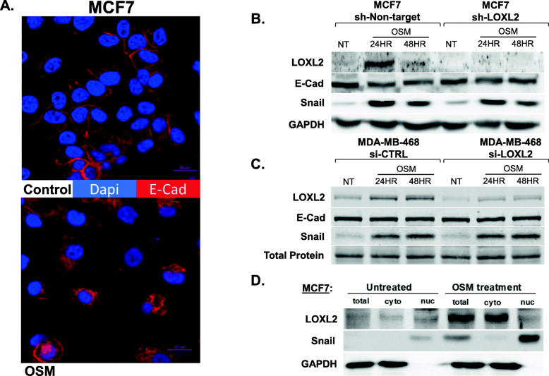
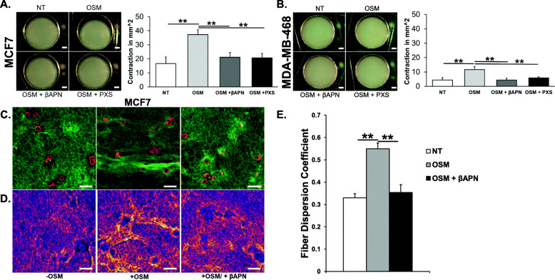
Results: Our studies demonstrate that IDC patients with high LOXL2 and OSM co-expression had worse rates of metastasis-free survival than those with high levels of either, individually, and LOXL2 expression is positively correlated to OSM/OSM receptor (OSMR) expression in IDC patients. Furthermore, human IDC cells treated with OSM resulted in a significant increase in LOXL2 mRNA, which led to upregulated protein expression of secreted, glycosylated, and enzymatically active LOXL2. The expression of LOXL2 in IDC cells did not affect OSM-promoted EMT, and LOXL2 was localized to the cytoplasm and/or secreted. OSM-induced LOXL2 promoted an increase in ECM collagen I fiber crosslinking, which led to significant fiber alignment between cells and increased IDC cell invasion.

Conclusions: Aligned collagen fibers in the ECM provide pathways for tumor cells to migrate more easily through the stroma to nearby vasculature and tissue. These results provide a new paradigm through which proinflammatory cytokine OSM promotes tumor progression. Understanding the nuances in IDC metastasis will lead to better potential therapeutics to combat against the possibility.

Keywords: Breast cancer; Collagen; Cytokines; Extracellular matrix; IL-6; Inflammation; LOXL2; Metastasis; OSM; Tumor microenvironment.


Conflict of interest statement

The authors declare that they have no competing interests.

Figures

Fig. 1
Fig. 1
Co-expression of OSM and LOXL2 leads to drastically decreased metastasis-free survival. a Distant metastasis-free survival (DMFS) plotted from de Vijver et al. [58] invasive ductal carcinoma patient microarray database comparing low OSM/ low LOXL2 to low OSM/high LOXL2, high OSM/low LOXL2, and high OSM/high LOXL2 mRNA expression (n = 295). We observed a stronger negative impact on DMFS with high OSM and LOXL2 co-expression compared to high expression of OSM or LOXL2 separately (log rank test). b LOXL2 mRNA expression Z-score is positively correlated as measured by Pearson correlation coefficient to the beta subunit expression of OSM receptor (OSMR) mRNA expression Z-score in cancer patients analyzed from The Cancer Genome Atlas (TCGA) RNA-Seq database, specifically: breast cancer (BRCA), glioblastoma (GBM), prostate cancer (PRAD), and ovarian cancer (OV). Scatter plot consists of Z-score mRNA expression and line of best fit as determined by linear regression; a summary of the data is found in the accompanying table. c The mRNA Z-score of several LOXL2 family members exhibit positive Pearson correlation to OSMR mRNA Z-score in the breast invasive carcinoma dataset from TCGA. d qRT-PCR analysis of MCF7 luminal A invasive ductal carcinoma cells treated with OSM shows LOXL2 mRNA induction starting at 12 h and peaking at 24 h; there is no induction with IL-6. e qRT-PCR analysis of MDA-MB-468 basal A invasive ductal carcinoma cells treated with OSM also shows an increase in LOXL2 mRNA expression starting at 4 h. f qRT-PCR analysis of MDA-MB-231 basal B breast cancer cells, that constitutively express high levels of LOXL2, show no significant induction of LOXL2 mRNA expression by either OSM or IL-6 signaling. (All qRT-PCR experiments n = 3+; n.s. p > 0.05, **p < 0.01, ***p < 0.001; two-way ANOVA)
Fig. 2
Fig. 2
OSM promotes LOXL2 protein expression. All experiments and results pertain to immunoblot assays run with 10–20 μg total protein. a MCF7 breast cancer cells were treated with OSM, IL-6, LIF, and IL-1β for 24 h. Our analysis showed that only OSM and IL-1β promoted a significant upregulation of LOXL2 protein expression. b In analyzing MDA-MB-468 breast cancer cells, the same treatments that are described above were utilized. We observed that only cells treated with OSM had significantly induced LOXL2 protein expression. c BT474 breast cancer cells, under the same conditions, showed a significant increase in LOXL2 expression with OSM and LIF treatments. d In Sk-Br-3 breast cancer cells, we observed a significant increase in LOXL2 expression with OSM and IL-6 treatment. e We again used the same treatments in MDA-MB-231 breast cancer cells. LOXL2 expression was not significantly affected by either OSM, IL-6, LIF, or IL-1β treatment after 24 h. f Relative LOXL2 protein expression was compared among three breast cancer cell lines treated with OSM. From least invasive (MCF7) to the most (MDA-MB-231), we observed a stepwise increase in LOXL2 protein expression. OSM treatment bridges LOXL2 expression between cells. g MCF7 cells were treated for 24 h with OSM; OSM, IL-6, and LIF for LOX expression. No changes are observed in lysyl oxidase expression; LOXL1 is constitutively expressed. (All experiments n = 3+; n.s. p > 0.05, **p < 0.01, ***p < 0.001; Students t test)
Fig. 3
Fig. 3
OSM signaling promotes an EMT that is independent of LOXL2 expression. a Confocal images of MCF7 cells depict a distinct loss of cell polarity and a transition from membrane localization to cytoplasmic localization of E-cadherin with 48-h OSM treatment, both hallmarks of EMT. E-cadherin (red) and nuclei (DAPI, blue). Magnification × 40 with digital zooming × 2; scale bar = 20 μm. b Immunoblot of MCF7-sh-non-target and MCF7-sh-LOXL2 cells treated with OSM for 24 and 48 h. Expression of EMT markers, E-cadherin (E-Cad), and Snail are compared between these cell lines. The absence of LOXL2 expression in MCF-7 cells had no effect on OSM-induced EMT. c Immunoblot of MDA-MB-468 breast cancer cells exposed to siCTRL and siLOXL2 for 48 h prior to OSM treatment for 24 and 48 h. Expression of EMT markers, E-Cad, and Snail are compared between siLOXL2 and siCTRL treatments. Inhibited LOXL2 expression in MDA-MB-468 cells had no effect on OSM-induced EMT. d Immunoblot of MCF7 cells treated with OSM for 24 h; post treatment cells are collected and subjected to nuclear-cytoplasmic protein fractionation. LOXL2 protein expression is not present in the nuclear fraction, only in the cytoplasmic fraction. GAPDH protein expression is used to confirm purity of cytoplasmic fraction, and Snail transcriptional factor expression is used to confirm nuclear fraction purity. (All experiments n = 3+)
Fig. 4
Fig. 4
OSM-induced LOXL2 is glycosylated, enzymatically active, and secreted from breast cancer cells. a MCF7 and b MDA-MB-468 cells were treated with OSM for 24 h to induce the expression of LOXL2. PNGase F, an N-linked glycosylase, is then added to cleave N-linked glycosylation sites. The immunoblot results confirm LOXL2 glycosylation as the LOXL2 protein band size goes from ~ 105 to ~ 87 kDa with PNGase F treatment. c Lysyl oxidase activity assay performed on MCF7 cell conditioned media (CM) is analyzed by using an Amplex red-based fluorometric assay. Immunoblot analysis is utilized to confirm siLOXL2 knockdown of LOXL2 expression. Results show that 24-h OSM treatment led to significantly increased lysyl oxidase activity, this is repressed with exposure to siLOXL2. d ELISA is used to quantify LOXL2 protein secreted into CM from MCF7 cells after 36 h with OSM treatment. The results confirm that OSM signaling induces the expression, and promotes the secretion, of LOXL2 protein. (All experiments n = 3+; *p < 0.05, **p < 0.01, ***p < 0.001; Students t test)
Fig. 5
Fig. 5
OSM-induced LOXL2 promotes ECM crosslinking and alignment of collagen I fibers. a Collagen contraction assay was performed using 1.5 mg/mL collagen I matrices or “pucks” seeded with MCF7 cells and treated for 48 h with OSM or OSM with βAPN (500 μM) or PXS-5120A (200 nM) [LOXL2 inhibitors]. After 48 h, dissecting microscope images of the collagen I matrix depict significantly more contraction in the OSM-treated samples, which is reversed in the presence of LOXL2 inhibitors. Scale bar = 2 mm. Graph quantifying the change in area of the matrix (in mm2) due to contraction. Collagen I fiber contraction correlates to fiber crosslinking and is reversed with the inhibition of LOXL2. b The same experiment as above was performed with MDA-MB-468 cells. Representative images after 48 h and accompanying graph are depicted using the same scale as above. As with MCF7 cells, LOXL2 inhibition prevented contraction due to OSM-induced LOXL2, but overall contraction is not as pronounced. Scale bar = 2 mm. c Confocal images of the collagen “pucks” depict the increase in collagen I fiber (green) alignment between MCF7 cells (red) in collagen I matrices that are treated with OSM for 36 h. Alignment is not observable with the addition of LOXL2 inhibition using βAPN (500 μM). Magnification × 63; scale bar = 50 μm. d Representative images with areas of greatest collagen I fiber density emphasized using ImageJ image processing. This clearly highlights the increase in fiber density and alignment present with OSM treatment, which reversed by LOXL2 inhibition. e Graph depicting the average fiber dispersion coefficient of collagen I fibers perpendicular to and bridging MCF7 cells in collagen I matrices. Confirms qualitative data that OSM treatment significantly increases alignment which is reversed with LOXL2 inhibition. (All experiments n = 3+; **p < 0.01, ***p < 0.001; Students t test and one-way ANOVA)
Fig. 6
Fig. 6
OSM-induced LOXL2 promotes invasion in 3D extracellular matrix. a MCF7-GFP cells were seeded into a 1.5 mg/mL collagen I solution and molded into wells of a 96-well plate containing “cell-free zones.” Cells were supplemented with estrogen (50 ng/mL) and treated with OSM, OSM with βAPN (500 μM), or OSM with PXS-5120A (200 nM). Fluorescent images were taken at day 0, and at the conclusion of the experiment on day 5. We observed an increase in 3D invasion of MCF7 cells treated with OSM that was limited by the inhibition of LOXL2 enzymatic function using βAPN or PXS-5120A. Scale bar = 1,000 μm. b Graph represents the total number of MCF7-GFP cells that invaded into the cell-free space by day 5, with the various treatments discussed above. OSM treatment significantly increases 3D invasion in MCF7 cells and that is significantly reversed by LOXL2 inhibition. (Experiment n = 3; ***p < 0.001; one-way ANOVA)
Fig. 7
Fig. 7
Mechanism by which OSM induces LOXL2 and promotes ECM remodeling. OSM binds to gp130, which recruits OSMRβ to form a heterodimer and allow the phosphorylation and activation of downstream signaling pathways, including STAT3, MAPK, and PI3K. OSM signaling promotes EMT in invasive ductal carcinomas and as the data shows LOXL2 expression in its ~ 105 kDa glycosylated form. The 105 kDa LOXL2 is enzymatically active and secreted into the ECM of the breast tumor microenvironment. In the ECM, LOXL2 promotes crosslinking of the main constituent of the stroma, collagen I, which leads to collagen I fiber alignment. The alignment of collagen I fibers in the stroma provides pathways for cancer cells that have undergone EMT to invade nearby tissue and vasculature [52, 53]. Therefore, these changes to the ECM of the tumor microenvironment likely play a functional role in invasive ductal carcinoma metastasis [27, 37]

References

    1. Kuukasjarvi T, Tanner M, Pennanen S, Karhu R, Kallioniemi OP, Isola J. Genetic changes in intraductal breast cancer detected by comparative genomic hybridization. Am J Pathol (Article) 1997;150:1465–1471. - PMC - PubMed
    1. Howlader N, Noone AM, Krapcho M, Miller D, Brest A, Yu M, et al. SEER Cancer Statistics Review. Natl Canc Inst. 1975–2016.
    1. Grenier A, Dehoux M, Boutten A, Arce-Vicioso M, Durand G, Gougerot-Pocidalo MA, et al. Oncostatin M production and regulation by human polymorphonuclear neutrophils. Blood. 1999;93(4):1413–1421. doi: 10.1182/blood.V93.4.1413. - DOI - PubMed
    1. Queen MM, Ryan RE, Holzer RG, Keller-Peck CR, Jorcyk CL. Breast cancer cells stimulate neutrophils to produce oncostatin M: potential implications for tumor progression. Cancer Res (Article) 2005;65(19):8896–8904. doi: 10.1158/0008-5472.CAN-05-1734. - DOI - PubMed
    1. Hurst SM, McLoughlin RM, Monslow J, Owens S, Morgan L, Fuller GM, et al. Secretion of oncostatin M by infiltrating neutrophils: regulation of IL-6 and chemokine expression in human mesothelial cells. J Immunol. 2002;169(9):5244–5251. doi: 10.4049/jimmunol.169.9.5244. - DOI - PubMed

Publication types

MeSH terms