. 2021 May 25;118(21):e2100686118.
doi: 10.1073/pnas.2100686118.

Chromatin remodeler Arid1a regulates subplate neuron identity and wiring of cortical connectivity

Affiliations

Chromatin remodeler Arid1a regulates subplate neuron identity and wiring of cortical connectivity

Daniel Z Doyle et al. Proc Natl Acad Sci U S A. .

Abstract

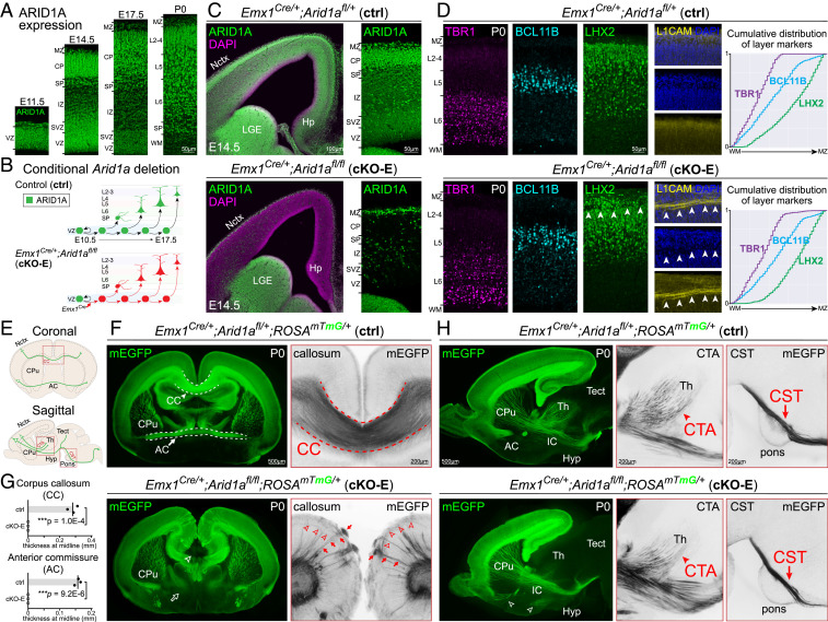
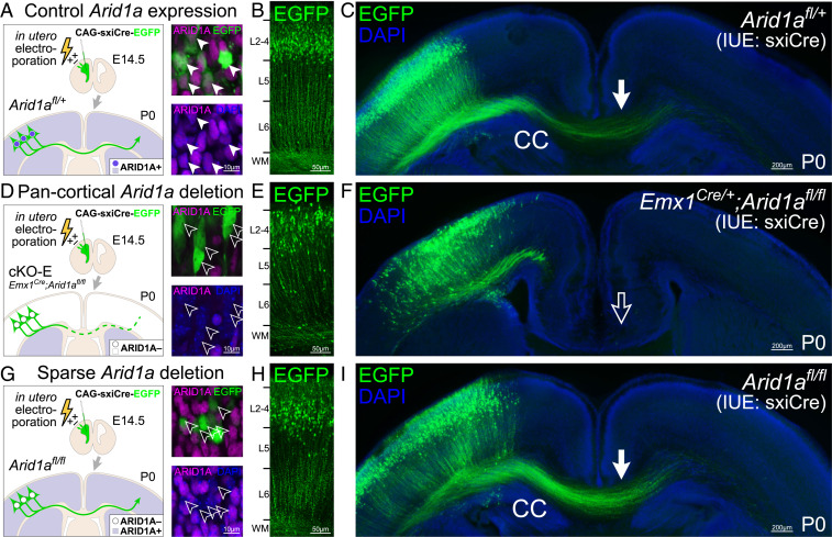
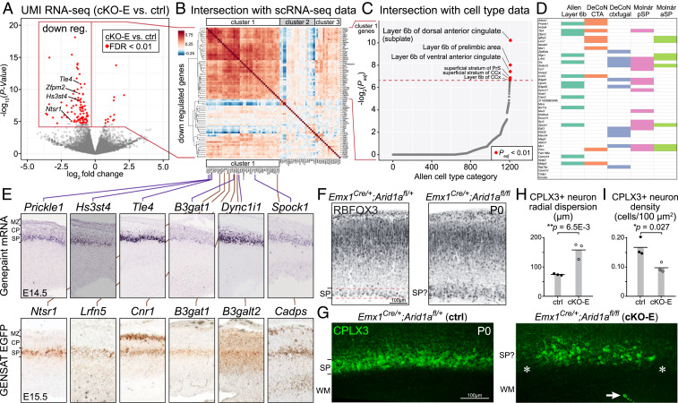
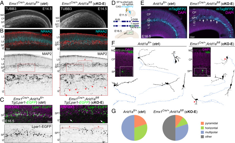
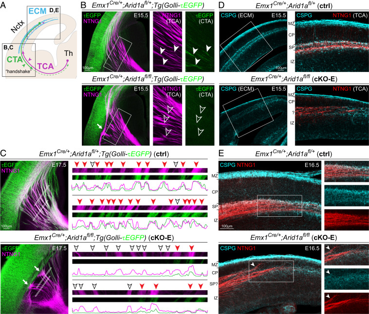
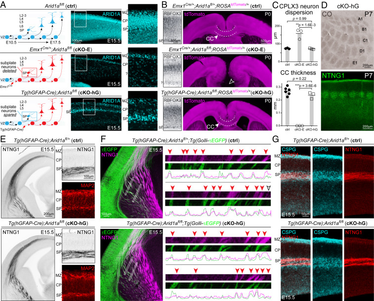
Loss-of-function mutations in chromatin remodeler gene ARID1A are a cause of Coffin-Siris syndrome, a developmental disorder characterized by dysgenesis of corpus callosum. Here, we characterize Arid1a function during cortical development and find unexpectedly selective roles for Arid1a in subplate neurons (SPNs). SPNs, strategically positioned at the interface of cortical gray and white matter, orchestrate multiple developmental processes indispensable for neural circuit wiring. We find that pancortical deletion of Arid1a leads to extensive mistargeting of intracortical axons and agenesis of corpus callosum. Sparse Arid1a deletion, however, does not autonomously misroute callosal axons, implicating noncell-autonomous Arid1a functions in axon guidance. Supporting this possibility, the ascending axons of thalamocortical neurons, which are not autonomously affected by cortical Arid1a deletion, are also disrupted in their pathfinding into cortex and innervation of whisker barrels. Coincident with these miswiring phenotypes, which are reminiscent of subplate ablation, we unbiasedly find a selective loss of SPN gene expression following Arid1a deletion. In addition, multiple characteristics of SPNs crucial to their wiring functions, including subplate organization, subplate axon-thalamocortical axon cofasciculation ("handshake"), and extracellular matrix, are severely disrupted. To empirically test Arid1a sufficiency in subplate, we generate a cortical plate deletion of Arid1a that spares SPNs. In this model, subplate Arid1a expression is sufficient for subplate organization, subplate axon-thalamocortical axon cofasciculation, and subplate extracellular matrix. Consistent with these wiring functions, subplate Arid1a sufficiently enables normal callosum formation, thalamocortical axon targeting, and whisker barrel development. Thus, Arid1a is a multifunctional regulator of subplate-dependent guidance mechanisms essential to cortical circuit wiring.

Keywords: axon pathfinding; cerebral cortex; chromatin regulation; development; neural circuits.


Conflict of interest statement

The authors declare no competing interest.

Figures

Fig. 1.
Fig. 1.
Tract-dependent misrouting of cortical axons following conditional Arid1a deletion. (A) ARID1A (green) immunostaining on coronal E11.5, E14.5, E17.5, and P0 brain sections revealed widespread ARID1A expression during cortical development. (B) Schematic illustration of conditional Arid1a deletion using Emx1Cre, which mediates recombination in cortical NPCs at E10.5, near the onset of neurogenesis. (C) ARID1A (green) and DAPI (magenta) staining of coronal E14.5 Emx1Cre/+;Arid1afl/fl (cKO-E) brain sections revealed loss of ARID1A from VZ and subventricular zone NPCs and CP neurons. ARID1A expression was unaffected in the ventral forebrain. (D) Layer marker immunostaining on coronal P0 brain sections. TBR1+ (L6, magenta), BCL11B+ (L5, cyan), and LHX2+ (L2 to 5, green) neurons were correctly ordered in cKO-E. Cumulative distribution of layer marker-expressing neurons from WM to marginal zone (MZ) revealed no disruption in cortical lamination in cKO-E (n = 3 animals). In all analyzed cKO-E brains (3/3 animals), but no littermate ctrl brains (0/3 animals), a stereotyped gap (arrowheads) in LHX2 and DAPI (blue) staining was present in upper cortical layers. This gap contained misrouted L1CAM+ (yellow) axons. (E) Schematic illustration of cortical axon tracts on coronal and sagittal brain sections. (F) Coronal sections of P0 brains. mEGFP (green) was expressed Cre dependently from ROSAmTmG, enabling visualization of cortical axons. Callosal agenesis (open arrowhead) was observed in cKO-E (3/3 animals). AC also failed to form (open arrow). cKO-E cortex was characterized by widespread axon misrouting (3/3 animals), including radially directed axons extending to the pia (red arrows) and tangentially directed axons traveling across the upper layers (red arrowheads). (G) Quantification of callosum and AC thickness at midline (data are mean, two-tailed unpaired t test, n = 3 animals). (H) Sagittal sections of P0 brains. In cKO-E, corticofugal axons innervated internal capsule. Corticothalamic axons (CTA, red arrowheads), corticotectal axons, and corticospinal tract (CST) axons (red arrows) were qualitatively reduced but followed normal trajectories without misrouting defects in cKO-E. AC axons were misrouted to the hypothalamus (Hyp, open arrowheads). VZ, ventricular zone; SVZ, subventricular zone; PP, preplate; IZ, intermediate zone; SP, subplate; CP, cortical plate; MZ, marginal zone; Ln, layer n; WM, white matter; Nctx, neocortex; Hp, hippocampus; LGE, lateral ganglionic eminence; CC, corpus callosum; AC, anterior commissure; CPu, caudate putamen; CTA, corticothalamic axons; CST, corticospinal tract; Th, thalamus; Tect, tectum; Hyp, hypothalamus.
Fig. 2.
Fig. 2.
Noncell-autonomous disruption of TCA pathfinding following Arid1a deletion. (A) Schematic illustration of TCA development. (B) Whisker barrels in P7 primary somatosensory cortex were visualized by CO staining (brown) on flattened cortices and NTNG1 immunostaining (green) on coronal sections. In cKO-E, barrel formation was severely disrupted (4/4 animals). Many barrels were missing, and the remaining barrels were distorted or disorganized. In cKO-E, NTNG1+ TCAs were defasciculated in cortical WM (open arrowheads). (C) NTNG1 immunostaining (green) on coronal P0 sections. In ctrl, NTNG1+ TCAs were present in WM, L6, and marginal zone (MZ). In cKO-E, NTNG1+ TCAs were markedly misrouted, extending dorsally from WM through the cortical layers (arrowheads). These aberrant axons then traveled tangentially across the upper layers and toward the midline. NTNG1+ TCAs did not contribute to radially directed axon bundles labeled by L1CAM (magenta, arrows) in cKO-E. (D) Analysis of TCA development at E15.5. In ctrl, NTNG1+ TCAs ascended across the PSB and paused within SP during the embryonic “waiting period.” In cKO-E, NTNG1+ TCAs did not cross the PSB along the normal trajectory (3/3 animals). They formed an aberrant bundle parallel to the PSB (red arrow) and entered the cortex via a narrow medial path. Notably, NTNG1+ TCAs prematurely invaded CP in the lateral (Lat) and medial (Med) cortex (red arrowheads) (3/3 animals). (E) Analysis of E16.5 cortex revealed an abundance of NTNG1+ TCAs prematurely invading CP in cKO-E (red arrowheads).
Fig. 3.
Fig. 3.
Correct callosal axon targeting following sparse deletion of Arid1a. A self-excising Cre expression EGFP reporter construct (CAG-sxiCre-EGFP or sxiCre) was transfected into dorsal cortical NPCs of Arid1afl/+ (ctrl, AC), Emx1Cre/+;Arid1afl/fl (cKO-E, DF), and Arid1afl/fl (without genetic Cre, GI) using IUE at E14.5. At P0, ARID1A expression (magenta) was analyzed in EGFP+ transfected cells by immunostaining. ARID1A was present in transfected ctrl EGFP+ cells (solid arrowheads, A) but lost following pancortical genetic Arid1a deletion (cKO-E, open arrowheads, D) or sparse Arid1a deletion (Arid1afl/fl, open arrowheads, G). EGFP+ cells migrated to upper cortical layers in each condition (B, E, and H). EGFP+ axons innervated CC in ctrl (solid arrow, C) but failed to do so following broad Arid1a deletion in cKO-E (open arrow, F). Remarkably, sparse deletion of Arid1a from Arid1afl/fl EGFP+ cells did not disrupt their innervation of CC (solid arrow, I). Loss of ARID1A expression following sparse Arid1a deletion (open arrowheads, G) did not cell-autonomously cause axon misrouting defects.
Fig. 4.
Fig. 4.
Selective disruption of SPN gene expression following Arid1a deletion. (A) Volcano plot of UMI RNA-seq data comparing E15.5 cortex of cKO-E to ctrl littermates (n = 6 animals). For each gene, P value was calculated with likelihood ratio tests, and false discovery rate (FDR) was calculated using the Benjamini-Hochberg procedure. Of 103 differentially expressed genes (FDR < 0.01, red dots), 91 were down-regulated and 12 were up-regulated in cKO-E. (B) Intersectional analysis of significantly down-regulated genes with scRNA-seq data from wild-type embryonic forebrain (52). Unsupervised hierarchical clustering revealed a cluster of 46 down-regulated genes (cluster 1) highly coexpressed at the level of single cells, suggesting that they may be expressed from one cell type. (C) Intersectional analysis of the 46 genes in cluster 1 with a spatiotemporal gene expression dataset covering over 1,200 brain subregions (54). Cluster 1 showed significant overrepresentation of genes selectively expressed in layer 6b (alternative nomenclature for SP). (D) Intersectional analyses with orthogonal datasets (31, 54, 56). (E) SP expression of cluster 1 genes was confirmed by E14.5 in situ hybridization data from the Genepaint database and E15.5 EGFP transgene expression data from the GENSAT consortium (57, 58). (F) RBFOX3 immunostaining (black) on coronal sections of P0 ctrl revealed a distinct and organized SP band positioned beneath the cortical layers. In cKO-E, the SP band was unclearly defined and not distinct from CP. (G) CPLX3 immunostaining (green) on coronal P0 sections. In ctrl, CPLX3+ SPNs were organized into a discrete, continuous band. In cKO-E, CPLX3+ SPNs were more dispersed, characterized by gaps (asterisks), and sometimes aberrantly positioned in WM (arrow). (H and I) Quantification of CPLX3+ SPN radial dispersion and density at P0 (data are mean, two-tailed unpaired t test, n = 3 animals). pSP, posterior subplate; aSP, anterior subplate.
Fig. 5.
Fig. 5.
Disrupted SP organization and SPN morphology following Arid1a deletion. (A) TUBB3 (TUJ1) immunostaining (white) on E14.5 sections. In ctrl, TUBB3+ processes were horizontally organized in IZ and SP and radially organized in CP. In cKO-E, TUBB3+ axons became defasciculated in IZ and invaded CP diagonally. (B) MAP2 (red, black) and NR4A2 (cyan) immunostaining on E14.5 sections. In ctrl, MAP2+/NR4A2+ SPNs were organized within a clearly delineated layer below CP. In cKO-E, SPNs were characterized by abnormal clustering and cell-sparse gaps (asterisk), and misoriented MAP2+ dendrites aberrantly projected ventrally into IZ (red arrowheads, Inset). (C) RBFOX3 (NEUN) immunostaining (magenta) on E16.5 brains carrying the Lpar1-EGFP transgene. In cKO-E, Lpar1-EGFP+ SPNs (green, black) were characterized by cell-sparse gaps (asterisks), and some Lpar1-EGFP+ neurons were aberrantly positioned in IZ (arrows). (D) Schematic illustration of sparse SPN labeling by in utero genome editing. A DNA break was induced by CRISPR-Cas9 within the coding region of Actb near the C terminus. A reporter repair template was designed such that correct repair would lead to ACTB-3xHA expression. CRISPR-Cas9, reporter repair, and mTagBFP2 expression constructs were cotransfected into cortical NPCs at E11.5 by IUE. Electroporated brains were analyzed at E16.5. (E) mTagBFP2 (cyan) successfully targeted SPNs in electroporated brains. In cKO-E, labeled SPNs showed disorganization with abnormal cell clusters (arrows) and cell-sparse gaps (asterisks). (F) HA immunostaining (green) revealed complete morphology of ACTB-3xHA-labeled SPNs. Neurons were reconstructed based on confocal Z-stacks. Dendrites are indicated in black. Axons are indicated in blue. In cKO-E, some SPNs were characterized by a dendrite ventrally directed into IZ (red arrowheads). (G) Quantification of SPN morphological subclasses.
Fig. 6.
Fig. 6.
Aberrant SPN axon projections and extracellular matrix following Arid1a deletion. (A) Schematic illustration of SPN functions. (B and C) NTNG1 immunostaining (magenta) on E15.5 (B) and E17.5 (C) brains carrying the Golli-τEGFP transgene. In E15.5 ctrl, τEGFP+ (green) descending axons from SPNs closely cofasciculated (solid arrowheads) with ascending NTNG1+ TCAs (magenta). In E15.5 cKO-E, τEGFP+ axons largely have not descended across the PSB. Ascending NTNG1+ TCAs, without cofasciculation with descending τEGFP+ axons (open arrowheads), did not cross the PSB along the normal trajectory and formed an aberrant bundle parallel to the boundary (solid arrow). In E17.5 ctrl, we found frequent cofasciculation (red arrowheads, Inset) of τEGFP+ and NTNG1+ axons, consistent with the “handshake hypothesis.” In E17.5 cKO-E, most NTNG1+ TCAs did not cofasciculate with τEGFP+ axons (open arrowheads) and were unable to cross the PSB (solid arrows). (D and E) CSPG (cyan) and NTNG1 (red) immunostaining on E15.5 (D) and E16.5 (E) brain sections. In ctrl, NTNG1+ TCAs tangentially traversed the embryonic cortex within a SP/IZ corridor neatly delineated by extracellular matrix (ECM) component CSPG. In cKO-E, CSPG expression was reduced and the CSPG corridor had collapsed. NTNG1+ TCAs were not confined within SP/IZ, deviated from their normal trajectory, and prematurely invaded CP (arrowhead). CTA, corticothalamic axon.
Fig. 7.
Fig. 7.
SP-spared CP deletion of Arid1a. (A) Schematic illustration of SP-spared CP deletion of Arid1a. Emx1Cre mediates Cre recombination in cortical NPCs starting at E10.5, prior to SPN genesis. ARID1A immunostaining (cyan) in E15.5 cKO-E revealed loss of ARID1A from SPNs and CP neurons. Tg(hGFAP-Cre) mediates Cre recombination in cortical NPCs starting at E12.5, after the majority of SPNs have been generated. In E15.5 Tg(hGFAP-Cre);Arid1afl/fl (cKO-hG), ARID1A was lost from CP neurons but present in SPNs. (B) SP and axon tract analyses on P0 brain sections. RBFOX3 immunostaining revealed in cKO-hG an organized, distinct SP band positioned just beneath CP. tdTomato (magenta) was expressed Cre dependently from ROSAtdTomato, enabling visualization of cortical axons. Callosal agenesis (open arrowhead) was observed in cKO-E. However, the CC formed without gross defect in cKO-hG (solid arrowhead, 3/3 animals). (C) Quantitative analyses revealed no significant changes in CPLX3+ SPN radial dispersion or callosal thickness at midline in cKO-hG compared to ctrl (data are mean, ANOVA with Tukey’s post hoc test, n ≥ 3 animals). (D) Whisker barrels in P7 primary somatosensory cortex were visualized by CO staining (brown) on flattened cortices and NTNG1 immunostaining (green) on coronal sections. Whisker barrels formed without defect in cKO-hG (3/3 animals). (E) Analysis of TCAs and SPNs in E15.5 cortex. In cKO-hG, NTNG1+ TCAs extended along a normal trajectory across the PSB, without forming an aberrant bundle parallel to the boundary. Upon reaching the cortex, NTNG1+ axons correctly paused within SP without prematurely invading CP in cKO-hG. MAP2 immunostaining (red) revealed that SPNs were organized within a continuous and clearly delineated layer below CP. (F) NTNG1 immunostaining (magenta) on E15.5 brains carrying the Golli-τEGFP transgene. In cKO-hG, τEGFP+ (green) descending axons from SPNs closely cofasciculated (red arrowheads) with ascending NTNG1+ TCAs (magenta). (G) CSPG (cyan) and NTNG1 (red) immunostaining on E15.5 brain sections. In cKO-hG, NTNG1+ TCAs traveled within an SP/IZ corridor neatly delineated by extracellular matrix component CSPG in a manner indistinguishable from ctrl.

Similar articles

Cited by

References

    1. Allendoerfer K. L., Shatz C. J., The subplate, a transient neocortical structure: Its role in the development of connections between thalamus and cortex. Annu. Rev. Neurosci. 17, 185–218 (1994). - PubMed
    1. McConnell S. K., Ghosh A., Shatz C. J., Subplate neurons pioneer the first axon pathway from the cerebral cortex. Science 245, 978–982 (1989). - PubMed
    1. McConnell S. K., Ghosh A., Shatz C. J., Subplate pioneers and the formation of descending connections from cerebral cortex. J. Neurosci. 14, 1892–1907 (1994). - PMC - PubMed
    1. Kostović I., Rakic P., Developmental history of the transient subplate zone in the visual and somatosensory cortex of the macaque monkey and human brain. J. Comp. Neurol. 297, 441–470 (1990). - PubMed
    1. Hoerder-Suabedissen A., Molnár Z., Development, evolution and pathology of neocortical subplate neurons. Nat. Rev. Neurosci. 16, 133–146 (2015). - PubMed

Publication types

MeSH terms

Supplementary concepts