. 2021 Jun;594(7861):111-116.
doi: 10.1038/s41586-021-03566-4. Epub 2021 May 19.

Ubiquitylation of lipopolysaccharide by RNF213 during bacterial infection

Affiliations

Ubiquitylation of lipopolysaccharide by RNF213 during bacterial infection

Elsje G Otten et al. Nature. 2021 Jun.

Abstract

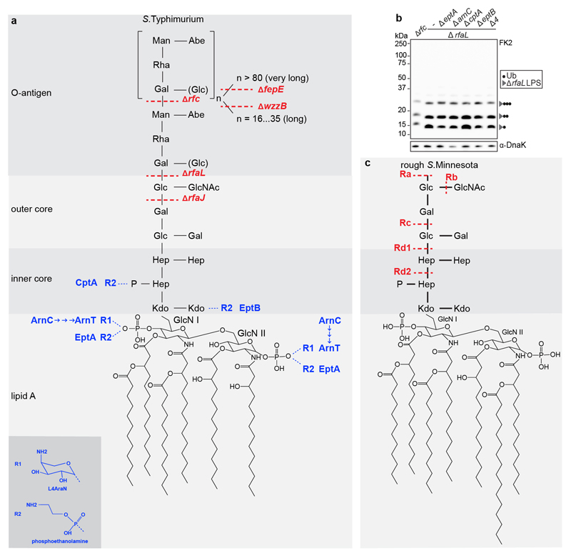
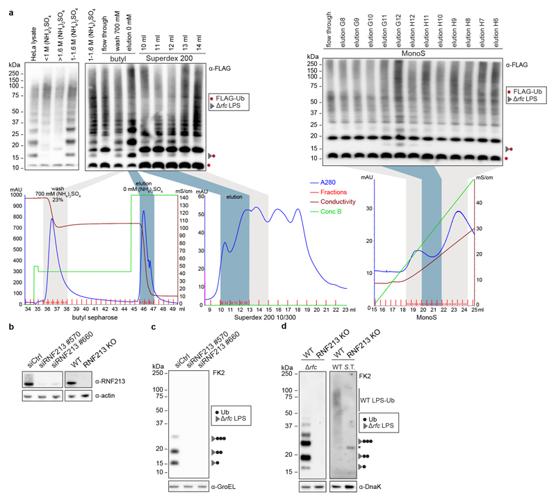
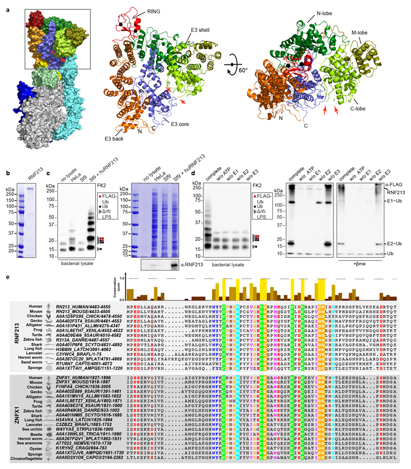
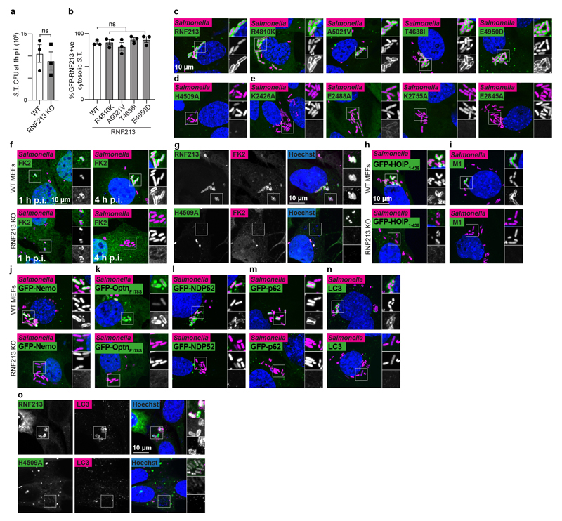
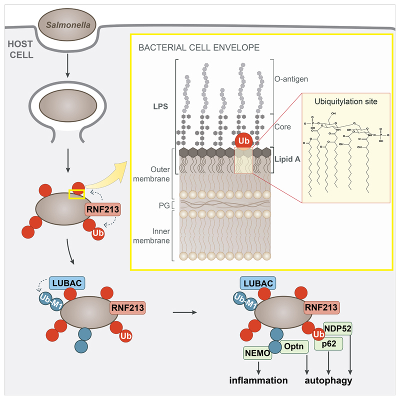
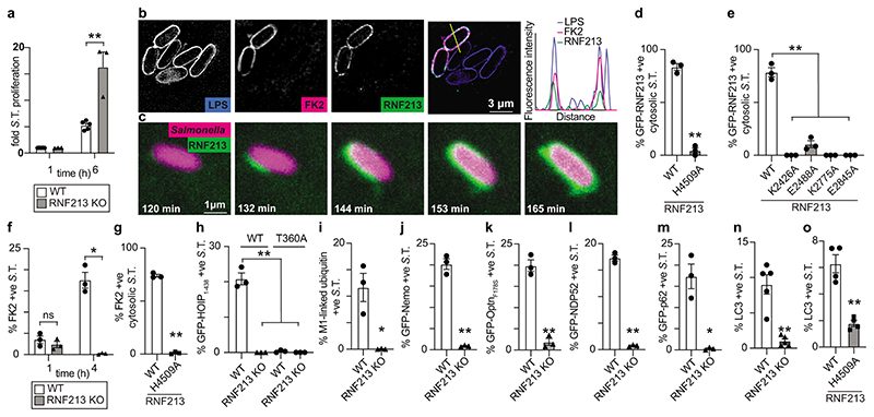
Ubiquitylation is a widespread post-translational protein modification in eukaryotes and marks bacteria that invade the cytosol as cargo for antibacterial autophagy1-3. The identity of the ubiquitylated substrate on bacteria is unknown. Here we show that the ubiquitin coat on Salmonella that invade the cytosol is formed through the ubiquitylation of a non-proteinaceous substrate, the lipid A moiety of bacterial lipopolysaccharide (LPS), by the E3 ubiquitin ligase ring finger protein 213 (RNF213). RNF213 is a risk factor for moyamoya disease4,5, which is a progressive stenosis of the supraclinoid internal carotid artery that causes stroke (especially in children)6,7. RNF213 restricts the proliferation of cytosolic Salmonella and is essential for the generation of the bacterial ubiquitin coat, both directly (through the ubiquitylation of LPS) and indirectly (through the recruitment of LUBAC, which is a downstream E3 ligase that adds M1-linked ubiquitin chains onto pre-existing ubiquitin coats8). In cells that lack RNF213, bacteria do not attract ubiquitin-dependent autophagy receptors or induce antibacterial autophagy. The ubiquitylation of LPS on Salmonella that invade the cytosol requires the dynein-like core of RNF213, but not its RING domain. Instead, ubiquitylation of LPS relies on an RZ finger in the E3 shell. We conclude that ubiquitylation extends beyond protein substrates and that ubiquitylation of LPS triggers cell-autonomous immunity, and we postulate that non-proteinaceous substances other than LPS may also become ubiquitylated.


Conflict of interest statement

Competing interests

The authors declare no competing interests.

Figures

Extended Data Figure 1
Extended Data Figure 1. LPS structure in S. Typhimurium and S.Minnesota
The composition of lipid A, inner core, outer core and O-antigen from S.Typhimurium (a) and rough variants of S.Minnesota (c). S.Typhimurium mutants deficient in specific steps of LPS biosynthesis and truncated LPS species from S.Minnesota are indicated in red. Substoichiometric modifications introducing amino groups and the enzymes responsible are indicated in blue. ArnC, the undecaprenyl-phosphate 4-deoxy-4-formamido-L-arabinose transferase, functions upstream of ArnT and is required for the ultimate incorporation of L4AraN into LPS. EptA, EptB and CptA incorporate phosphoethanolamine into LPS. b, Immunoblot analysis of the indicated S. Typhimurium strains, extracted from HeLa cells. Blots were probed with the indicated antibodies. DnaK, loading controls for bacterial lysates. A4 AeptA AarnC AcptA AeptB in ArfaL background. Representative of 3 biological repeats. For gel source data, see Supplementary Fig. 1.
Extended Data Figure 2
Extended Data Figure 2. RNF213 is required for LPS ubiquitylation
a, In vitro ubiquitylation (IVU) of S. Typhimurium Arfc extracted from infected HeLa cells. The reaction comprised FLAG-ubiquitin, E1 enzyme (UBE1), E2 enzyme (UBCH5C), and fractionated HeLa cell lysate as indicated. In the chromatograms depicted below each blot, light grey indicates fractions with little or no LPS-ubiquitylating activity, whereas blue indicates fractions with LPS-ubiquitylating activity used for further fractionation or mass spectrometry. b,Immunoblot analysis of HeLa cells transfected with the indicated siRNAs and WT HeLa and RNF213KO HeLa cells c, d, Immunoblot analysis of S. Typhimurium Arfc (c,d left panel) or WT (d right panel) extracted from HeLa cells transfected with the indicated siRNAs (c) or extracted from WT or RNF213KO MEFs (d). Representative of 3 biological repeats. Blots were probed with the indicated antibodies. Actin and GroEL or DnaK, loading controls for mammalian and bacterial lysates, respectively. *, non-specific band. For gel source data, see Supplementary Fig. 1.
Extended Data Figure 3
Extended Data Figure 3. LPS ubiquitylation by RNF213 is a RING-independent, RZ finger-mediated reaction
a, RNF213 domain structure (pdb 6TAX) (left panel) with domain colours corresponding to Fig.3a and zoom in on the E3 module (middle and right panel) indicating N-terminal (N), middle (M) and C-terminal (C) lobes of the E3 shell domain. Red arrows point at the position of the unresolved RZ finger. b, Coomassie-stained gel of purified RNF213. c left panel, d left panel, Immunoblot analysis of S.Typhimurium Arfc extracted from HeLa cells and subjected to in vitro ubiquitylation using HeLa, Sf9 or Sf9 expressing huRNF213 lysates (c) or purified RNF213 (d) corresponding to Fig.3c,d. c right panel, Coomassie gel and immunoblot analysis of lysates used in Fig.3c. d right panel, Immunoblot analysis of IVU reaction supernatants, separated under non-reducing and reducing (+pme) conditions corresponding to Fig.3d. c,d, Blots were probed with indicated antibodies. e, Alignment and conservation scores of RZ fingers in RNF213 and ZNFX1 from the indicated species. Colour and height signify degree of conservation (from Jalview) (upper panel). RZ finger alignment (lower panel). b,d, Representative of 3 biological repeats. c, n=1. For gel source data, see Supplementary Fig. 1.
Extended Data Figure 4
Extended Data Figure 4. RNF213 provides cell-autonomous immunity
a, S. Typhimurium CFU 1 h p.i. extracted from WT and RNF213KO MEFs. Bacteria were counted by serial dilution of cell lysate on LB agar plates. Data are expressed as the mean ±s.e.m. of three experiments. b, Percentage of cytosolic S. Typhimurium positive for FLAG-GFP-RNF213 at 3 h p.i. in RNF213KO MEFs stably expressing the indicated GFP- RNF213 alleles. c-o, representative confocal micrographs for Fig.4d–o. c-e, WT or RNF213KO MEFs stably expressing the indicated GFP-RNF213 alleles infected with mCherry-expressing S. Typhimurium, fixed 3 h p.i. f-o, WT and RNF213KO MEFs (f,g,n), RNF213KO MEFs stably expressing GFP-RNF213 H4509A (g,o) or WT and RNF213KO MEFs stably expressing GFP- HOIP1-438 WT or T360A (h), GFP-Nemo (j), GFP-OptineurinF178S (k), GFP-NDP52 (l), GFP- p62 (m), infected with mCherry-expressing (c-f, h-n) or BFP-expressing (g,o) S. Typhimurium, fixed 3 h p.i. and stained for FK2 (ubiquitin) (f,g), M1-linked linear ubiquitin chains (i) or LC3 (n,o). g,o, For better visibility of bacteria, Hoechst/BFP channel has been depicted in white in the zoomed sections. c-n, Scale bar, 10 pm. Statistical significance was assessed by twotailed unpaired Student’s t-test (a) or one-way anova (b). ns, not significant. Data are expressed as the mean ± s.e.m. of 3 independent experiments (a,b, source data Extended Data Fig.4) and micrographs are representative of 3 biological repeats (c-o)
Extended Data Figure 5
Extended Data Figure 5. Model of RNF213-mediated LPS ubiquitylation during bacterial infection.
Damage of Salmonella-containing vacuoles releases S.Typhimurium into the host cytosol, where RNF213 associates with the bacterial surface and ubiquitylates LPS, resulting in LUBAC recruitment and the deposition of M1-linked ubiquitin chains linked to an unidentified substrate. Recruitment of the autophagy cargo receptors NDP52 and p62 requires RNF213 but not LUBAC, while that of Optn and the IKK subunit NEMO relies on the activity of both RNF213 and LUBAC. Yellow insert: The structure of the Gram-negative cell envelope. PG peptidoglycan. Red insert: Lipid A, the minimal substrate for RNF213-mediated ubiquitylation of LPS. Ubiquitylation of lipid A is predicted to target its hydroxy or phosphate groups. Note that the C6’ OH function is not available as a potential ubiquitylation site when core is present.
Figure 1
Figure 1. Ubiquitylation of lipopolysaccharide (LPS)
a, Structured illumination micrograph of HeLa cells at 1 h and 4 h p.i. with S. Typhimurium and immunostained for LPS, FK2 and Galectin-8. Scale bar, 3 pm. Lines indicate the plot profiles adjacent. b-f, Immunoblot analysis of the indicated S. Typhimurium strains, extracted from HeLa cells (b,c,d,f) or MEFs stably expressing HA-Ubiquitin or HA-Ubiquitin AGG (e). GroEL, loading controls for bacterial lysates. b, Bacterial lysates left at room temperature (RT) or incubated at 90°C for 15 minutes. c, right panel, Emerald300 stain of LPS extracted from indicated S. Typhimurium strains grown in Luria Broth (LB). d, f, S. Typhimurium Arfc extracted from HeLa cells 4 h p.i. and treated with 0 nM, 10 nM or 2 pM recombinant USP2 catalytic domain for 30 min (d) or lysed and incubated with 100 or 200 mM NaOH for 20 min (f). K48 Ub2 provides an amide-linked control. a-f, Representative of 3 biological repeats. For gel source data, see Supplementary Fig. 1.
Figure 2
Figure 2. RNF213 is required for LPS ubiquitylation
a, In vitro ubiquitylation (IVU) of indicated S. Typhimurium strains extracted from infected HeLa cells. The IVU reaction was comprised of FLAG-ubiquitin, E1 enzyme (UBE1), E2 enzyme (UBCH5C), and HeLa lysate as E3 ligase donor, and was terminated at the indicated time points. b, Purification scheme for the LPS ubiquitylating activity. c, IVU of Hela-extracted S. Typhimurium Arfc with fractions eluted from MonoS column as E3 donor. Graph indicates the exclusive unique peptide count for RNF213 detected by mass spectrometry. d, Immunoblot analysis of indicated S. Typhimurium strains extracted from WT and RNF213KO HeLa cells. a,c,d, Blots were probed with the indicated antibodies. GroEL, loading control for bacterial lysates. S.T. Salmonella Typhimurium. a-c, n=1. d, Representative of 3 biological repeats. For gel and graph source data, see Supplementary Fig. 1 and source data Fig. 2.
Figure 3
Figure 3. LPS ubiquitylation by RNF213 is a RING-independent, RZ finger-mediated reaction
a, RNF213 domain structure. Critical residues in Walker A (K2426 and K2775) and Walker B motifs (E2488 and E2845) are indicated with red and yellow lines, respectively. RZ finger RNF213-ZNFX1 finger, CTD C-terminal domain. b, h, immunoblot analysis of S. Typhimurium Arfc extracted from WT or RNF213KO MEFs complemented with GFP or GFP-RNF213 alleles as indicated. GFP blots present the upper and lower part of continuous blots from which the middle parts have been removed as indicated by the dashed line. c-e, Immunoblot analysis of S.Typhimurium Arfc extracted from HeLa cells (c,d), or purified LPS from indicated S.Minnesota strains (e) and subjected to in vitro ubiquitylation using HeLa, Sf9 or Sf9 expressing huRNF213 lysates (c) or purified RNF213 (d,e). f, Immunoblot analysis of an IVU reaction using GST-tagged RNF213 fragments expressed in E. coli to assess autoubiquitylating activity. Representative of 3 experiments. b-f, h, Blots were probed with the indicated antibodies. Actin and GroEL, loading controls for mammalian and bacterial lysates respectively. g, RZ finger consensus amongst RNF213 and ZNFX1 proteins, human RNF213 protein sequence is depicted below. b, *, non-specific band. b,d-f,h, Representative of at least 3 biological repeats. c, n=1 For gel source data, see Supplementary Fig. 1.
Figure 4
Figure 4. RNF213 provides cell-autonomous immunity
a, Fold replication of intracellular S. Typhimurium in WT and RNF213KO MEFs normalised to 1 h p.i. time point. Bacteria were counted by serial dilution of cell lysate on LB agar plates. b, Structured illumination micrograph of HeLa cells at 4 h p.i. with S. Typhimurium and immunostained for LPS, ubiquitin (FK2) and RNF213. Scale bar, 3 pm. Line indicates the plot profile adjacent. c, Still images from Video 1: Instant structured illumination microscopy (iSIM) of MEFs expressing FLAG-GFP-RNF213 infected with mCherry-expressing S. Typhimurium. Time p.i. as indicated. Scale bar, 1 pm d-e, Percentage of cytosolic S. Typhimurium positive for FLAG-GFP-RNF213 at 3 h p.i. in WT or RNF213KO MEFs stably expressing the indicated GFP-RNF213 alleles. f-o, Percentage of S. Typhimurium positive at 3h p.i. for FK2 (ubiquitin) (f,g), GFP- HOIP1-438 WT or T360A (h), M1-linked linear ubiquitin chains (i), GFP-Nemo (j), GFP-OptineurinF178S (k), GFP-NDP52 (l), GFP-p62 (m) and LC3 (n,o) in WT and RNF213KO MEFs (f,h-n) or RNF213KO MEFs stably expressing the indicated GFP-RNF213 alleles (g,o). Statistical significance was assessed by two-tailed unpaired Student’s t-test (a,d,f,h,i-o) or one-way anova (e,h). *P < 0.05, **P < 0.01. Data are expressed as the mean ± s.e.m. of 3 (a- m),4 (o) or 5 (n) independent biological repeats (see source data Fig. 4).

Comment in

References

    1. Huang J, Brumell JH. Bacteria–autophagy interplay: a battle for survival. Nat Rev Microbiol. 2014;12:101–114. - PMC - PubMed
    1. Perrin AJ, Jiang X, Birmingham CL, So NSY, Brumell JH. Recognition of 666 Bacteria in the Cytosol of Mammalian Cells by the Ubiquitin System. Curr Biol. 2004;14:806–811. - PubMed
    1. Deretic V, Saitoh T, Akira S. Autophagy in infection, inflammation and immunity. Nat Rev Immunol. 2013;13:722–737. - PMC - PubMed
    1. Kamada F, et al. A genome-wide association study identifies RNF213 as the first Moyamoya disease gene. J Hum Genet. 2011;56:34–40. - PubMed
    1. Liu W, et al. Identification of RNF213 as a Susceptibility Gene for Moyamoya Disease and Its Possible Role in Vascular Development. Plos One. 2011;6:e22542. - PMC - PubMed

Publication types