. 2021 May 13:14:141-154.
doi: 10.2147/MDER.S307775. eCollection 2021.

A Novel Non-Invasive Device for the Assessment of Central Venous Pressure in Hospital, Office and Home

Affiliations

A Novel Non-Invasive Device for the Assessment of Central Venous Pressure in Hospital, Office and Home

Emanuela Marcelli et al. Med Devices (Auckl). .

Abstract

Background: Venous congestion can be quantified by central venous pressure (CVP) and its monitoring is crucial to understand and follow the hemodynamic status of patients with cardio-respiratory diseases. The standard technique for CVP measurement is invasive, requiring the insertion of a catheter into a jugular vein, with potential complications. On the other hand, the current non-invasive methods, mainly based on ultrasounds, remain operator-dependent and are unsuitable for use in the home environment. In this paper, we will introduce a novel, non-invasive device for the hospital, office and home assessment of CVP.

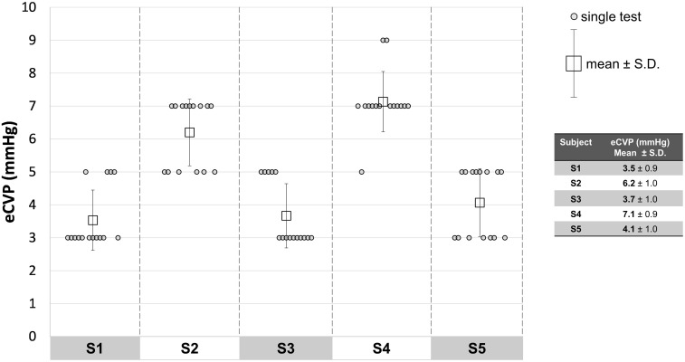
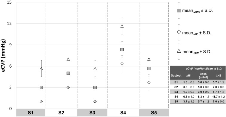
Methods: After describing the measurement concept, we will report a preliminary experimental study enrolling 5 voluntary healthy subjects to evaluate the VenCoM measurements' repeatability, and the system's capability in measuring small elicited venous pressure variations (2 mmHg), as well as an induced venous hypertension within a pathological range (12÷20 mmHg).

Results: The experimental measurements showed a repeatability of ±1mmHg. The VenCoM device was able to reliably detect the elicited venous pressure variations and the simulated congestive status.

Discussion and conclusion: The proposed non-invasive VenCoM device is able to provide a fast and repeatable CVP estimate, having a wide spectrum of potential clinical applications, including the monitoring of venous congestion in heart failure patients and in subjects with renal and hepatic dysfunction, as well as pulmonary hypertension (PH) that can be extended to pneumonia COVID-19 patients even after recovery. The device needs to be tested further on a large sample size of both healthy and pathological subjects, to systematically validate its reliability and impact in clinical setting.

Keywords: COVID-19; cardiovascular measurements; central venous pressure; heart failure; home monitoring; non-invasive device; pulmonary artery pressure.


Conflict of interest statement

E.M., L.C., B.B. have served as consultants for TRE ESSE Progettazione Biomedica srl. within the consultancy agreement between DIMES Department of Alma Mater Studiorum - Università di Bologna, and TRE ESSE Progettazione Biomedica srl. G.P. is the Scientific Director of TRE ESSE Progettazione Biomedica srl; in addition, G.P. has patents pending: 1 Italian Patent Application (IT201900010248, Filed: 27 June 2019); Dispositivo per la rilevazione della pressione venosa; Applicant: TRE ESSE Progettazione Biomedica srl; Inventor: Gianni Plicchi; 1 PCT Application (PCT/IB2020/055060, Filed 28 May 2020): Device for venous pressure sensing; Applicant: TRE ESSE Progettazione Biomedica srl; Inventor: Gianni Plicchi. The authors reported no other potential conflicts of interest for this work.

Figures

Figure 1
Figure 1
Schematic representation of the prototypal VenCoM device (A) and a photograph of the overall device assembly during a measuring session (B).
Figure 2
Figure 2
Block diagram of the main components of VenCoM device.
Figure 3
Figure 3
Example of VOP principle: (B, C) rapid volume change (RVC) in the forearm occurring when an occlusive (OC) pressure exceeding the actual central venous pressure (CVP) is applied to the arm; (A) no RVC is observed if OC pressure is below the actual CVP.
Figure 4
Figure 4
Relation between central venous pressure (CVP) and peripheral venous pressure (PVP).
Figure 5
Figure 5
The multi-step measuring algorithm used in VenCoM to obtain a quite accurate estimate of CVP (eCVP) by consecutive approximations is illustrated here through an hydraulic scheme. PVP stands for “peripheral venous pressure” (directly correlated to CVP). Blue bars represent the applied occlusive (OC) pressure at each step. The red bar represents the forearm volume, therefore the rapid volume change (RVC) that occurs when an OC pressure exceeds the actual venous pressure.
Figure 6
Figure 6
Scheme of the experimental set-up to elicit known venous pressure variations (∆P1, ∆P2) by changing the height (∆H1, ∆H2) between the center of the occlusive cuff and the level of the right atrium (B, C), starting from no height difference (A).
Figure 7
Figure 7
Results of repeatability tests, performed in 5 subjects.
Figure 8
Figure 8
Results collected when eliciting small venous pressure variations by changing the relative position between the center of the occlusive cuff and the heart (∆H1, ∆H2).
Figure 9
Figure 9
Results collected with elicited venous hypertension (VH).
Figure 10
Figure 10
Example of CVP trend assessed with VenCoM device in a subject with congestive heart failure before (measurement (A) and after successfully diuretic treatment (B–F measurements).
Figure 11
Figure 11
Example of use of VenCoM measurements for categorizing HF patient history according to the four clinical profiles of congestion dry/wet and perfusion warm/cold: transition from profile II (point 1) to profile I (point 2), after the infusion of a drug for treating pulmonary hypertension, ie, reducing CVP.
Figure 12
Figure 12
Scheme depicting the supposed CVP-based stratification of population in normal/grey zone/congestion that could be achieved by measuring CVP in a simple and non-invasive way, for preventive medicine purposes.

Similar articles

Cited by

References

    1. Magder S. Central venous pressure monitoring. Current Opinion in Critical Care. 2006;12(3):219–227. doi:10.1097/01.ccx.0000224866.01453.43. - DOI - PubMed
    1. Monge García MI, Santos Oviedo A. Why should we continue measuring central venous pressure? Medicina Intensiva. 2017;41(8):483–486. doi:10.1016/j.medin.2016.12.006. - DOI - PubMed
    1. Dellinger RP, Levy MM, Rhodes A, et al. Surviving sepsis campaign: international guidelines for management of severe sepsis and septic shock: 2012. Crit Care Med. 2013;41(2):580–637. doi:10.1097/CCM.0b013e31827e83af. - DOI - PubMed
    1. Hariri RJ, et al. Traumatic brain injury, hemorrhagic shock, and fluid resuscitation: effects on intracranial pressure and brain compliance. J Neurosurg. 1993;79(3):421–427. doi:10.3171/jns.1993.79.3.0421. - DOI - PubMed
    1. Isaacson IJ, Lowdon JD, Berry AJ, et al. The value of pulmonary artery and central venous monitoring in patients undergoing abdominal aortic reconstructive surgery: a comparative study of two selected, randomized groups. J Vasc Surg. 1990;12(6):754–760. doi:10.1067/mva.1990.24456. - DOI - PubMed

LinkOut - more resources