

Case Reports
doi: 10.1155/2021/9940304.
eCollection 2021.
Clinical, Imaging and Histopathology of Angioleiomyoma of the Buccal Cheek
Affiliations
- PMID: 34012685
- PMCID: PMC8105109
- DOI: 10.1155/2021/9940304
Item in Clipboard
Case Reports
Clinical, Imaging and Histopathology of Angioleiomyoma of the Buccal Cheek
Case Rep Dent.
.
Abstract
Angioleiomyoma is a benign neoplasia originating from vascular smooth muscle and very uncommon in the oral cavity. In this report, we present a rare case of angioleiomyoma in oral cavity in a 46-year-old female buccal cheek and discuss the clinical, histological, and immunohistochemical characteristics. As the treatment of choice is the complete excision, the lesion was excised under local anesthesia with no further complications. In addition, a brief update on other reported cases of angiomyoma in the oral cavity is further discussed.
Copyright © 2021 Mohammad Jafarian et al.
Conflict of interest statement
The authors declare no conflict of interest.
Figures

Ultrasonography shows a well-defined hypoechoic heterogenous mass lying between the skin and buccinator muscle.
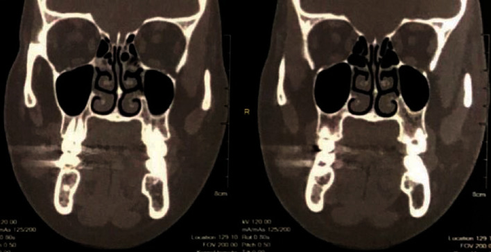
Coronal MDCT image shows a circular well-defined homogenous mass in the right buccal area.

(a) Axial T1 MRI shows a low signal homogenous mass in the right lying over the buccinator muscle and under the skin. (b) Axial T2 MRI shows the lesion as high signal and homogenous.

Intraoral incision on the buccal mucosa revealed a spherical lesion just underneath the buccinator muscle.

Image shows gross specimen, a spherical brown-gray firm tissue, measuring 1 cm in diameter.

(a) Thin blood vessels with papillary projections in the fibrous stroma (×40). (b) Paralleling pattern of fascicle and glomus cells around the blood vessels (×100).

(a) ×40 and (b) ×100 positive IHC staining for SMA. (c) ×40 and (d) ×100 positive for desmin.
References
-
- Cepeda L. G., Rivera D. Q., Rocha F. T., Huerta E., Sanchez E. Vascular leiomyoma of the oral cavity. Clinical, histopathological and immunohistochemical characteristics. Presentation of five cases and review of the literature. Medicina Oral, Patología Oral y Cirugía Bucal. 2008;13(8):e483–e488. - PubMed
Publication types
LinkOut - more resources
Full Text Sources
Research Materials
