

Pigmented Villonodular Synovitis of the Glenohumeral Joint and Biceps Tendon Sheath
- PMID: 34012737
- PMCID: PMC8127026
- DOI: 10.7759/cureus.14529
Pigmented Villonodular Synovitis of the Glenohumeral Joint and Biceps Tendon Sheath
Abstract
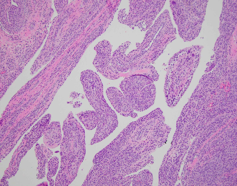
A 34-year-old woman presented with paroxysmal, insidious shoulder pain with effusion. MRI demonstrated a permeative, intermediate-signal lesion on T1 and T2 sequences involving the glenohumeral joint and biceps tendon sheath. The patient was treated with arthroscopic synovectomy, debridement, and subpectoral biceps tenodesis, with histopathology demonstrating pigmented villonodular synovitis (PVNS). PVNS is an extremely rare lesion of the glenohumeral joint and surrounding extra-articular structures. Awareness of this condition is paramount for timely diagnosis and intervention before joint destruction occurs. Arthroscopic treatment with meticulous attention to surgical technique is a feasible treatment strategy in the absence of end-stage chondral damage.
Keywords: arthroscopy; biceps tendon sheath; glenohumeral joint; pigmented villonodular synovitis; pvns; shoulder.
Copyright © 2021, Sayegh et al.
Conflict of interest statement
The authors have declared that no competing interests exist.
Figures


References
-
- Pigmented villonodular synovitis and tenosynovitis: a clinical epidemiologic study of 166 cases and literature review. Myers BW, Masi AT. https://pubmed.ncbi.nlm.nih.gov/7412554/ Medicine (Baltimore) 1980;59:223–238. - PubMed
-
- Pigmented villonodular synovitis of the shoulder: radiologic-pathologic assessment. Dorwart RH, Genant HK, Johnston WH, Morris JM. AJR Am J Roentgenol. 1984;143:886–888. - PubMed
-
- Pigmented villonodular synovitis. Tyler WK, Vidal AF, Williams RJ, Healey JH. J Am Acad Orthop Surg. 2006;14:376–385. - PubMed
-
- Diagnostic features of diffuse pigmented villonodular synovitis of the knee. Flandry F, Hughston JC, McCann SB, Kurtz DM. https://pubmed.ncbi.nlm.nih.gov/8118978/ Clin Orthop Relat Res. 1994:212–220. - PubMed
-
- Pigmented villonodular synovitis: radiologic-pathologic correlation. Murphey MD, Rhee JH, Lewis RB, Fanburg-Smith JC, Flemming DJ, Walker EA. Radiographics. 2008;28:1493–1518. - PubMed
Publication types
LinkOut - more resources
Full Text Sources
Other Literature Sources