Radiomics Nomograms Based on Multi-Parametric MRI for Preoperative Differential Diagnosis of Malignant and Benign Sinonasal Tumors: A Two-Centre Study
- PMID: 34012922
- PMCID: PMC8127839
- DOI: 10.3389/fonc.2021.659905
Radiomics Nomograms Based on Multi-Parametric MRI for Preoperative Differential Diagnosis of Malignant and Benign Sinonasal Tumors: A Two-Centre Study
Abstract
Objectives: To investigate the efficacy of multi-parametric MRI-based radiomics nomograms for preoperative distinction between benign and malignant sinonasal tumors.
Methods: Data of 244 patients with sinonasal tumor (training set, n=192; test set, n=52) who had undergone pre-contrast MRI, and 101 patients who underwent post-contrast MRI (training set, n=74; test set, n=27) were retrospectively analyzed. Independent predictors of malignancy were identified and their performance were evaluated. Seven radiomics signatures (RSs) using maximum relevance minimum redundancy (mRMR), and the least absolute shrinkage selection operator (LASSO) algorithm were established. The radiomics nomograms, comprising the clinical model and the RS algorithms were built: one based on pre-contrast MRI (RNWOC); the other based on pre-contrast and post-contrast MRI (RNWC). The performances of the models were evaluated with area under the curve (AUC), calibration, and decision curve analysis (DCA) respectively.
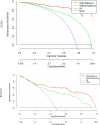
Results: The efficacy of the clinical model (AUC=0.81) of RNWC was higher than that of the model (AUC=0.76) of RNWOC in the test set. There was no significant difference in the AUC of radiomic algorithms in the test set. The RS-T1T2 (AUC=0.74) and RS-T1T2T1C (RSWC, AUC=0.81) achieved a good distinction efficacy in the test set. The RNWC and the RNWOC showed excellent distinction (AUC=0.89 and 0.82 respectively) in the test set. The DCA of the nomograms showed better clinical usefulness than the clinical models and radiomics signatures.
Conclusions: The radiomics nomograms combining the clinical model and RS can be accurately, safely and efficiently used to distinguish between benign and malignant sinonasal tumors.
Keywords: benign; differential diagnosis; magnetic resonance imaging; malignant; radiomics; sinonasal.
Copyright © 2021 Bi, Zhang, Wang, Ge, Zhang, Wang and Hao.
Conflict of interest statement
Author Y-QG was employed by company GE Healthcare China. The remaining authors declare that the research was conducted in the absence of any commercial or financial relationships that could be construed as a potential conflict of interest.
Figures


Similar articles
-
Multi-parametric MRI-based radiomics signature for preoperative prediction of Ki-67 proliferation status in sinonasal malignancies: a two-centre study.Eur Radiol. 2022 Oct;32(10):6933-6942. doi: 10.1007/s00330-022-08780-w. Epub 2022 Jun 10. Eur Radiol. 2022. PMID: 35687135
-
Bi-parametric magnetic resonance imaging based radiomics for the identification of benign and malignant prostate lesions: cross-vendor validation.Phys Eng Sci Med. 2021 Sep;44(3):745-754. doi: 10.1007/s13246-021-01022-1. Epub 2021 Jun 1. Phys Eng Sci Med. 2021. PMID: 34075559
-
Automated Prediction of Early Recurrence in Advanced Sinonasal Squamous Cell Carcinoma With Deep Learning and Multi-parametric MRI-based Radiomics Nomogram.Acad Radiol. 2023 Oct;30(10):2201-2211. doi: 10.1016/j.acra.2022.11.013. Epub 2023 Mar 14. Acad Radiol. 2023. PMID: 36925335
-
Additional value of metabolic parameters to PET/CT-based radiomics nomogram in predicting lymphovascular invasion and outcome in lung adenocarcinoma.Eur J Nucl Med Mol Imaging. 2021 Jan;48(1):217-230. doi: 10.1007/s00259-020-04747-5. Epub 2020 May 25. Eur J Nucl Med Mol Imaging. 2021. PMID: 32451603
-
Radiomics Analysis in Characterization of Salivary Gland Tumors on MRI: A Systematic Review.Cancers (Basel). 2023 Oct 10;15(20):4918. doi: 10.3390/cancers15204918. Cancers (Basel). 2023. PMID: 37894285 Free PMC article. Review.
Cited by
-
Multi-parametric MRI-based radiomics signature for preoperative prediction of Ki-67 proliferation status in sinonasal malignancies: a two-centre study.Eur Radiol. 2022 Oct;32(10):6933-6942. doi: 10.1007/s00330-022-08780-w. Epub 2022 Jun 10. Eur Radiol. 2022. PMID: 35687135
-
Artificial intelligence and MRI in sinonasal tumors discrimination: where do we stand?Eur Arch Otorhinolaryngol. 2025 Mar;282(3):1557-1566. doi: 10.1007/s00405-024-09169-9. Epub 2024 Dec 24. Eur Arch Otorhinolaryngol. 2025. PMID: 39719474
-
MRI radiomics may predict early tumor recurrence in patients with sinonasal squamous cell carcinoma.Eur Radiol. 2024 May;34(5):3151-3159. doi: 10.1007/s00330-023-10389-6. Epub 2023 Nov 6. Eur Radiol. 2024. PMID: 37926740
-
Towards Precision Medicine in Sinonasal Tumors: Low-Dimensional Radiomic Signature Extraction from MRI.Diagnostics (Basel). 2025 Jun 30;15(13):1675. doi: 10.3390/diagnostics15131675. Diagnostics (Basel). 2025. PMID: 40647674 Free PMC article.
-
A Clinical-Radiomics Nomogram Based on the Apparent Diffusion Coefficient (ADC) for Individualized Prediction of the Risk of Early Relapse in Advanced Sinonasal Squamous Cell Carcinoma: A 2-Year Follow-Up Study.Front Oncol. 2022 May 16;12:870935. doi: 10.3389/fonc.2022.870935. eCollection 2022. Front Oncol. 2022. PMID: 35651794 Free PMC article.
References
-
- Harnsberger HR, Wiggins HR, Hudgins AP, Al E. Diagnostic Imaging: Head and Neck. Am Assoc Neurol Surgeons (2011) 104(6):167–7. 10.3171/jns.2006.104.1.167a - DOI
LinkOut - more resources
Full Text Sources
Other Literature Sources

