Review
. 2021 May 3:9:660145.
doi: 10.3389/fbioe.2021.660145. eCollection 2021.

Hydrogel Scaffolds to Deliver Cell Therapies for Wound Healing

Affiliations
Review

Hydrogel Scaffolds to Deliver Cell Therapies for Wound Healing

Dharshan Sivaraj et al. Front Bioeng Biotechnol. .

Abstract

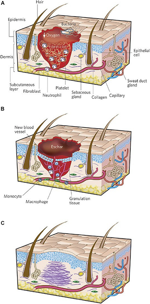
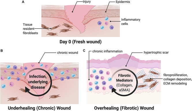
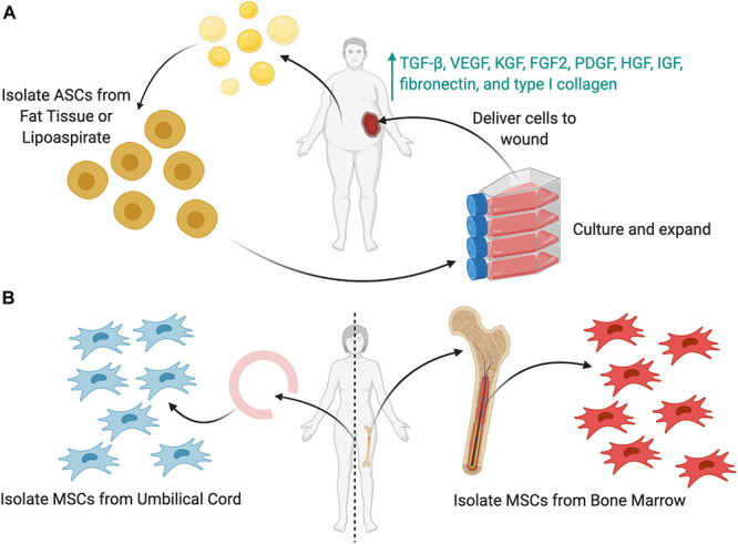
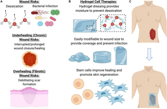
Cutaneous wounds are a growing global health burden as a result of an aging population coupled with increasing incidence of diabetes, obesity, and cancer. Cell-based approaches have been used to treat wounds due to their secretory, immunomodulatory, and regenerative effects, and recent studies have highlighted that delivery of stem cells may provide the most benefits. Delivering these cells to wounds with direct injection has been associated with low viability, transient retention, and overall poor efficacy. The use of bioactive scaffolds provides a promising method to improve cell therapy delivery. Specifically, hydrogels provide a physiologic microenvironment for transplanted cells, including mechanical support and protection from native immune cells, and cell-hydrogel interactions may be tailored based on specific tissue properties. In this review, we describe the current and future directions of various cell therapies and usage of hydrogels to deliver these cells for wound healing applications.

Keywords: cell therapy; fibrosis; hydrogel; stem cell; wound healing.


Conflict of interest statement

The authors declare that the research was conducted in the absence of any commercial or financial relationships that could be construed as a potential conflict of interest.

Figures

FIGURE 1
FIGURE 1
Three stages of wound repair. The three phases of wound repair consist of (A) inflammation, (B) new tissue formation, and (C) remodeling. (A) The inflammatory phase lasts until about 48 h after injury. Depicted is a skin wound at about 24–48 h after injury. The wound is characterized by a hypoxic (ischemic) environment in which a fibrin clot has formed. Bacteria, neutrophils, and platelets are abundant in the wound. Normal skin appendages (such as hair follicles and sweat duct glands) are still present in the skin outside the wound. (B) New tissue formation occurs about 2–10 days after injury. Depicted is a skin wound at about 5–10 days after injury. The majority of cells from the previous stage of repair have migrated from the wound, and new blood vessels now populate the area. An eschar (scab) has formed on the surface of the wound, and the migration of epithelial cells can be observed under the eschar. (C) Remodeling lasts for a year or longer. Depicted is a skin wound about 1–12 months after repair. Disorganized collagen has been laid down by fibroblasts that have migrated into the wound and contracted the wound. The re-epithelialized wound is slightly higher than the surrounding surface, and the healed region does not contain normal skin appendages. Figure adapted with permission from Figure 1 of Gurtner et al. (2008).
FIGURE 2
FIGURE 2
Overview of wound healing. (A) After an initial wound, both circulating and tissue resident cells are recruited to the wound, including fibroblasts and inflammatory cells. (B) Chronic wounds are characterized by interruptions and subsequent prolongation of the wound healing process. (C) Fibrotic wounds are characterized by upregulation of myofibroblast differentiation and increased collagen production and may be driven by mechanical signaling. aSMA, alpha smooth muscle actin.
FIGURE 3
FIGURE 3
Sources of stem cells to be used as cellular therapies. (A) Adipose-derived stem cells (ASCs) can be harvested from either lipoaspirate or fat tissue. These cells (or MSCs) can then be cultured in vitro, expanded in number, and then delivered to the wound as a cell therapy. To enhance the cell delivery, several delivery techniques (shown later) may be used. (B) Mesenchymal stromal cells (MSCs) can be harvested from either the umbilical cord or the bone marrow.
FIGURE 4
FIGURE 4
Benefits of using a hydrogel dressing to deliver cellular therapies. (A) Open wounds are at risk for desiccation (loss of moisture) and bacterial infection, as well either underhealing or overhealing. (B) To address these major risk factors, hydrogel dressings provide coverage and moisture, as well as a beneficial ECM environment for cells to grow in. (C) Once the hydrogel has been seeded with cells, it can be laid on the wound to promote healing and regeneration.
FIGURE 5
FIGURE 5
Pullulan-collagen hydrogel can be easily modified across a wide range of factors and provides biocompatibility with stem cells. (A–F) Scanning electron microscopy (SEM) imaging demonstrates that varying collagen and KCl concentrations significantly alters the porosity of the hydrogels. Hydrogels fabricated with KCl showed increased porosity, while increasing collagen concentrations decreased porosity. Scale bar 100 μm. (G,H) Brightfield imaging and fluorescent imaging of live (green) dead (red/yellow) stain shows successful in vitro cellular incorporation of MSCs in the hydrogels. Scale bar 50 μm. (I) SEM shows MSCs (arrows) viably incorporated within the pullulan-collagen hydrogels. Scale bar 25 μm. Figures adapted with permission from Figures 2, 6 of Wong et al. (2011b).
FIGURE 6
FIGURE 6
Hydrogel with cellular therapy promotes wound healing. Hydrogel delivery system serves as a physiologic milieu to encapsulate cells to ensure efficient delivery to wound site. Cells such as MSCs or ASCs may be seeded into the hydrogels. Upon application to the wound, cells migrate into the wound to initiate a cascade of beneficial phenotypes.

Similar articles

Cited by

References

    1. Aderibigbe B. A., Buyana B. (2018). Alginate in wound dressings. Pharmaceutics 10:42. 10.3390/pharmaceutics10020042 - DOI - PMC - PubMed
    1. Adli M. (2018). The CRISPR tool kit for genome editing and beyond. Nat. Commun. 9:1911. - PMC - PubMed
    1. Ahmed T. A., Griffith M., Hincke M. (2007). Characterization and inhibition of fibrin hydrogel-degrading enzymes during development of tissue engineering scaffolds. Tissue Eng. 13 1469–1477. 10.1089/ten.2006.0354 - DOI - PubMed
    1. Alison L., Menasce S., Bouville F., Tervoort E., Mattich I., Ofner A., et al. (2019). 3D printing of sacrificial templates into hierarchical porous materials. Sci. Rep. 9:409. - PMC - PubMed
    1. Atiyeh B. S., Gunn S. W., Hayek S. N. (2007). Military and civilian burn injuries during armed conflicts. Annals Burns Fire Disasters 20 203–215. - PMC - PubMed

LinkOut - more resources