. 2021 Mar 31;2(1):180-192.
doi: 10.1089/neur.2020.0058. eCollection 2021.

Repetitive Blast Exposure Produces White Matter Axon Damage without Subsequent Myelin Remodeling: In Vivo Analysis of Brain Injury Using Fluorescent Reporter Mice

Affiliations

Repetitive Blast Exposure Produces White Matter Axon Damage without Subsequent Myelin Remodeling: In Vivo Analysis of Brain Injury Using Fluorescent Reporter Mice

Donald V Bradshaw Jr et al. Neurotrauma Rep. .

Abstract

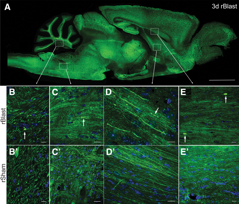
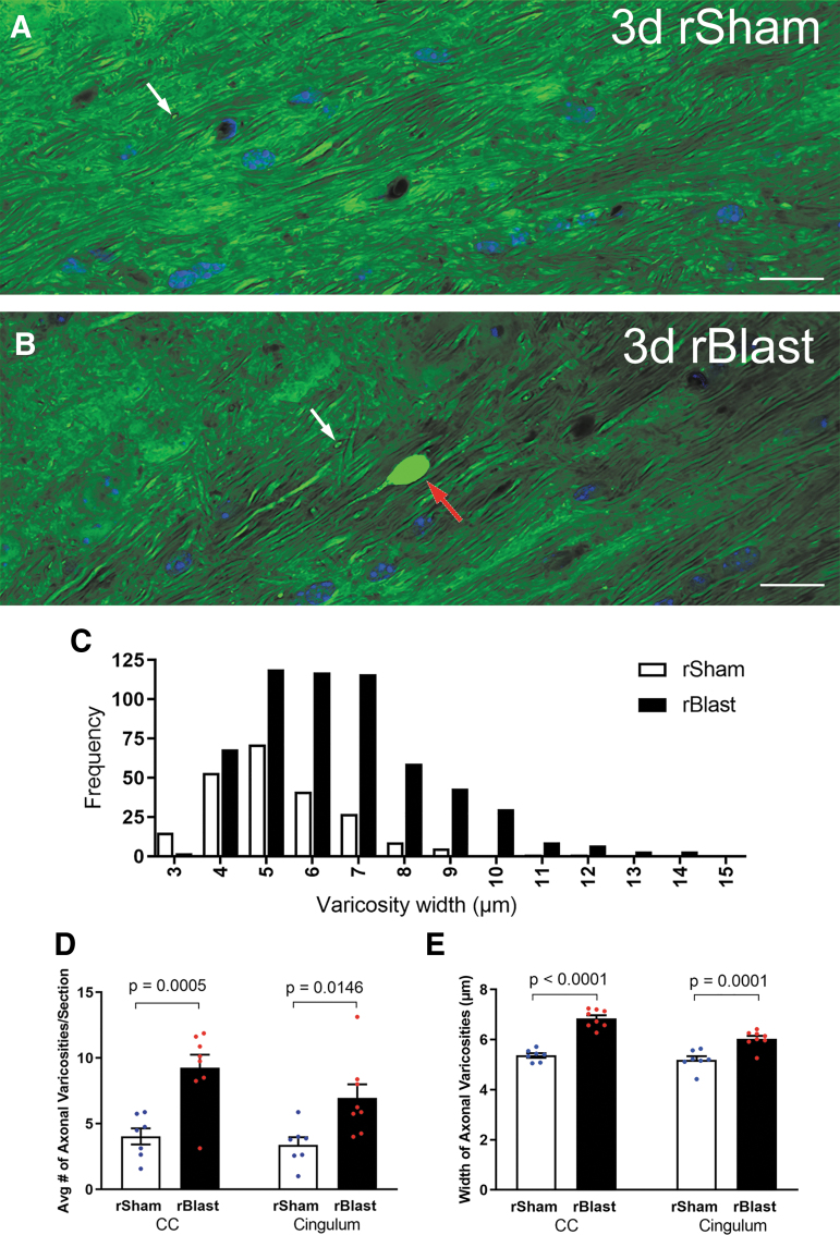
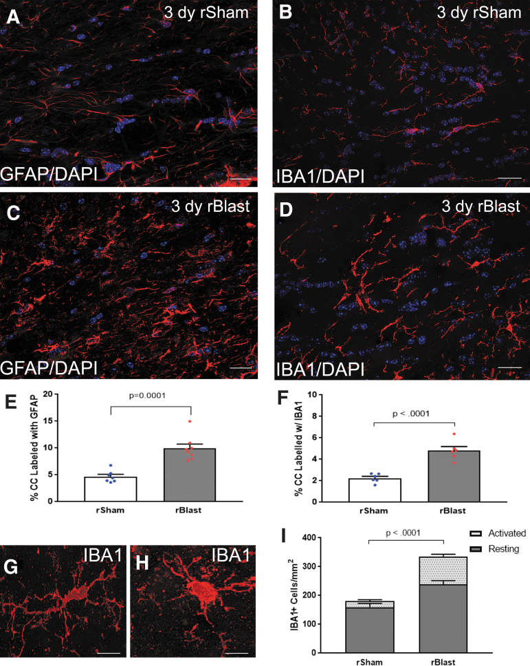
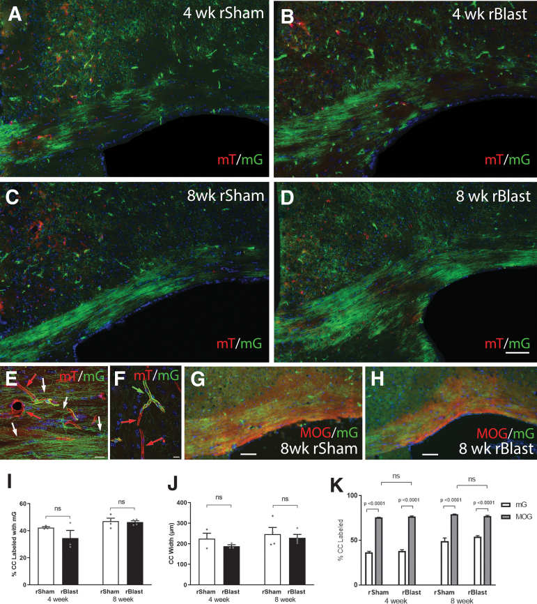
The potential effects of blast exposure on the brain health of military personnel have raised concerns and led to increased surveillance of blast exposures. Neuroimaging studies have reported white matter abnormalities in brains of service members with a history of blast exposure. However, blast effects on white matter microstructure remain poorly understood. As a novel approach to screen for white matter effects, transgenic mice that express fluorescent reporters to sensitively detect axon damage and myelin remodeling were exposed to simulated repetitive blasts (once/day on 5 consecutive days). Axons were visualized using Thy1-YFP-16 reporter mice that express yellow fluorescent protein (YFP) in a broad spectrum of neurons. Swelling along damaged axons forms varicosities that fill with YFP. The frequency and size of axonal varicosities were significantly increased in the corpus callosum (CC) and cingulum at 3 days after the final blast exposure, versus in sham procedures. CC immunolabeling for reactive astrocyte and microglial markers was also significantly increased. NG2CreER;mTmG mice were given tamoxifen (TMX) on days 2 and 3 after the final blast to induce fluorescent labeling of newly synthesized myelin membranes, indicating plasticity and/or repair. Myelin synthesis was not altered in the CC over the intervening 4 or 8 weeks after repetitive blast exposure. These experiments show the advantages of transgenic reporter mice for analysis of white matter injury that detects subtle, diffuse axon damage and the dynamic nature of myelin sheaths. These results show that repetitive low-level blast exposures produce infrequent but significant axon damage along with neuroinflammation in white matter.

Keywords: axon damage; blast exposure; myelin; transgenic reporter mice; traumatic brain injury; white matter.


Conflict of interest statement

No competing financial interests exist.

Figures

FIG. 1.
FIG. 1.
Broad, sensitive screening for axon damage in brain sections after repetitive blast exposure using Thy1-YFP-16 mice. (A) Sagittal section from a Thy1-YFP-16 mouse at 3 days after rBlast exposure shows widely distributed neuron cell bodies and processes expressing yellow fluorescent protein (YFP; shown in green). (B–D’) Sagittal high-magnification confocal microscope images of axons in white matter tracts in distributed brain regions of rBlast mice (B–D) and matching regions in rSham mice (B’–D’). YFP shown in green. Nuclear stain 4′,6-diamidino-2-phenylindole (DAPI) shown in blue. (B) Example of YFP accumulated in damaged axon to illustrate a terminal end bulb (arrow; transverse view) in the cerebellum of an rBlast mouse. Note that other YFP-filled oval and elongated structures are axons shown in angled cuts in both rBlast (B) and rSham (B’) mice. The intense YFP of damaged axons is distinct from larger, more diffuse labeling in neuron cell bodies (adjacent to B’ label). Examples from rBlast mice of YFP in an axonal thickening (arrow; longitudinal view) in the pons region of the brainstem (C) and in fragmented axons (arrows; longitudinal view) in the column of the fornix (D). (E,E’) Coronal confocal image showing corpus callosum axons in longitudinal view with two examples of intense YFP labeling in large terminal end bulbs in an rBlast section (E; arrows), in comparison with an rSham section (E’). Low-power image (A) is representative of images from three Thy1-YFP-16 mice examined in full sagittal sections. Higher-power images (B–E’) were acquired from sections of these or additional Thy1-YFP-16 mice examined in coronal sections for quantification in Figure 2. Scale bars = 2 mm (A), 20 μm (B–E and B’–E’).
FIG. 2.
FIG. 2.
Repetitive blast exposure increases axon damage in the corpus callosum (CC) and adjacent cingulum. (A,B) Confocal images of coronal sections of the CC from Thy1-YFP-16 mice at 3 days after repetitive sham procedures (rSham; A) or repetitive blast exposure (rBlast; B) showing high magnification of yellow fluorescent protein signal (YFP) within axons and showing 4′,6-diamidino-2-phenylindole (DAPI) nuclear stain in blue. The rSham mice exhibit relatively uniform YFP signal filling of axons viewed longitudinally (A). There are small variations in diameter along the length of the axons. In addition, YFP signal is visible in axons of crossing fibers cut transversely (A; white arrow). An example in an rBlast mouse of an axon with a large, intensely fluorescent YFP-filled enlargement indicative of axon damage (B; red arrow) that is distinct from crossing fiber axons (B; white arrow). (C) Analysis of the frequency distribution indicates an increase in both the number and diameter of YFP accumulations in the CC and cingulum of rBlast mice compared with rSham controls. (D) The rBlast significantly increases the incidence of damaged axons with abnormal YFP localization (thickenings, varicosities, and terminal end bulbs) in the CC and cingulum, compared with rSham mice. (E) YFP accumulations in axonal varicosities are significantly larger in rBlast compared with rSham mice (B). Values are mean ± standard error of the mean (SEM). Two-way analysis of variance (ANOVA) was used with repeated measures, for CC and cingulum within same subjects, and Sidak's post hoc multiple comparisons test. For the procedures, mouse numbers were: rSham group, n = 7 and rBlast group, n = 8. Scale bars = 20 μm (A and B).
FIG. 3.
FIG. 3.
Neuroinflammation is significantly increased in the corpus callosum (CC) after blast exposure. (A–D) Immunohistochemistry to detect glial fibrillary acidic protein (GFAP) in astrocytes and ionized calcium binding adaptor molecule 1 (IBA1) as a marker of microglia/macrophages. Confocal microscope images of coronal CC sections from Thy1-YFP-16 mice at 3 days after repetitive sham procedures (rSham; A,B) or repetitive blast exposure (rBlast; C,D). Astrocytes (pseudocolored white) and microglia/macrophages (red) have thicker processes with more intense immunoreactivity in rBlast mice as compared with rShams. Nuclear stain 4′,6-diamidino-2-phenylindole (DAPI) shown in blue. (E,F) The rBlast exposure significantly increased immunoreactivity for GFAP (E) and IBA1 (F). (G,H) Representative confocal images showing IBA1+ cells as examples of resting state morphology with an elongated cell body and numerous processes (G) or activated morphology with a more rounded and intensely immunolabeled cell body and processes (H). (I) rBlast exposure significantly increases the number of IBA1+ cells in the CC. Values are mean ± standard error of the mean (SEM). Student's t test was used to compare between groups. GFAP groups include rSham, n = 7; rBlast, n = 8. IBA1 groups include rSham, n = 6; rBlast, n = 7. Scale bars = 20 μm (A–D) and 5 μm (G,H).
FIG. 4.
FIG. 4.
Myelin membrane synthesis in the corpus callosum (CC) after repetitive blast exposure in NG2CreER;mTmG reporter mice. NG2CreER;mTmG mice were given tamoxifen (TMX) on days 2–3 after repetitive blast (rBlast) or sham (rSham) procedures. TMX induces recombination to stop expression of membrane-localized tdTomato (mT) and initiate expression of membrane-localized green fluorescent protein (mG) driven from the NG2 promoter (NG2mG). NG2 expression drives genetic mG fate-labeling of oligodendrocyte progenitors that is maintained in newly generated oligodendrocytes and their myelin membranes. (A–D) Representative images of mG (green) labeled membranes in coronal sections through the CC at 4 weeks (A,B) or 8 weeks (C,D) after rSham (A,C) or rBlast (B,D). (E,F) High-magnification confocal microscope images within the CC (E) and cortex (F). Constitutive mT (red) labeling is visible in membranes of non-recombined cells, including blood vessels (E; red arrows). NG2 fate-labeled oligodendrocytes extend myelin-like membranes (E; white arrows) along axons. NG2 fate-labeled pericyte (F; green arrow) extend processes around blood vessels (F; red arrows). Nuclear stain 4′,6-diamidino-2-phenylindole (DAPI) shown in blue. (G,H) Representative images of mG fluorescence (green) in newly synthesized myelin membranes along with immunolabeling for myelin oligodendrocyte glycoprotein (MOG) in new and persisting myelin membranes (red). The mG membranes represents only a subset of the total myelinated area of the CC. (I) rBlast did not significantly alter the proportion of mG-labeled myelin membranes synthesized in the CC at either time point. (J) rBlast exposure did not result in significant CC atrophy at either time-point. (K) Quantification of mG labeling relative to total MOG immunolabeled myelin in the same sections. The myelinated fraction in the CC is significantly greater than the mG new myelin and is not changed by rBlast exposure. Values are mean ± standard error of the mean (SEM). Two-way analysis of variance (ANOVA) with Sidak's post hoc multiple comparisons test. Four-week groups include rSham, n = 3; rBlast, n = 3. Eight-week groups include rSham, n = 4; rBlast, n = 4. Scale bars = 100 μm (A–D, shown in D), 20μm (E–H).

References

    1. Mac Donald, C.L., Barber, J., Patterson, J., Johnson, A.M., Dikmen, S., Fann, J.R., and Temkin, N. (2019). Association between 5-year clinical outcome in patients with nonmedically evacuated mild blast traumatic brain injury and clinical measures collected within 7 days postinjury in combat. JAMA Netw. Open 2, e186676. - PMC - PubMed
    1. Aldag, M., Armstrong, R.C., Bandak, F., Bellgowan, P.S.F., Bentley, T., Biggerstaff, S., Caravelli, K., Cmarik, J., Crowder, A., DeGraba, T.J., Dittmer, T.A., Ellenbogen, R.G., Greene, C., Gupta, R.K., Hicks, R., Hoffman, S., Latta, R.C., 3rd, Leggieri, M.J.Jr., Marion, D., Mazzoli, R., McCrea, M., O'Donnell, J., Packer, M., Petro, J.B., Rasmussen, T.E., Sammons-Jackson, W., Shoge, R., Tepe, V., Tremaine, L.A., and Zheng, J. (2017). The biological basis of chronic traumatic encephalopathy following blast injury: a literature review. J. Neurotrauma 34, S26–S43 - PMC - PubMed
    1. Regasa, L.E., Agimi, Y., and Stout, K.C. (2019). Traumatic brain injury following military deployment: evaluation of diagnosis and cause of injury. J. Head Trauma Rehabil. 34, 21–29 - PubMed
    1. Engel, C.C., Hoch, E., Simmons, M.M. (2019). The neurological effects of repeated exposure to military occupational levels of blast: a review of scientific literature. RAND Conference Proceedings
    1. Carr, W., Stone, J.R., Walilko, T., Young, L.A., Snook, T.L., Paggi, M.E., Tsao, J.W., Jankosky, C.J., Parish, R.V., and Ahlers, S.T. (2016). Repeated low-level blast exposure: a descriptive human subjects study. Mil Med. 181, 28–39 - PubMed

LinkOut - more resources