[Preprint]. 2021 Apr 23:rs.3.rs-448370.
doi: 10.21203/rs.3.rs-448370/v1.

In vivo monoclonal antibody efficacy against SARS-CoV-2 variant strains

Affiliations

In vivo monoclonal antibody efficacy against SARS-CoV-2 variant strains

Michael Diamond et al. Res Sq. .

Update in

  • In vivo monoclonal antibody efficacy against SARS-CoV-2 variant strains.
    Chen RE, Winkler ES, Case JB, Aziati ID, Bricker TL, Joshi A, Darling TL, Ying B, Errico JM, Shrihari S, VanBlargan LA, Xie X, Gilchuk P, Zost SJ, Droit L, Liu Z, Stumpf S, Wang D, Handley SA, Stine WB Jr, Shi PY, Davis-Gardner ME, Suthar MS, Knight MG, Andino R, Chiu CY, Ellebedy AH, Fremont DH, Whelan SPJ, Crowe JE Jr, Purcell L, Corti D, Boon ACM, Diamond MS. Chen RE, et al. Nature. 2021 Aug;596(7870):103-108. doi: 10.1038/s41586-021-03720-y. Epub 2021 Jun 21. Nature. 2021. PMID: 34153975 Free PMC article.

Abstract

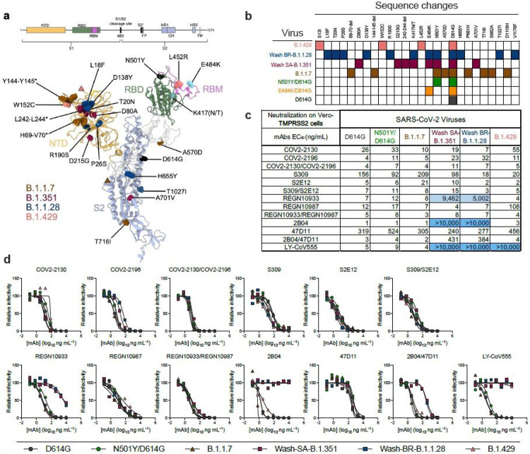
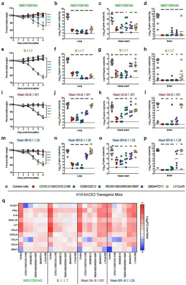
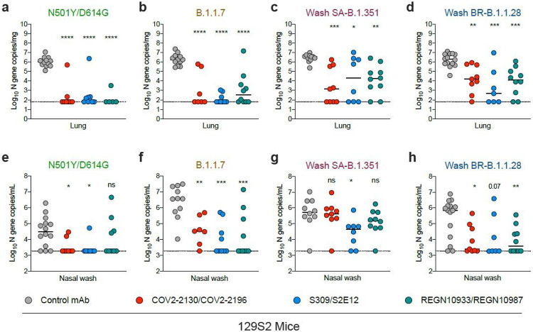
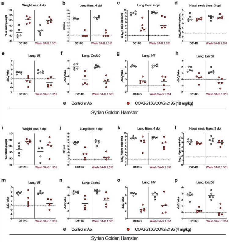
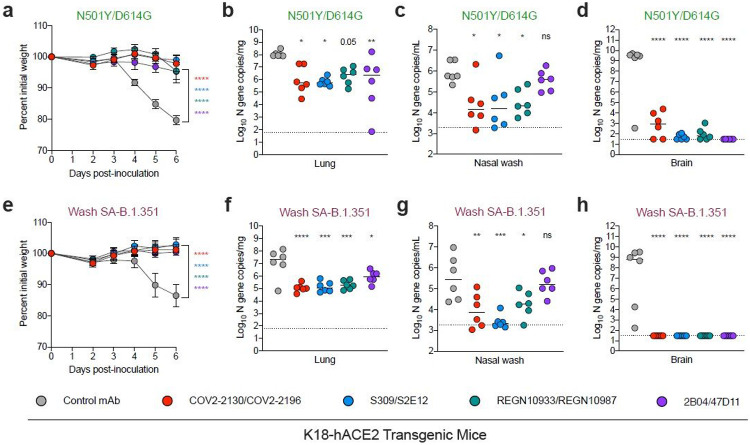
Rapidly-emerging variants jeopardize antibody-based countermeasures against SARS-CoV-2. While recent cell culture experiments have demonstrated loss of potency of several anti-spike neutralizing antibodies against SARS-CoV-2 variant strains1-3, the in vivo significance of these results remains uncertain. Here, using a panel of monoclonal antibodies (mAbs) corresponding to many in advanced clinical development by Vir Biotechnology, AbbVie, AstraZeneca, Regeneron, and Lilly we report the impact on protection in animals against authentic SARS-CoV-2 variants including WA1/2020 strains, a B.1.1.7 isolate, and chimeric strains with South African (B.1.351) or Brazilian (B.1.1.28) spike genes. Although some individual mAbs showed reduced or abrogated neutralizing activity against B.1.351 and B.1.1.28 viruses with E484K spike protein mutations in cell culture, low prophylactic doses of mAb combinations protected against infection in K18-hACE2 transgenic mice, 129S2 immunocompetent mice, and hamsters without emergence of resistance. Two exceptions were mAb LY-CoV555 monotherapy which lost all protective activity in vivo, and AbbVie 2B04/47D11, which showed partial loss of activity. When administered after infection as therapy, higher doses of mAb cocktails protected in vivo against viruses displaying a B.1.351 spike gene. Thus, many, but not all, of the antibody products with Emergency Use Authorization should retain substantial efficacy against the prevailing SARS-CoV-2 variant strains.


Figures

Figure 1
Figure 1
Neutralization of SARS-CoV-2 variant strains by clinically relevant mAbs. (a) SARS-CoV-2 variant substitutions mapped onto the structure of the spike protein. Schematic layout of the spike protein monomer is depicted at the top. Structure of spike monomer (PDB: 7C2L with RBD from PDB: 6W41) is depicted as a cartoon, with NTD, RBD, RBM, and S2 colored orange, green, magenta, and light blue, respectively. Substitutions for each variant (B.1.1.7: 69-70 deletion, 144-145 deletion, N501Y, A570D, D614G, P681H,and T716I; B.1.351: 242-244 deletion, D80A, D215G, K417N, E484K, N501Y, D614G, and A701V; B.1.1.28: L18F, T20N, P26S, D138Y, R190S, K417T, E484K, N501Y, D614G, H655Y, T1027I; B.1.429: S13I, W152C, L452R) are shown as spheres and colored accordingly. Substitutions shown in cyan (E484K and K417[N/T]) are shared by B.1.351 and B.1.1.28. Substitutions shown in black (D614G and N501Y) are shared by B.1.1.7, B.1.351, and B.1.1.28. Pink and brown triangles show approximate locations of S13 and P681, which were not modelled in the original structures. Structural figure generated using UCSF ChimeraX45. (b) Viruses used with indicated mutations in the spike protein. (c) Summary of EC50 values (ng/mL) of neutralization of SARS-CoV-2 viruses performed in Vero-TMPRSS2 cells. Blue shading of cells indicates a partial (EC50 > 1,000 ng/mL) or complete (EC50 > 10,000 ng/mL) loss of neutralizing activity. (d) Neutralization curves comparing the sensitivity of SARS-CoV-2 strains to the indicated individual or combinations of mAbs. Data are representative of two to five experiments, each performed in technical duplicate.
Figure 2
Figure 2
Antibody prophylaxis against SARS-CoV-2 variants in K18-hACE2 mice. (a-q) 8-10-week-old female and male K18-hACE2 transgenic mice received 40 g (~2 mg/kg) of the indicated mAb treatment by intraperitoneal injection one day before intranasal inoculation with 103 FFU of the indicated SARS-CoV-2 strain. Tissues were collected at 6 dpi. (a, e, i, m) Weight change following infection with SARS-CoV-2 (mean ± SEM; n = 6-12 mice per group, two experiments; one-way ANOVA with Dunnett’s test of area under the curve: ns, not significant, **** P < 0.0001). Viral RNA levels in the lung (b, f, j, n), nasal washes (c, g, k, o), and brain (d, h, l, p) were measured (n = 6-12 mice per group, two experiments; one-way ANOVA with Dunnett’s test with comparison to control mAb: ns, not significant, * P < 0.05, ** P < 0.01, *** P < 0.001, **** P < 0.0001). Dotted line indicates the limit of detection of the assay. Heat map of cytokine and chemokine protein expression levels in lung homogenates collected at 6 dpi from the indicated groups (q). Data are presented as log2 fold-change over naive animals. Blue, reduction; red, increase.
Figure 3
Figure 3
Antibody-mediated protection against SARS-CoV-2 variants in 129S2 mice. 6-7-week-old female and male immunocompetent 129S2 mice received 40 mg (~ 2 mg/kg) of the indicated mAb treatment by intraperitoneal injection one day before intranasal inoculation with 103 FFU of WA1/2020 N501Y/D614G, Wash SA-B.1.351, or Wash BR-B.1.1.28 and 105 FFU of B.1.1.7. Tissues were collected at 3 dpi. Viral RNA levels in the lung (a-d) or nasal washes (e-h) were determined (n = 7-12 mice per group, pooled from two experiments; one-way ANOVA with Dunnett’s test with comparison to control mAb: ns, not significant, * P < 0.05, ** P < 0.01, *** P < 0.001). Dotted line indicates the limit of detection of the assay.
Figure 4
Figure 4
COV2-2130/COV2-2196 antibody cocktail protects hamsters against historical and variant SARS-CoV-2 strains. Six-week-old male Syrian golden hamsters received a single 800 mg (~10 mg/kg) (a-h) or 320 mg (~4 mg/kg) dose (i-p) of COV2-2130/COV2196 mAb cocktail or control mAb by intraperitoneal injection one day before intranasal inoculation with 5 x 10⁵ FFU of WA1/2020 D614G or Wash SA-B.1.351 viruses. Nasal swabs and lung tissues were collected at 3 and 4 dpi, respectively. (a, i) Weight change following infection with SARS-CoV-2 (mean ± SEM; n = 5 animals per group, one experiment). Infectious virus in the lung (b, j) or viral RNA levels in the lung (c, k) and nasal swabs (d, l) were determined (n = 5 animals per group, one experiment). Dotted line indicates the limit of detection of the assay. (e-h, m-p) Cytokine and inflammatory gene expression in lung homogenates collected at 6 dpi from indicated groups. Values were calculated using the DDCt method compared to a naive control group. Because data were obtained from a single experiment (even with multiple animals), statistical analysis was not performed.
Figure 5
Figure 5
Post-exposure antibody therapy against SARS-CoV-2 variants in K18-hACE2 mice. (a-h) 8-10-week-old female and male K18-hACE2 transgenic mice were administered 103 FFU of the indicated SARS-CoV-2 strain by intranasal inoculation. One day later, mice received 200 mg (~10 mg/kg) of the indicated mAb treatment by intraperitoneal injection. Tissues were collected at 6 dpi. (a, e) Weight change following infection with SARS-CoV-2 (mean ± SEM; n = 6 mice per group, two experiments; one-way ANOVA with Dunnett’s test of area under the curve: **** P < 0.0001). Viral RNA levels in the lung (b, f), nasal wash (c, g), and brain (d, h) (n = 6 mice per group, two experiments; one-way ANOVA with Dunnett’s test with comparison to control mAb: ns, not significant, * P < 0.05, ** P < 0.01, *** P < 0.001, **** P < 0.0001). Dotted line indicates the limit of detection of the assay.

Similar articles

References

    1. Chen R.E., et al. Resistance of SARS-CoV-2 variants to neutralization by monoclonal and serum-derived polyclonal antibodies. Nat Med(2021). - PMC - PubMed
    1. Wang P., et al. Antibody Resistance of SARS-CoV-2 Variants B. 1.351 and B.1.1.7. Nature(2021). - PubMed
    1. Wang Z., et al. mRNA vaccine-elicited antibodies to SARS-CoV-2 and circulating variants. Nature(2021). - PMC - PubMed
    1. Sempowski G.D., Saunders K.O., Acharya P., Wiehe K.J. & Haynes B.F. Pandemic Preparedness: Developing Vaccines and Therapeutic Antibodies For COVID-19. Cell 181, 1458–1463 (2020). - PMC - PubMed
    1. Letko M., Marzi A. & Munster V. Functional assessment of cell entry and receptor usage for SARS-CoV-2 and other lineage B betacoronaviruses. Nature microbiology 5, 562–569 (2020). - PMC - PubMed

Publication types

LinkOut - more resources