[Preprint]. 2021 May 10:2021.05.09.443331.
doi: 10.1101/2021.05.09.443331.

SARS-CoV-2 ferritin nanoparticle vaccines elicit broad SARS coronavirus immunogenicity

Affiliations

SARS-CoV-2 ferritin nanoparticle vaccines elicit broad SARS coronavirus immunogenicity

M Gordon Joyce et al. bioRxiv. .

Update in

  • SARS-CoV-2 ferritin nanoparticle vaccines elicit broad SARS coronavirus immunogenicity.
    Joyce MG, Chen WH, Sankhala RS, Hajduczki A, Thomas PV, Choe M, Martinez EJ, Chang WC, Peterson CE, Morrison EB, Smith C, Chen RE, Ahmed A, Wieczorek L, Anderson A, Case JB, Li Y, Oertel T, Rosado L, Ganesh A, Whalen C, Carmen JM, Mendez-Rivera L, Karch CP, Gohain N, Villar Z, McCurdy D, Beck Z, Kim J, Shrivastava S, Jobe O, Dussupt V, Molnar S, Tran U, Kannadka CB, Soman S, Kuklis C, Zemil M, Khanh H, Wu W, Cole MA, Duso DK, Kummer LW, Lang TJ, Muncil SE, Currier JR, Krebs SJ, Polonis VR, Rajan S, McTamney PM, Esser MT, Reiley WW, Rolland M, de Val N, Diamond MS, Gromowski GD, Matyas GR, Rao M, Michael NL, Modjarrad K. Joyce MG, et al. Cell Rep. 2021 Dec 21;37(12):110143. doi: 10.1016/j.celrep.2021.110143. Epub 2021 Dec 8. Cell Rep. 2021. PMID: 34919799 Free PMC article.

Abstract

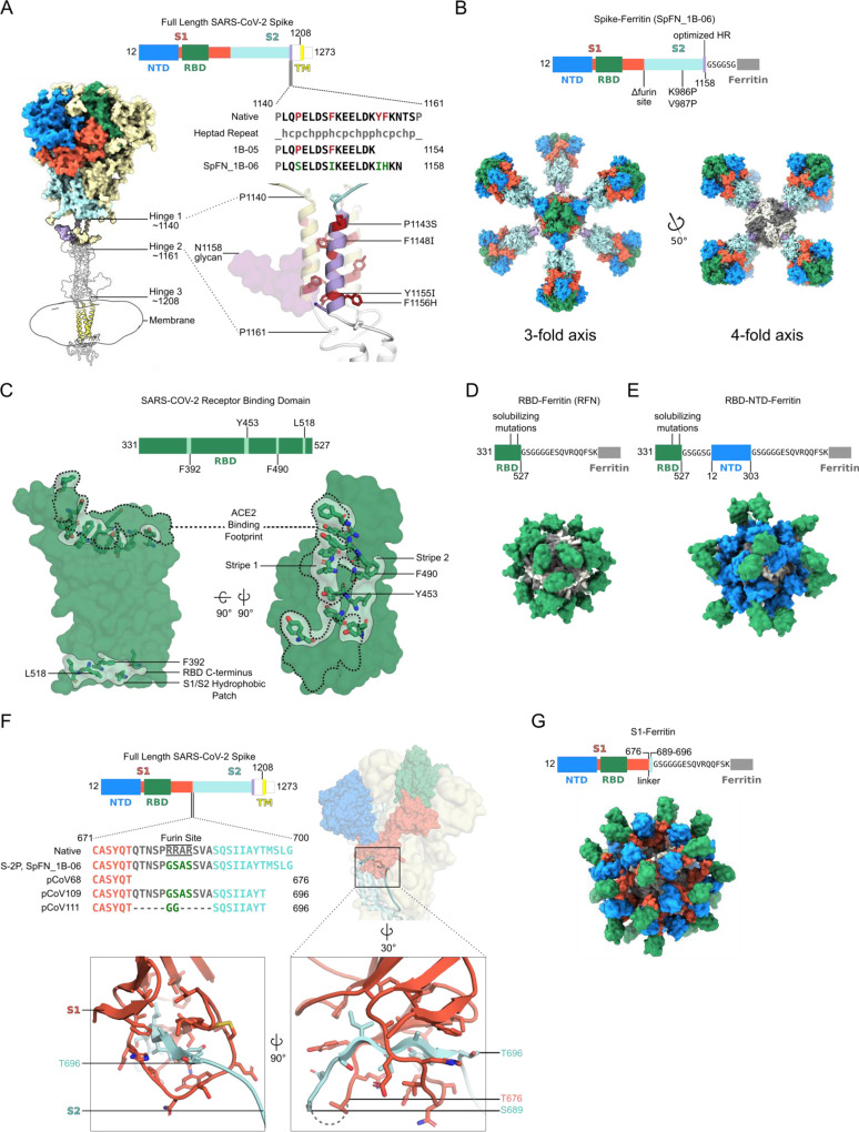
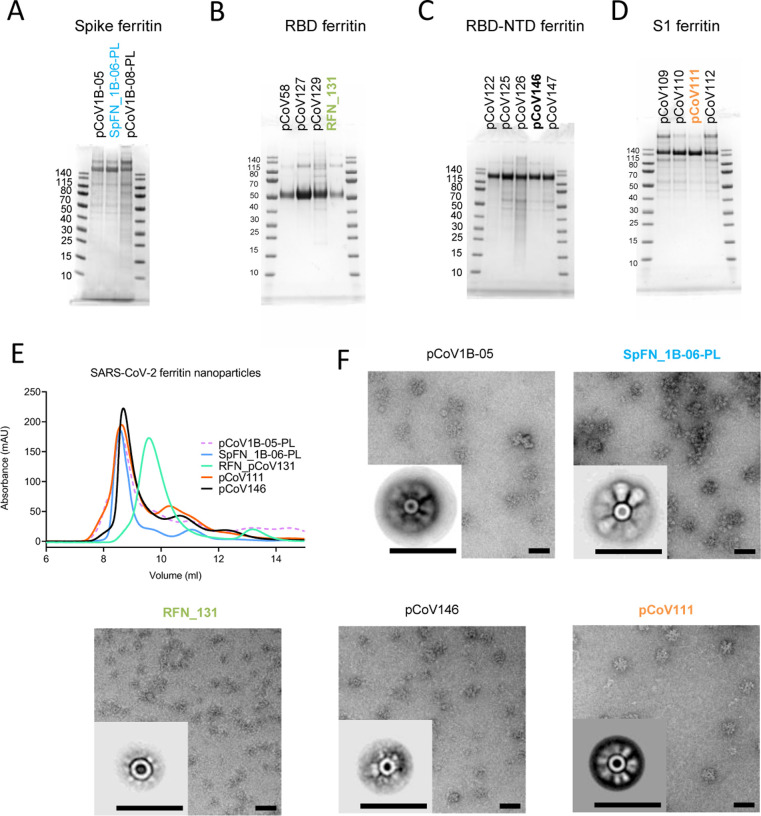
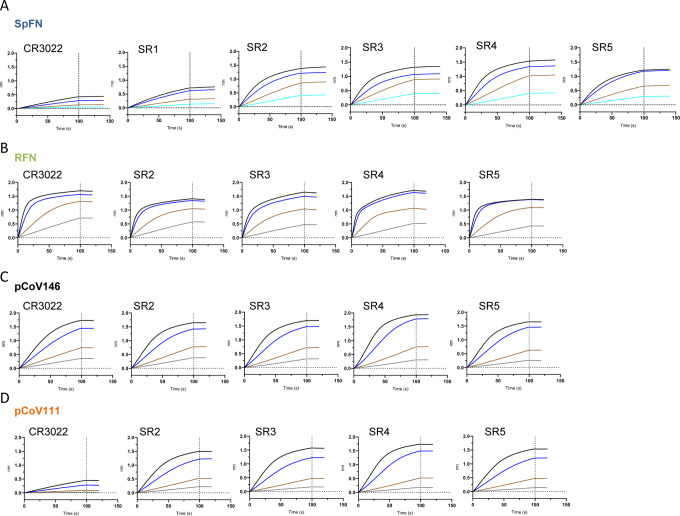
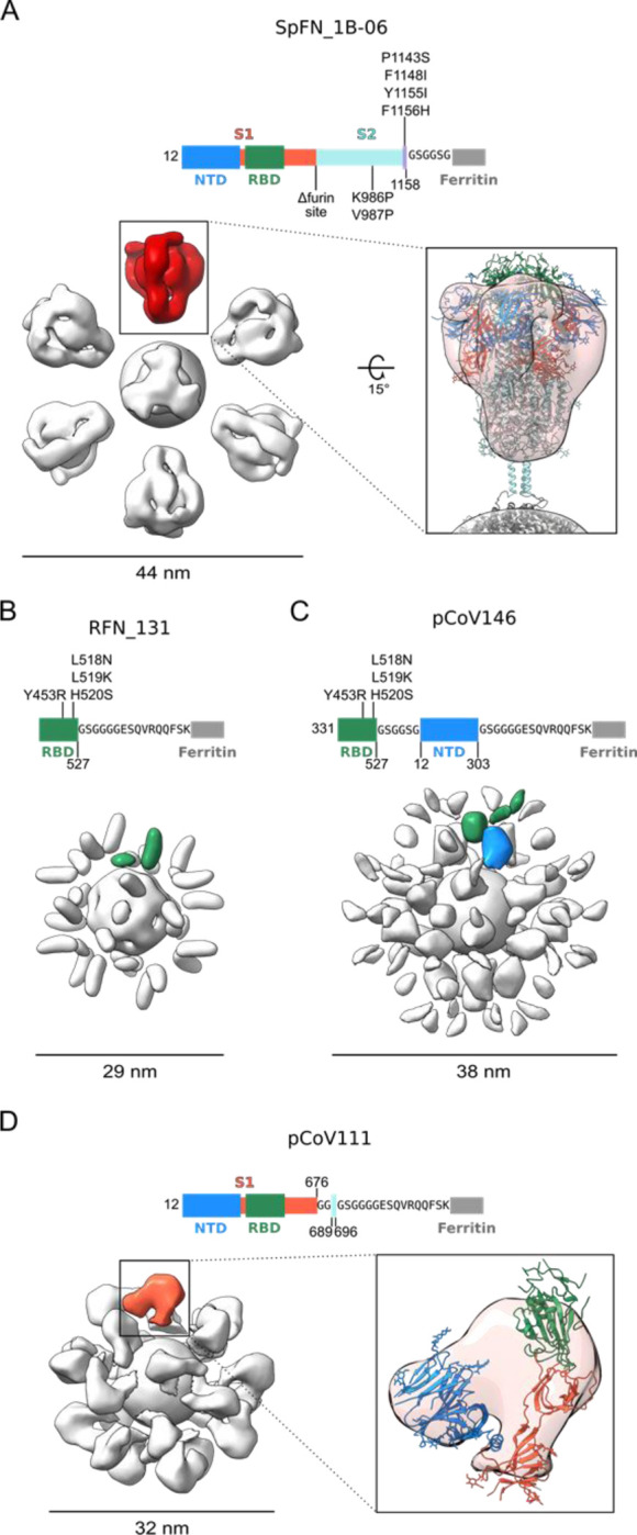
The need for SARS-CoV-2 next-generation vaccines has been highlighted by the rise of variants of concern (VoC) and the long-term threat of other coronaviruses. Here, we designed and characterized four categories of engineered nanoparticle immunogens that recapitulate the structural and antigenic properties of prefusion Spike (S), S1 and RBD. These immunogens induced robust S-binding, ACE2-inhibition, and authentic and pseudovirus neutralizing antibodies against SARS-CoV-2 in mice. A Spike-ferritin nanoparticle (SpFN) vaccine elicited neutralizing titers more than 20-fold higher than convalescent donor serum, following a single immunization, while RBD-Ferritin nanoparticle (RFN) immunogens elicited similar responses after two immunizations. Passive transfer of IgG purified from SpFN- or RFN-immunized mice protected K18-hACE2 transgenic mice from a lethal SARS-CoV-2 virus challenge. Furthermore, SpFN- and RFN-immunization elicited ACE2 blocking activity and neutralizing ID50 antibody titers >2,000 against SARS-CoV-1, along with high magnitude neutralizing titers against major VoC. These results provide design strategies for pan-coronavirus vaccine development.

Highlights: Iterative structure-based design of four Spike-domain Ferritin nanoparticle classes of immunogensSpFN-ALFQ and RFN-ALFQ immunization elicits potent neutralizing activity against SARS-CoV-2, variants of concern, and SARS-CoV-1Passively transferred IgG from immunized C57BL/6 mice protects K18-hACE2 mice from lethal SARS-CoV-2 challenge.


Conflict of interest statement

DECLARATIONS OF INTERESTS

M.G.J. and K.M. are named as inventors on International Patent Application No. WO/2021/21405 entitled “Vaccines against SARS-CoV-2 and other coronaviruses.” M.G.J. is named as an inventor on International Patent Application No. WO/2018/081318 and U.S. patent 10,960,070 entitled “Prefusion Coronavirus Spike Proteins and Their Use.” Z.B. is named as an inventor on U.S. patent 10,434,167 entitled “Non-toxic adjuvant formulation comprising a monophosphoryl lipid A (MPLA)-containing liposome composition and a saponin.” Z.B. and G.R.M are named inventors on “Compositions And Methods For Vaccine Delivery”, US Patent Application: 16/607,917. M.S.D. is a consultant for Inbios, Vir Biotechnology, NGM Biopharmaceuticals and Carnival Corporation and on the Scientific Advisory Boards of Moderna and Immunome. The Diamond laboratory has received funding support in sponsored research agreements from Moderna, Vir Biotechnology and Emergent BioSolutions. S.R., P.M.M., and M.T.E. are employees of AstraZeneca and currently hold AstraZeneca stock or stock options. Zoltan Beck is currently employed at Pfizer.

Figures

Figure 1.
Figure 1.. Structure-based design of SARS-CoV-2 S-based ferritin nanoparticle immunogens
(A) Full length SARS-CoV-2 S schematic and 3D-structure. S hinges identified by molecular dynamics simulations and electron cryotomography are labeled on the 3D- model ((Turonova et al., 2020). The structured trimeric ectodomain is colored according to the schematic with the N-terminal domain (NTD) and Receptor-Binding Domain (RBD) of the S1 polypeptide and the C-terminal coiled coil N-terminal to hinge 1 colored blue, green, and purple, respectively. Remaining portions of the S1 and S2 polypeptides are colored in red and cyan with regions membrane-proximal from hinge 2 colored in white. The transmembrane domain of all chains is depicted in yellow. To design a Spike-Ferritin molecule, the C-terminal heptad repeat (residues 1140 to 1161) between Hinge 1 and 2 were aligned to an ideal heptad repeat sequence. Residues in the native S sequence which break this pattern are highlighted in red. These residues are also labeled and highlighted in red on the 3D-structure. Two engineered designs (1B-05 and 1B-06) are shown, with S end residue used to link to Ferritin, and heptad-repeat mutations colored green. (B) Schematic and 3D model of Spike Ferritin nanoparticle (SpFN). Differences between the native S sequence and the engineered nanoparticle are indicated on the schematic. A 3D-model of SpFN displaying eight trimeric Spikes was created using PDB ID 6VXX and 3EGM with the ferritin molecule shown in alternating grey and white. The nanoparticle is depicted along the 4-fold and the 3-fold symmetry axes of the ferritin. (C) RBD–Ferritin nanoparticle design and optimization. The RBD of SARS-CoV-2 (PDB ID:6MOJ) is shown in surface representation, with the ACE2 binding site outlined in dashed lines. Three hydrophobic regions of the RBD which were mutated for nanoparticle immunogen design are shown in light green surface, with residues in stick representation. The ACE2 binding site contains two of these regions, while a third hydrophobic patch near the C-terminus of the RBD is typically buried by S2 and part of S1 in the context of the trimer molecule. (D) Schematic and 3D model of an RBD–Ferritin nanoparticle. A modeled 24-mer nanoparticle displaying the RBD domain is depicted at the 3-fold symmetry axis of ferritin and colored green. Truncation points, linkers, and alterations made to the RBD sequence are indicated on the schematic. (E) Schematic and 3D model of an RBD–NTD–Ferritin nanoparticle. A modeled nanoparticle displaying RBD and NTD epitopes is depicted and colored according to the schematic. Truncation points, linkers, and alterations made to the native S sequence are indicated on the primary structure. (F) S1-Ferritin immunogen design. The SARS-CoV-2 S1 forms a hydrophobic collar around the N-terminal β-sheet of S2 (residues 689–676). S1-ferritin immunogen design required inclusion of this short stretch of S2 (colored cyan) attached by a linker. Terminal residues of the structured portions of S1 and S2 are labeled. (G) Schematic and 3D model of an S1–Ferritin nanoparticle. A modeled nanoparticle displaying RBD and NTD domains is depicted and colored according to the S1–ferritin schematic with truncation points and domain linkers indicated. See also Figure S1 and Table S1.
Figure 2.
Figure 2.. Antigenic and biophysical characterization of SARS-CoV-2 Spike-based ferritin nanoparticle vaccine candidates
SDS-PAGE of (A) Spike-Ferritin nanoparticle designs, (B) Receptor-binding domain-Ferritin nanoparticle designs, (C) S1-ferritin nanoparticle, and (D) RBD-NTD-Ferritin nanoparticles. Molecular weight standards are indicated in kDa. (E) Size-exclusion chromatography on a Superdex S200 10/300 column of representative SARS-CoV-2 S-based ferritin nanoparticles. (F) Negative-stain electron microscopy 2D class averages of purified nanoparticles. The black bars represent 50 nm. See also Figure S2 and S3.
Figure 3.
Figure 3.. Antigenic characterization of select SARS-CoV-2 Spike-based ferritin nanoparticle vaccine candidates
Binding response of SARS-CoV-2 neutralizing antibodies to each of the lead candidates from the four design categories measured by biolayer interferometry with two-fold serial dilution of each antibody starting at 30 μg/ml). (A) Spike-Ferritin nanoparticle SpFN_1B-06-PL. (B) RBD-Ferritin pCoV131. (C) RBD-NTD-Ferritin nanoparticle pCoV146. (D) S1-Ferritin nanoparticle pCoV111. See also Figure S3.
Figure 4.
Figure 4.. Negative-Stain Electron Microscopy 3D Reconstructions of SARS-CoV-2 Spike Domain-Ferritin Nanoparticles
Modifications made to native sequence and linkers used for each construct are shown in schematic diagrams. (A) Negative-stain 3D reconstructions with applied octahedral symmetry are shown with an asymmetric unit of non-ferritin density colored and the size of each particle indicated in nanometers. Spike trimer density, is colored in red, and a model of a SpFN trimer based on PDB 6VXX is shown docked into the negative-stain map and colored according to the sequence diagram. (B) Two non-ferritin densities per asymmetric unit were observed for RFN and are highlighted in green. These densities putatively correspond to the receptor-binding domain (RBD) but lack low resolution distinguishing features due to the small, globular shape of these domains. The presence of two densities is likely due to flexibility in the linker and heterogeneity in the RBD pose. (C) Two layers of densities were distinguishable for pCoV146, with the putative N-terminal domain (NTD) density of an asymmetric unit colored blue, proximal to the ferritin and two smaller, more flexible densities corresponding to RBD distal to the ferritin and colored green. (D) An asymmetric unit of non-ferritin density for pCoV111 is colored in orange and a monomer of S1 in the closed trimer state from PDB 6VXX is shown docked into the density with domains colored as in the sequence diagram. See also Figure S2 and Table S2.
Figure 5
Figure 5. SARS-CoV-2 Spike-domain nanoparticle vaccine candidates elicit robust binding and neutralizing antibody responses in mice.
(A) Biolayer interferometry binding of mouse sera to SARS-COV-2 RBD. Study week is indicated on the base of the graph. Mean value is indicated by a horizontal line. Statistical comparison at each timepoint was carried out using a a Kruskal-Wallis test followed by a Dunn’s post-test. (B) ELISA binding of mouse sera to SARS-COV-2 S-2P or RBD. Study week is indicated on the base of the graph. Geometric mean value is indicated by a horizontal line. Statistical comparison at each timepoint was carried out using a a Kruskal-Wallis test followed by a Dunn’s post-test. (C) SARS-COV-2 pseudovirus neutralization ID50 and ID80 values. Geometric mean value is indicated by a horizontal line. Statistical comparisons at each given timepoint was carried out using a Kruskal-Wallis test followed by a Dunn’s post-test. (D) Binding and pseudovirus neutralization of sera from mice immunized with 0.08 μg SpFN + ALFQ. (E) Authentic SARS-CoV-2 virus strain 2019-nCoV/USA_WA1/2020 neutralization ID50 and ID80 are shown for mice immunized with 10 or 0.08 μg SpFN + ALFQ. Geometric mean value is indicated by a horizontal line. Comparisons between dose group at each time point were carried out using a Mann-Whitney unpaired two-tailed non-parametric test, n=10 mice/group. In panels A – C, all immunogen groups at a given study timepoint were compared to each other. Only groups with statistically significant differences are indicated by a bar; all other groups did not show statistically significant differences. P values <0.0001 (****), <0.001 (***), <0.01 (**), or <0.05 (*). See also Figure S4 and S5, and Table S3 and S4.
Figure 6.
Figure 6.. SpFN- and RBD-FN protective immunity in K18-hACE2 transgenic mice.
(A) IgG was purified from SPFN- or RFN-vaccinated mouse sera and passively transferred at specific IgG amounts ranging from 4 – 470 μg/mouse in a final volume of 200 μl. Control naïve mouse IgG was formulated at 2 mg/ml. (n=10/group, 5 female, 5 male). (B) Mouse challenge study schematic. K18-hACE2 mice (n=10/group, 5 female, 5 male) received control IgG (black), PBS (gray), and purified IgG, one day prior to challenge with 1.25 × 10⁴ PFU of SARS-CoV-2. (C) SARS-CoV-2 pseudovirus neutralization ID50 titers of mouse sera at study day 0. (D) Percentage of initial weight of K18-hACE2 mice for the 8 study groups. Legend is shown in panel E. (E) Survival curves of K18-hACE2 mice with groups indicated based on animal vaccination group and the pseudovirus ID50 neutralization values. Statistical comparisons were carried out using Mantel-Cox test followed by Bonferroni correction. See also Figure S6.
Figure 7.
Figure 7.. SARS-CoV-2 Spike-domain nanoparticle vaccine candidates elicit robust antibody binding responses and neutralizing activity against SARS-CoV-2 VoC and SARS-CoV-1.
(A) Biolayer Interferometry binding of study week 10 immunized C57BL/6 mouse serum to SARS-CoV-2 RBD, and SARS-CoV-2 RBD variants. Immunogens are indicated at the top left of each graph. Mean values are indicated by a horizontal line, n=10, Significance was assessed using a Kruskal-Wallis test followed by a Dunn’s post-test. (B) Pseudovirus neutralization (ID50 values) of study week 10 immunized C57BL/6 and BALB/c mouse serum to SARS-CoV-2 Wuhan-1, B.1.1.7, and B.1.351 pseudotyped viruses. Immunogens are indicated at the base of each graph. Geometric mean values are indicated by a horizontal line, n=5, statistical significance for each immunogen was assessed using a Kruskal-Wallis test followed by a Dunn’s post-test. (C) Biolayer Interferometry binding of study week 10 immunized C57BL/6 and BALB/c mouse serum to SARS-CoV-1 RBD. Immunogens are indicated at the base of each graph. Mean values are indicated by a horizontal line, n=10, Significance was assessed using a Kruskal-Wallis test followed by a Dunn’s post-test. (D) Pseudovirus neutralization (ID50 values) of study week 10 immunized C57BL/6 and BALB/c mouse serum to SARS-CoV-1 Urbani strain pseudotyped viruses. Data related to SpFN and RFN are colored blue and green respectively. Statistical comparisons between SpFN and RFN responses at each time point were carried out using a Mann-Whitney unpaired two-tailed non-parametric test. Immunogens are indicated at the base of each graph. Geometric mean values are indicated by a horizontal line, n=10, P values <0.0001 (****), <0.01 (**) or <0.05 (*). See also Figure S4 and S5.

References

    1. Bangaru S., Ozorowski G., Turner H.L., Antanasijevic A., Huang D., Wang X., Torres J.L., Diedrich J.K., Tian J.H., Portnoff A.D., et al. (2020). Structural analysis of full-length SARS-CoV-2 spike protein from an advanced vaccine candidate. Science 370, 1089–1094. - PMC - PubMed
    1. Barnes C.O., West A.P. Jr., Huey-Tubman K.E., Hoffmann M.A.G., Sharaf N.G., Hoffman P.R., Koranda N., Gristick H.B., Gaebler C., Muecksch F., et al. (2020). Structures of Human Antibodies Bound to SARS-CoV-2 Spike Reveal Common Epitopes and Recurrent Features of Antibodies. Cell 182, 828–842 e816. - PMC - PubMed
    1. Boyarsky B.J., Werbel W.A., Avery R.K., Tobian A.A.R., Massie A.B., Segev D.L., and Garonzik-Wang J.M. (2021). Immunogenicity of a Single Dose of SARS-CoV-2 Messenger RNA Vaccine in Solid Organ Transplant Recipients. JAMA. - PMC - PubMed
    1. Boyington J.C., Joyce M.G., Sastry M., Stewart-Jones G.B., Chen M., Kong W.P., Ngwuta J.O., Thomas P.V., Tsybovsky Y., Yang Y., et al. (2016). Structure-Based Design of Head-Only Fusion Glycoprotein Immunogens for Respiratory Syncytial Virus. PLoS One 11, e0159709. - PMC - PubMed
    1. Boyoglu-Barnum S., Ellis D., Gillespie R.A., Hutchinson G.B., Park Y.-J., Moin S.M., Acton O., Ravichandran R., Murphy M., Pettie D., et al. (2020). Elicitation of broadly protective immunity to influenza by multivalent hemagglutinin nanoparticle vaccines. bioRxiv, 2020.2005.2030.125179.

Publication types