Review
. 2021 Jun;187(2):224-234.
doi: 10.1002/ajmg.c.31917. Epub 2021 May 20.

Neurocutaneous syndromes in art and antiquities

Affiliations
Review

Neurocutaneous syndromes in art and antiquities

Martino Ruggieri et al. Am J Med Genet C Semin Med Genet. 2021 Jun.

Erratum in

Abstract

Neurocutaneous syndromes are a group of genetic disorders affecting the skin, the central and peripheral nervous system, and the eye with congenital abnormalities and/or tumors. Manifestations may also involve the heart, vessels, lungs, kidneys, endocrine glands and bones. When people with these disorders are portrayed in works of art, physicians have speculated on possible diagnoses. In particular, many figures have been labeled as possibly having a neurocutaneous disorder, sometimes distorting the popular conception of these diseases. We review numerous documents, drawings, prints, lithographs, xylographs, and portraits which span the ages from antiquity to the era of the pioneers behind the eponyms, depicting a large spectrum of neurocutaneous disorders.

Keywords: artwork; disorders; neurocutaneous; painting; phacomatosis; print.


Conflict of interest statement

None.

Figures
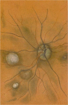
FIGURE 1
FIGURE 1
The original color print used by Jan van der Hoeve to depict the retinal “phakomas” seen as whitish/gray oval lesions in the central and left retina in the fundus of a patient with tuberous sclerosis. Copyright © The British Medical Council Library, London, UK
FIGURE 2
FIGURE 2
(a) Hellenistic statuette, representing a partial adult male body covered with multiple, smooth and sessile nodules, interpreted as neurofibromas. Copyright © 1994 Nature Publishing Group; (b,c) coins of the Parthian empire reproducing the left face of king Orodes II (b) [50 BC], and king Phraates IV (c) [20 BC] each represented with a large and round nodule, supposedly a neurofibroma, in their forehead (black arrows). Copyright © Archives of www.parthia.com (photographs by Douglas Mudd and Bart Lewis)
FIGURE 3
FIGURE 3
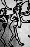
Close view‐up of a xylograph contained in the 12th Tome of Konrad von Magenberg's “Buch der Natur” [1475] representing a woman with large goiter, presumably a plexiform neurofibroma. Copyright © The Library of Congress, Washington, DC
FIGURE 4
FIGURE 4
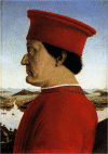
Portrait of the Duke Federico da Montefeltro by Piero della Francesca: note the multiple warts on his left face interpreted either as fibrous nodules or neurofibromas. Copyright © Galleria degli Uffizi, Firenze, Italy
FIGURE 5
FIGURE 5
(a) The imaginative “infant of Chieri” by Ambroise Paré: the fleshy mass hanging down along the back was interpreted as a plexiform neurofibroma. Copyright © Wellcome Library London, UK; (b) Aldrovandi's “homuncio,” with enormous, flabby masses of flesh, hanging from his right side, likely representing a plexiform neurofibroma in mosaic neurofibromatosis type 1 (NF1). Copyright © Biblioteca Universitaria di Bologna, Italy
FIGURE 6
FIGURE 6
(a) Lithograph depicting Maria Herig with multiple dark leaf‐shaped areas of hyper pigmentation of the skin (likely café‐au‐lait spots) in the limbs and in the trunk and a large, raised “pigskin” encircling lesion over her trunk (likely a cutaneous plexiform neurofibroma or an epidermal nevus). Copyright © Wellcome Library London, UK; (b) von Tilenau's “Wart Man”.© Authors' personal collection
FIGURE 7
FIGURE 7
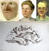
(a) A young man's face dotted with clusters of small, erythematous papules [végétations vasculaires] interpreted by Rayer as typical facial angiofibromas: Copyright © Wellcome Library, London, UK; (b,c) two 20th century prints depicting facial “adenoma sebaceum” (i.e., the old term used for the facial angiofibromas): Copyright © Wellcome Library, London, UK; (d) Drawings of the brain of the 15‐year‐old Marie: note the “sclerotic” cortical “tubers” and the “subependymal tumors” (nodules) of the lateral walls of the ventricles: Copyright © Wellcome Library, London, UK
FIGURE 8
FIGURE 8
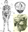
(a) One of the original drawings by Alfred Blaschko, depicting a child with the typical epidermal nevus lesions arranged according to specific lines of distribution of pigment, since called after his name and (b) Alfred Blaschko's bookplate, showing Hygieia, the Greek goddess of health, with the Blaschko lines superimposed. Copyright © The Cushing‐Whitney Medical Library, Yale University; (c) a print from a bookTreatise of Medicine [1889], depicting a man with multiple lipomas likely representing encephalocraniocutaneous lipomatosis

References

    1. Ahn, M. S. , Jackler, R. K. , & Lustig, L. R. (1996). The early history of the neurofibromatoses. Evolution of the concept of neurofibromatosis type 2. Archives of Otolaryngology ‐ Head & Neck Surgery, 122, 1240–1249. - PubMed
    1. Aldrovandi, U. (1642). Monstruorum Historia: Cum Paralipomenis Historiae omnium Animalium (p. 1642). Bologna: Typis Nocolai Tibaldini.
    1. Ashrafian, H. (2011). Limb gigantism, neurofibromatosis and royal heredity in the Ancient World 2500 years ago: Achaemenids and Parthians. Journal of Plastic, Reconstructive and Aesthetic Surgery, 64, 557. - PubMed
    1. Ashrafian, H. (2017). Leonardo da Vinci's Virgin of the Rocks: Perhaps the first depiction of congenital vascular malformation syndrome. Italian Journal of Anatomy and Embryology, 3, 155–156. 10.13128/IJAE-22974 - DOI
    1. Ashwal, S. (1990). The founders of child neurology. San Francisco, CA: Norman Publishing.

LinkOut - more resources