Association of Structural Changes in the Brain and Retina After Long-Duration Spaceflight
- PMID: 34014272
- PMCID: PMC8138750
- DOI: 10.1001/jamaophthalmol.2021.1400
Association of Structural Changes in the Brain and Retina After Long-Duration Spaceflight
Abstract
Importance: Long-duration spaceflight induces structural changes in the brain and eye. Identification of an association between cerebral and ocular changes could help determine if there are common or independent causes and inform targeted prevention strategies or treatments.
Objective: To determine if there is an association between quantitative changes in intracranial compartment volumes and peripapillary total retinal thickness after spaceflight.
Design, setting, and participants: This cohort study included healthy International Space Station crew members before and immediately after long-duration spaceflight. Data on race were not collected. Analysis was conducted from September to November 2020.
Exposures: Long-duration spaceflight (mean [SD], 191 [55] days).
Main outcomes and measures: Optical coherence tomography-derived peripapillary total retinal thickness as a quantitative assessment and early sign of optic disc edema and magnetic resonance imaging-derived measures of lateral ventricle volume, white matter volume, and whole brain plus cerebrospinal fluid volume.
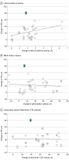
Results: In 19 healthy crew members included in this study (5 women [26.3%], 14 men [73.7%]; mean [SD] age, 45.2 [6.4] years), analyses revealed a positive, although not definitive, association between spaceflight-induced changes in total retinal thickness and lateral ventricle volume (4.7-μm increase in postflight total retinal thickness [95% CI, -1.5 to 10.8 μm; P = .13] per 1-mL postflight increase in lateral ventricle volume). Adjustments for mission duration improved the strength of association (5.1 μm; 95% CI, -0.4 to 10.5 μm; P = .07). No associations were detected between spaceflight-induced changes in total retinal thickness and white matter volume (0.02 μm; 95% CI, -0.5 to 0.5 μm; P = .94) or brain tissue plus cerebrospinal fluid volume, an estimate of intracranial volume (0.02 μm; 95% CI, -0.6 to 0.6 μm; P = .95).
Conclusions and relevance: These results help characterize spaceflight-associated neuro-ocular syndrome and the physiologic associations of headward fluid shifts with outcomes during spaceflight on the central nervous system. The possibly weak association between increased total retinal thickness and lateral ventricle volume suggest that while weightlessness-induced fluid redistribution during spaceflight may be a common stressor to the brain and retina, the development of optic disc edema appears to be uncoupled with changes occurring in the intracranial compartment.
Conflict of interest statement
Figures


Comment in
-
Increased Ventricular Cerebrospinal Fluid Volumes in Spaceflight-Associated Neuroocular Syndrome-A Curse or a Blessing?JAMA Ophthalmol. 2021 Nov 1;139(11):1245-1246. doi: 10.1001/jamaophthalmol.2021.3654. JAMA Ophthalmol. 2021. PMID: 34529027 No abstract available.
-
The Importance of the Intracranial Compartment in the Development of Spaceflight-Associated Neuro-ocular Syndrome-Reply.JAMA Ophthalmol. 2022 Jan 1;140(1):99-100. doi: 10.1001/jamaophthalmol.2021.4863. JAMA Ophthalmol. 2022. PMID: 34817558 No abstract available.
-
The Importance of the Intracranial Compartment in the Development of Spaceflight-Associated Neuro-ocular Syndrome.JAMA Ophthalmol. 2022 Jan 1;140(1):98-99. doi: 10.1001/jamaophthalmol.2021.4860. JAMA Ophthalmol. 2022. PMID: 34817563 No abstract available.
References
-
- Nathan Kline Institute (NKI)/Rockland sample. Accessed April 14, 2021. https://fcon_1000.projects.nitrc.org/indi/pro/nki.html
Publication types
MeSH terms
LinkOut - more resources
Full Text Sources
Other Literature Sources

