

Endothelial cells and coagulation
- PMID: 34014399
- PMCID: PMC8975780
- DOI: 10.1007/s00441-021-03471-2
Endothelial cells and coagulation
Abstract
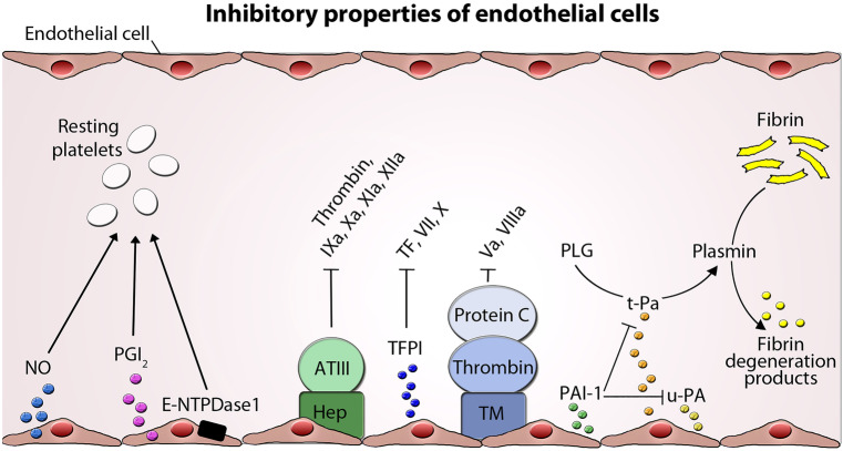
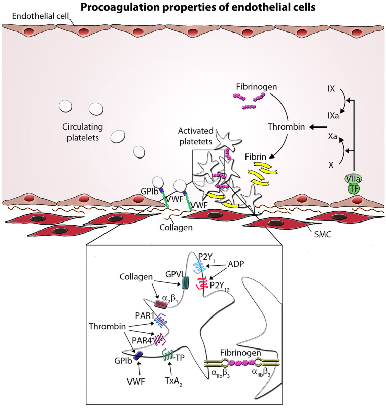
Endothelial cells form a monolayer, which lines blood vessels. They are crucially involved in maintaining blood fluidity and providing controlled vascular hemostasis at sites of injury. Thereby endothelial cells facilitate multiple mechanisms, including both procoagulant and anticoagulant, which must be kept in balance. Under physiological conditions, endothelial cells constitute a nonadhesive surface preventing activation of platelets and the coagulation cascade. Multiple fibrinolytic and antithrombotic properties act on their cell surface contributing to the maintenance of blood fluidity. These include platelet inhibition, the heparin-antithrombin III system, tissue factor pathway inhibition, thrombomodulin/protein C system, and fibrinolytic qualities. At sites of vascular damage, platelets react immediately by adhering to the exposed extracellular matrix, followed by platelet-platelet interactions to form a clot that effectively seals the injured vessel wall to prevent excessive blood loss. For solid thrombus formation, functional platelets are essential. In this process, endothelial cells serve as a support surface for formation of procoagulant complexes and clotting. This review gives an overview about the central role of the endothelium as a dynamic lining which controls the complex interplay of the coagulation system with the surrounding cells.
Keywords: Endothelial injury; Hemostasis; Platelets; Thrombosis; Von Willebrand factor.
© 2021. The Author(s).
Conflict of interest statement
None of the authors has any competing interests to declare. Since this article is a literature review, no ethical approval or declaration of informed consent is required.
Figures
References
-
- Adams TE, Huntington JA. Thrombin-cofactor interactions: structural insights into regulatory mechanisms. Arterioscler Thromb Vasc Biol. 2006;26:1738–1745. - PubMed
-
- Agarwal KC, Clarke E, Rounds S, Parks RE, Jr, Huzoor A. Platelet-activating factor (PAF)-induced platelet aggregation. Modulation by plasma adenosine and methylxanthines. Biochem Pharmacol. 1994;48:1909–1916. - PubMed
-
- Aird WC. Endothelium and haemostasis. Hamostaseologie. 2015;35:11–16. - PubMed
-
- Anastasiou G, Gialeraki A, Merkouri E, Politou M, Travlou A. Thrombomodulin as a regulator of the anticoagulant pathway: implication in the development of thrombosis. Blood coagulation & fibrinolysis : an international journal in haemostasis and thrombosis. 2012;23:1–10. - PubMed
-
- Bauer KA, Rosenberg RD. Role of antithrombin III as a regulator of in vivo coagulation. Semin Hematol. 1991;28:10–18. - PubMed
Publication types
MeSH terms
LinkOut - more resources
Full Text Sources
Other Literature Sources
Medical
