

The value of sex-specific abdominal visceral fat as measured via CT as a predictor of clear renal cell carcinoma T stage
- PMID: 34014795
- PMCID: PMC8143252
- DOI: 10.1080/21623945.2021.1924957
The value of sex-specific abdominal visceral fat as measured via CT as a predictor of clear renal cell carcinoma T stage
Abstract
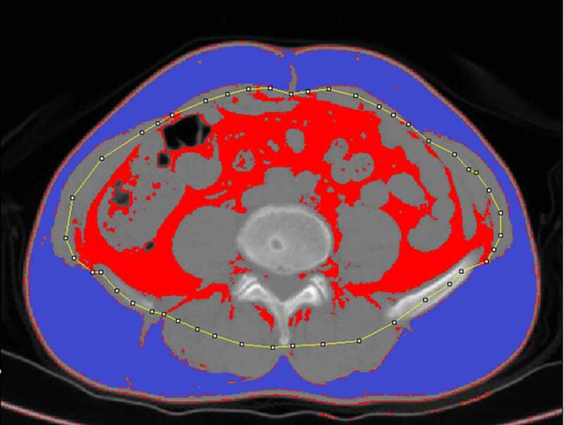
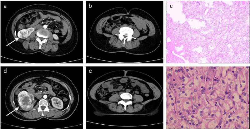
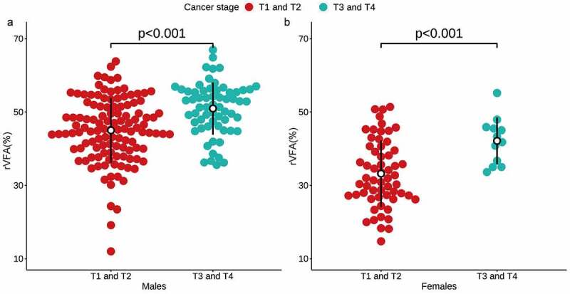
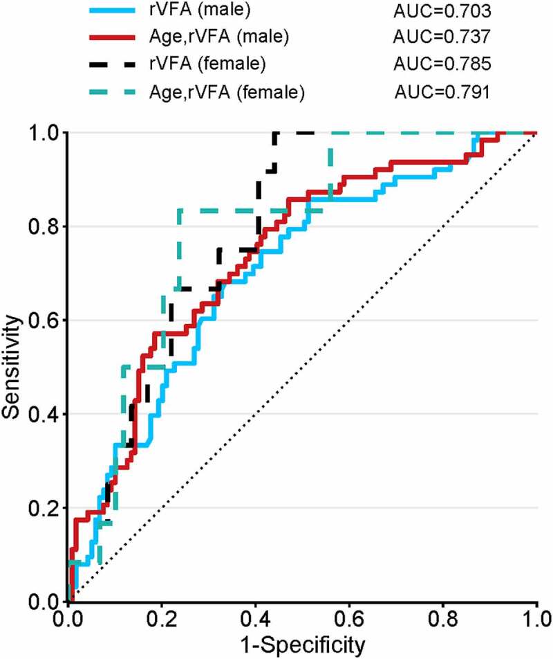
Although much is known about how adipose tissue affects the development of clear cell renal carcinoma (ccRCC), little information is available for the utility of sex-specific abdominal visceral fat composition as a predictor of clear cell renal carcinoma (ccRCC) T stage. We conducted CT-based sex-specific abdominal fat measurements in ccRCC patients to assess whether VFA distribution could predict the ccRCC T stage. In total, 253 patients (182 males and 71 females) from our hospital with pathologically confirmed ccRCC (178 low T-stage and 75 high T-stage) were retrospectively reviewed for the present study. Computed tomography (CT) scans were assessed using ImageJ to differentiate between the visceral and subcutaneous fat areas (VFA and SFA), after which the relative VFA (rVFA) and total fat area (TFA) were computed. The relationships between these fat area-related variables, patient age, sex, and BMI, and ccRCC T stage were then evaluated through univariate and multivariate logistic regression analysis to clarify the association between general or sex-specific abdominal visceral fat and T stage. Following adjustment for age, males with high T stage ccRCC exhibited an increased rVFA as compared to males with low T stage ccRCC, with the same relationship being observed among females. This association between rVFA and high T stage was confirmed through both univariate and multivariate models. As thus, sex-specific visceral fat composition is a reliable independent predictor that can identify both male and female patients with high T stage ccRCC.
Keywords: Clear renal cell carcinoma; T stage; abdominal; computed tomography; obesity.
Conflict of interest statement
No potential conflict of interest was reported by the authors.
Figures
Similar articles
-
Clear cell renal cell carcinoma: the value of sex-specific abdominal visceral fat measured on CT for prediction of Fuhrman nuclear grade.Eur Radiol. 2020 Jul;30(7):3977-3986. doi: 10.1007/s00330-020-06747-3. Epub 2020 Mar 6. Eur Radiol. 2020. PMID: 32144457
-
Can Gender-Specific Renal and Visceral Fat Be Evaluated by CT Predict Fuhrman Nuclear Classification of Clear Cell Renal Cell Carcinoma?Curr Med Imaging. 2024;20:e15734056295913. doi: 10.2174/0115734056295913240514020603. Curr Med Imaging. 2024. PMID: 38798222
-
T-stage-specific abdominal visceral fat, haematological nutrition indicators and inflammation as prognostic factors in patients with clear renal cell carcinoma.Adipocyte. 2022 Dec;11(1):133-142. doi: 10.1080/21623945.2022.2048546. Adipocyte. 2022. PMID: 35285399 Free PMC article.
-
Effect of sex-specific abdominal fat tissue composition on WHO/ISUP nuclear grade of clear cell renal cell carcinoma.Turk J Med Sci. 2024 Mar 4;54(4):784-791. doi: 10.55730/1300-0144.5850. eCollection 2024. Turk J Med Sci. 2024. PMID: 39295621 Free PMC article.
-
Does chromophobe renal cell carcinoma have better survival than clear cell renal cell carcinoma? A clinical-based cohort study and meta-analysis.Int Urol Nephrol. 2016 Feb;48(2):191-9. doi: 10.1007/s11255-015-1161-3. Epub 2015 Nov 20. Int Urol Nephrol. 2016. PMID: 26589610 Review.
Cited by
-
Cohort profile - the Renal cell cancer: Lifestyle, prognosis and quality of life (ReLife) study in the Netherlands.BMJ Open. 2023 Mar 27;13(3):e066909. doi: 10.1136/bmjopen-2022-066909. BMJ Open. 2023. PMID: 36972960 Free PMC article.
-
Evaluation of the relations between reproduction-related pituitary and ovarian hormones and abdominal fat area-related variables determined with computed tomography in overweight or obese women who have undergone bariatric surgery: a cross-sectional study.Quant Imaging Med Surg. 2023 Oct 1;13(10):7065-7076. doi: 10.21037/qims-22-1283. Epub 2023 Sep 14. Quant Imaging Med Surg. 2023. PMID: 37869350 Free PMC article.
-
Association of visceral and subcutaneous adiposity with tumor stage and Fuhrman grade in renal cell carcinoma.Sci Rep. 2022 Oct 6;12(1):16718. doi: 10.1038/s41598-022-20877-2. Sci Rep. 2022. PMID: 36202890 Free PMC article.
-
Development of a CT-assessed adiposity nomogram for predicting outcome in localized ccRCC.Abdom Radiol (NY). 2024 Oct;49(10):3485-3495. doi: 10.1007/s00261-024-04403-8. Epub 2024 Jun 6. Abdom Radiol (NY). 2024. PMID: 38842727
-
Association between CT-based adipose variables, preoperative blood biochemical indicators and pathological T stage of clear cell renal cell carcinoma.Heliyon. 2024 Jan 13;10(2):e24456. doi: 10.1016/j.heliyon.2024.e24456. eCollection 2024 Jan 30. Heliyon. 2024. PMID: 38268833 Free PMC article.
References
-
- Sung H, Siegel RL, Torre LA, et al. Global patterns in excess body weight and the associated cancer burden. CA Cancer J Clin. 2019;69:88–112. - PubMed
-
- Budny A, Grochowski C, Kozłowski P, et al. Obesity as a tumour development triggering factor. Ann Agric Environ Med. 2019;26:13–23. - PubMed
-
- Lycett D. The Association of religious affiliation and body mass index (BMI): an analysis from the health survey for England. J Relig Health. 2015;54:2249–2267. - PubMed
-
- Vrieling A, Kampman E, Knijnenburg NC, et al. Body composition in relation to clinical outcomes in renal cell cancer: a systematic review and meta-analysis. Eur Urol Focus. 2018;4:420–434. - PubMed
Publication types
MeSH terms
LinkOut - more resources
Full Text Sources
Other Literature Sources
Medical
Research Materials