. 2021 May 20;16(5):e0239793.
doi: 10.1371/journal.pone.0239793. eCollection 2021.

Structural coordinates: A novel approach to predict protein backbone conformation

Affiliations

Structural coordinates: A novel approach to predict protein backbone conformation

Vladislava Milchevskaya et al. PLoS One. .

Abstract

Motivation: Local protein structure is usually described via classifying each peptide to a unique class from a set of pre-defined structures. These classifications may differ in the number of structural classes, the length of peptides, or class attribution criteria. Most methods that predict the local structure of a protein from its sequence first rely on some classification and only then proceed to the 3D conformation assessment. However, most classification methods rely on homologous proteins' existence, unavoidably lose information by attributing a peptide to a single class or suffer from a suboptimal choice of the representative classes.

Results: To alleviate the above challenges, we propose a method that constructs a peptide's structural representation from the sequence, reflecting its similarity to several basic representative structures. For 5-mer peptides and 16 representative structures, we achieved the Q16 classification accuracy of 67.9%, which is higher than what is currently reported in the literature. Our prediction method does not utilize information about protein homologues but relies only on the amino acids' physicochemical properties and the resolved structures' statistics. We also show that the 3D coordinates of a peptide can be uniquely recovered from its structural coordinates, and show the required conditions under various geometric constraints.


Conflict of interest statement

The authors have declared that no competing interests exist.

Figures
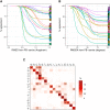
Fig 1
Fig 1. Relation between assignments based on RMSDA and RMSD.
A shows the percentage (Y-axis) of matching RMSDA- and RMSD assignments among all the RMSD assignments for a given cluster that lie within a certain distance (X-axis) from the cluster centre, i.e. the corresponding protein block (PB); B shows the reverse of A, namely, the percentage of matching RMSDA- and RMSD assignments among all the RMSDA assignments for a given cluster that lie within a certain RMSDA-distance (X-axis) from the centre; C shows the percentage of matching assignment for each RMSD-cluster among all fragments. The full set of fragments for this investigation is PDB30 (see Methods for details).
Fig 2
Fig 2. Discrepancy examples between RMSDA and RMSD.
Structural alignment of a 5-residue protein fragments (green) against the standard alpha-helical structure (red) with dihedral angles ϕi = −57, ψi = −47, i = 1, …, 5. The examples (A)-(C) are chosen such that to highlight that one can find a (green) fragment with the same RMSDA distance value to the reference (red) fragment but varying RMSD. In (D), on the contrary, we show an example of a (green) fragment that has RMSDA of 0 to the reference (red), but a large RMSD. Dihedral angles of the fragment (A) are ϕi = −61.1, ψi = −42.8, i = 1, …, 5, its RMSDA to the standard helix is 14.1° and its RMSD to the standard helix is 0.09 Å; Dihedral angles of the fragment (B) are ϕi = −32.9, ψi = −42.9, i = 1, …, 5, its RMSDA to the standard helix is 14.1421 and its RMSD to the standard helix is 1.0065; Dihedral angles of the fragment (C) are all ϕi = −57, ψi = −47, except for ϕ3 = −17, its RMSDA to the standard helix is again 14.1421 and its RMSD to the standard helix is 0.74. Dihedral angles of the fragment (D) are all ϕi = −57, ψi = −47, except ω3 = 0 instead of the usual 180.
Fig 3
Fig 3. Generation of predictors based on RMSD statistics between a protein fragment and the 16 basic protein blocks, schematic representation.
Fig 4
Fig 4. Generation of predictors based on physico-chemical properties of amino acids comprising a protein fragment, schematic representation.

References

    1. Torrisi M, Pollastri G, Le Q. Deep learning methods in protein structure prediction. Computational and Structural Biotechnology Journal. 2020. 10.1016/j.csbj.2019.12.011 - DOI - PMC - PubMed
    1. Joseph AP, Agarwal G, Mahajan S, Gelly JC, Swapna LS, Offmann B, et al.. A short survey on protein blocks. Biophysical Reviews. 2010;2(3):137–145. 10.1007/s12551-010-0036-1 - DOI - PMC - PubMed
    1. Yousef A, Charkari NM. A novel method based on physicochemical properties of amino acids and one class classification algorithm for disease gene identification. Journal of biomedical informatics. 2015;56:300–306. 10.1016/j.jbi.2015.06.018 - DOI - PubMed
    1. Deng H, Jia Y, Zhang Y. Protein structure prediction. International Journal of Modern Physics B. 2018;32(18):1840009. 10.1142/S021797921840009X - DOI - PMC - PubMed
    1. Kabsch W, Sander C. Dictionary of protein secondary structure: pattern recognition of hydrogen-bonded and geometrical features. Biopolymers: Original Research on Biomolecules. 1983;22(12):2577–2637. 10.1002/bip.360221211 - DOI - PubMed

Publication types

LinkOut - more resources