. 2021 May 20;12(1):2739.
doi: 10.1038/s41467-021-22977-5.

Epidermal chloroplasts are defense-related motile organelles equipped with plant immune components

Affiliations

Epidermal chloroplasts are defense-related motile organelles equipped with plant immune components

Hiroki Irieda et al. Nat Commun. .

Abstract

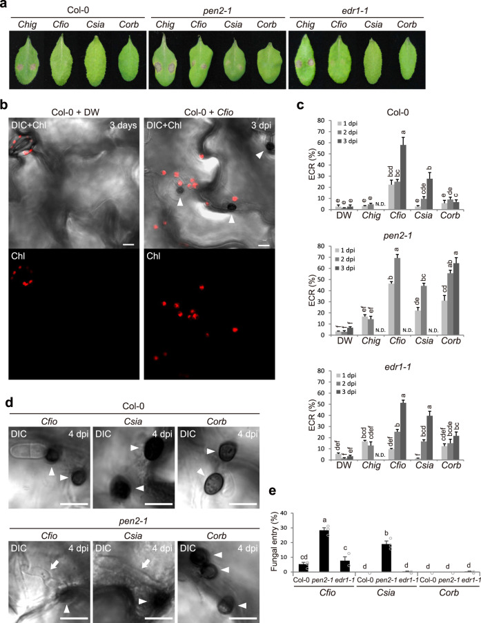
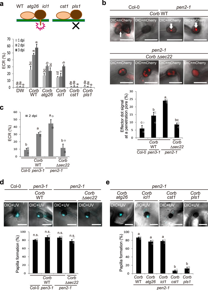
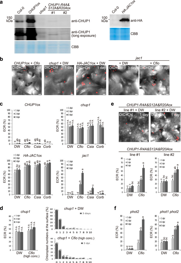
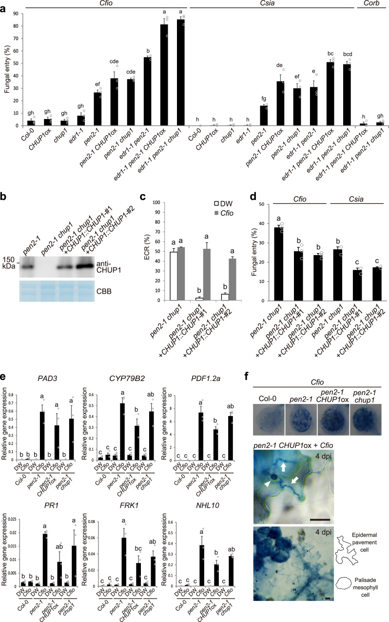
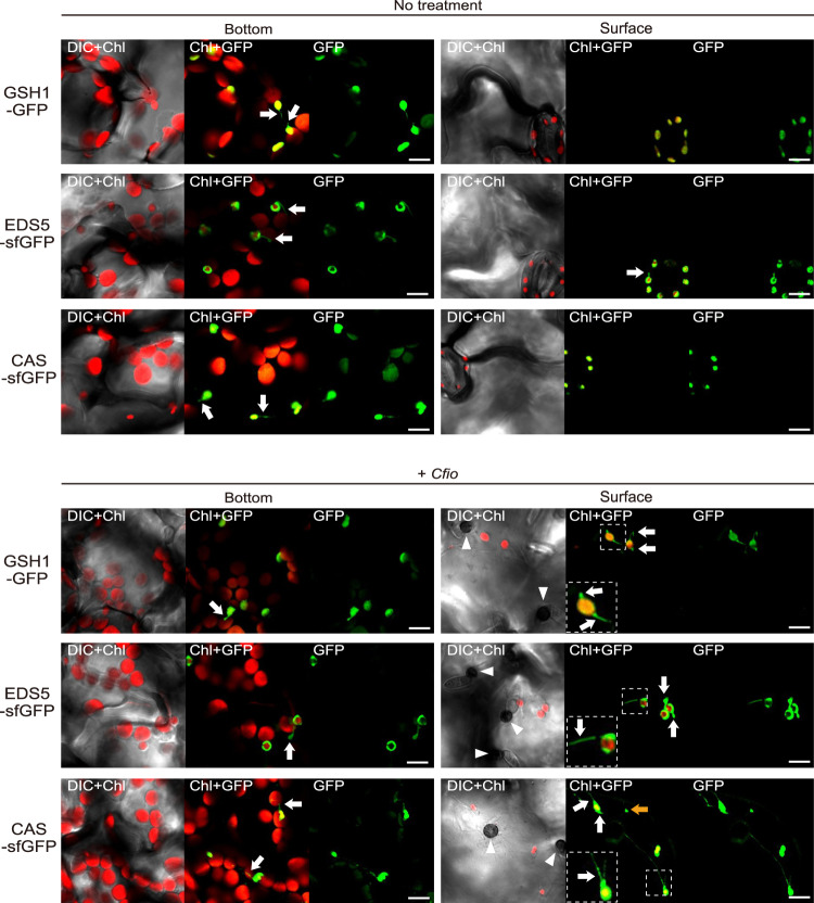
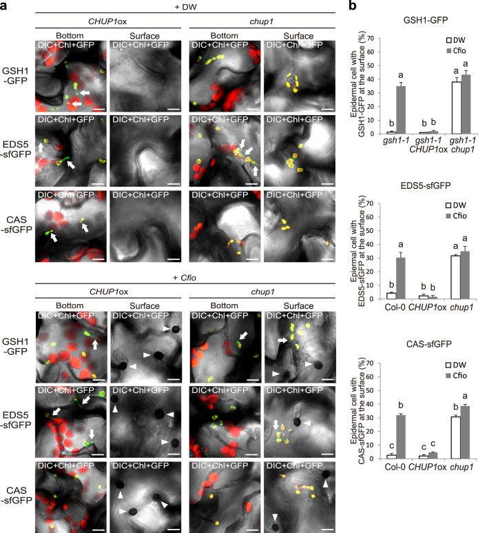
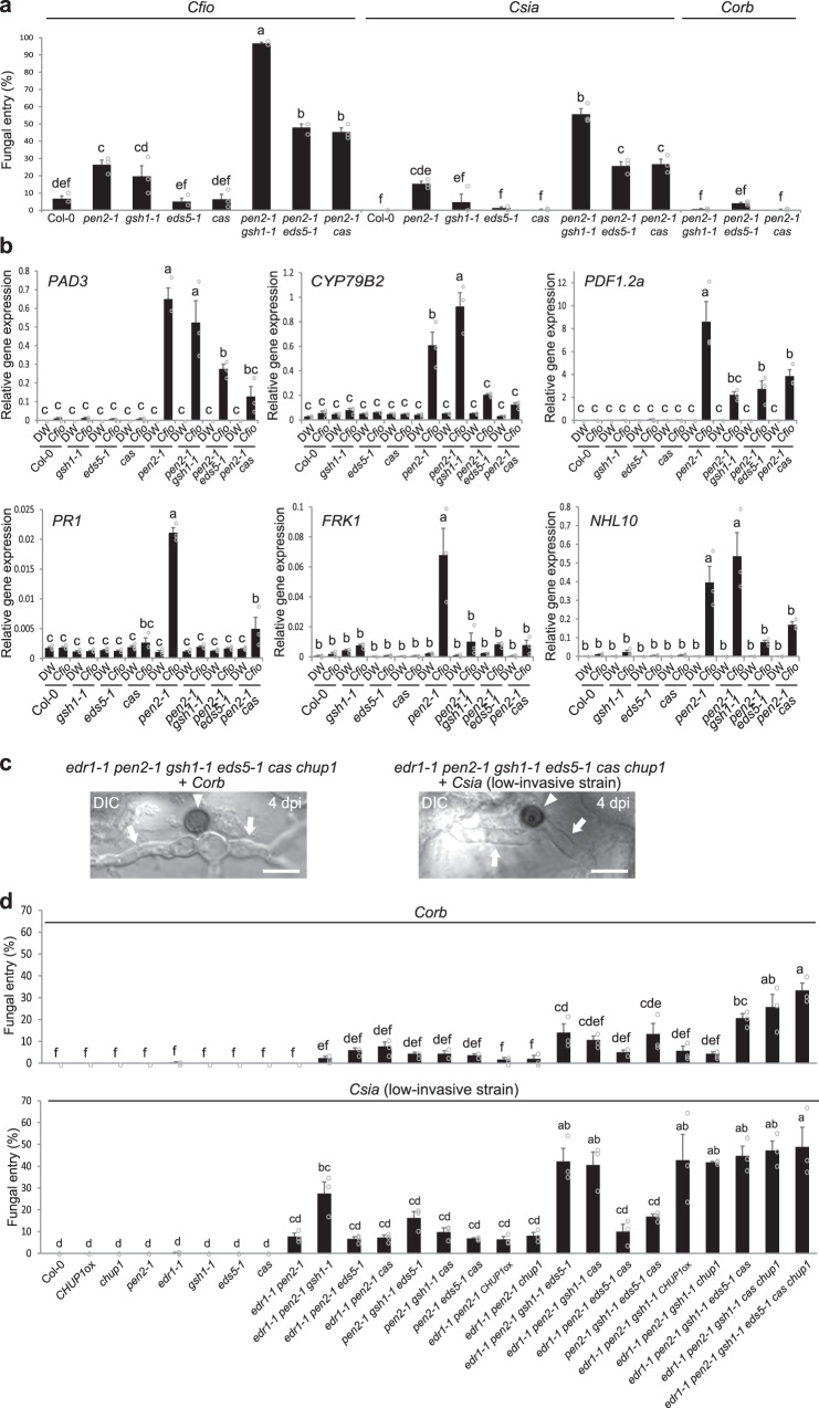
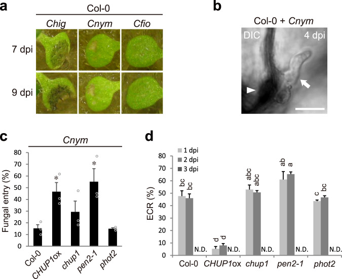
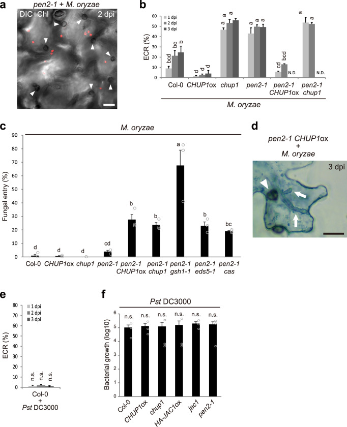
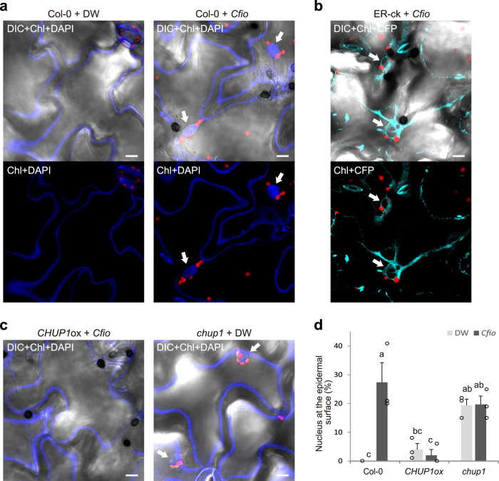
In addition to conspicuous large mesophyll chloroplasts, where most photosynthesis occurs, small epidermal chloroplasts have also been observed in plant leaves. However, the functional significance of this small organelle remains unclear. Here, we present evidence that Arabidopsis epidermal chloroplasts control the entry of fungal pathogens. In entry trials, specialized fungal cells called appressoria triggered dynamic movement of epidermal chloroplasts. This movement is controlled by common regulators of mesophyll chloroplast photorelocation movement, designated as the epidermal chloroplast response (ECR). The ECR occurs when the PEN2 myrosinase-related higher-layer antifungal system becomes ineffective, and blockage of the distinct steps of the ECR commonly decreases preinvasive nonhost resistance against fungi. Furthermore, immune components were preferentially localized to epidermal chloroplasts, contributing to antifungal nonhost resistance in the pen2 background. Our findings reveal that atypical small chloroplasts act as defense-related motile organelles by specifically positioning immune components in the plant epidermis, which is the first site of contact between the plant and pathogens. Thus, this work deepens our understanding of the functions of epidermal chloroplasts.


Conflict of interest statement

The authors declare no competing interests.

Figures

Fig. 1
Fig. 1. The ECR occurs at the lower layer of PEN2-related immunity and is critical for NHR against Colletotrichum fungi.
a Pathogenicity of Colletotrichum fungi on Arabidopsis mutants. A conidial suspension of adapted Chig and nonadapted Cfio, Csia, and Corb was inoculated onto leaves of wild-type Arabidopsis (Col-0), pen2-1, and edr1-1 and incubated for 7 days. b ECR after Cfio inoculation. The epidermal surface of the Cfio-inoculated cotyledon of Col-0 was investigated at 3 days post-inoculation (dpi). The chloroplasts were visualized based on chlorophyll autofluorescence. The DIC images were captured by confocal microscopy. DW was used as a control. c The ratio of epidermal cells with surface chloroplasts was investigated at 1, 2, and 3 dpi. A total of 100 cells in contact with the melanized appressorium were observed. N.D.: not determined due to damages of epidermal cell by fungal invasion. d, e Fungal invasion of Arabidopsis epidermis. Cfio, Csia, and Corb were inoculated onto cotyledons of the indicated Arabidopsis. The entry ratio was quantified at 4 dpi. A total of 100 melanized appressoria were investigated. The arrowheads and arrows indicate melanized appressoria and invasive hyphae, respectively. Scale bar, 10 µm. The means and SE were calculated from three independent plants. Means not sharing the same letter are significantly different (P < 0.05, two-way ANOVA with Tukey’s HSD test).
Fig. 2
Fig. 2. Fungal peg formation and penetration pore-mediated secretion correlate with plant ECR during entry trial.
a ECR after inoculation of Corb mutants. Wild-type (WT) Corb, atg26, icl1, cst1, and pls1 were inoculated onto cotyledons of pen2-1 plants. The ECR was quantified at 1, 2, and 3 dpi. DW was used as a control. b Focal accumulation of Corb effector at the penetration pore was abolished by disruption of the SEC22 gene in Corb. Corb expressing CoDN3-mCherry was inoculated onto the indicated Arabidopsis. At 2 dpi, the images were captured by confocal microscopy, and the ratio of the effector signals at the penetration pore was quantified. The arrows indicate effector accumulation at the penetration pore. c ECR after inoculation of Corb WT and Δsec22 at 2 dpi. d, e Papilla formation at the entry trial site of Corb WT, Δsec22, atg26, icl1, cst1, and pls1. Corb-inoculated plants were subjected to staining with aniline blue fluorochrome for visualization of callose deposition. At 1 dpi, the images were captured, and the ratio of papillary callose deposition was investigated. For all experiments, 100 melanized appressoria were observed, and the means and SE were calculated from three independent plants. Scale bar, 10 µm. Means not sharing the same letter are significantly different (P < 0.05, one-way (be) or two-way (a) ANOVA with Tukey’s HSD test). n.s. not significant.
Fig. 3
Fig. 3. CHUP1 and JAC1 regulate the ECR in a phototropin-independent manner.
a Levels of CHUP1 and HA-JAC1 proteins in the indicated Arabidopsis. Protein extracts were analyzed by immunoblot with anti-CHUP1 and anti-HA antibodies. CBB staining was used as a loading control. b, e Effects of CHUP1 and JAC1 on Cfio-induced ECR. Cfio was inoculated onto cotyledons of CHUP1ox and jac1, and the surface of the epidermis was observed. DW was spotted onto chup1, CHUP1-R4A&S12A&R20Aox lines, HA-JAC1ox, and jac1 as a control. The epidermal chloroplasts were visualized based on chlorophyll autofluorescence. The DIC images were captured by confocal microscopy at the indicated time point. Scale bar, 10 µm. ce Quantification of ECR in CHUP1ox, chup1, HA-JAC1ox, jac1, and CHUP1-R4A&S12A&R20Aox plants. Cfio, Csia, and Corb (5 × 105 conidia/mL) were inoculated onto cotyledons of the indicated Arabidopsis and examined at 1, 2, and 3 dpi (c, e). For more fungal inoculum on the chup1 mutant, Cfio (1.5 × 106 conidia/mL) was inoculated (d). A total of 100 cells in contact with the melanized appressorium were observed. As a control, DW was spotted. f Cfio-induced ECR in phot2 and phot1 phot2 mutants. As a control, DW was spotted. The means and SE were calculated from three independent plants. Means not sharing the same letter are significantly different (P < 0.05, two-way ANOVA with Tukey’s HSD test). n.s. not significant.
Fig. 4
Fig. 4. The ECR contributes to Arabidopsis NHR against Colletotrichum fungi.
a Fungal entry rate into the epidermis of ECR-defective Arabidopsis. A total of 100 melanized appressoria were examined at 4 dpi. b CHUP1 protein levels in the indicated plants. Protein extracts were analyzed by immunoblot with anti-CHUP1 antibody. CBB staining was used as a loading control. c ECR of pen2-1 chup1 and CHUP1-complementation lines. A total of 100 cells in contact with the melanized appressorium were examined at 2 dpi. DW was used as a control. d Entry rate of Cfio and Csia into the epidermis of pen2-1 chup1 and CHUP1-complementation lines at 4 dpi. e Induced gene expression of PAD3, CYP79B2, PDF1.2a, PR1, FRK1, and NHL10 by inoculation of Cfio. Cfio was inoculated onto cotyledons of the indicated Arabidopsis. Gene expression was assayed at 24 hpi by RT-qPCR. DW was used as a control. f Epidermal and mesophyll cell death caused by appressorium-mediated entry of Cfio at 4 dpi. The inoculated plants were subjected to TB staining and observed macroscopically (upper) and microscopically using x40 (middle) and x10 (lower) objective lenses. The arrowhead and arrows indicate melanized appressorium and invasive hyphae, respectively. Scale bar, 20 µm. For all quantification analyses except RT-qPCR, the means and SE were calculated from three independent plants. The means and SE of RT-qPCR results were calculated from three independent experiments. Means not sharing the same letter are significantly different (P < 0.05, two-way ANOVA with Tukey’s HSD test).
Fig. 5
Fig. 5. Epidermal chloroplasts preferentially position immune components GSH1, EDS5, and CAS.
Localization patterns of GSH1, EDS5, and CAS proteins in Arabidopsis. The transgenic gsh1 mutant expressing GSH1-GFP under its own promoter and Col-0 plants expressing EDS5-sfGFP and CAS-sfGFP under the CaMV 35S promoter were incubated with or without Cfio and observed at 2 dpi. Chloroplasts were visualized using chlorophyll autofluorescence. The lower left dashed-line square regions show a magnified image of the dashed box. The white arrowheads and arrows indicate melanized appressoria and stromules, respectively. The orange arrow indicates CAS localized in the epidermal plastid without chlorophyll. Scale bar, 10 µm. Similar results were obtained from three independent experiments.
Fig. 6
Fig. 6. Localization pattern of GSH1, EDS5, and CAS tightly linked to epidermal chloroplasts and the ECR.
a Localization pattern of GSH1, EDS5, and CAS proteins in CHUP1ox and chup1 plants. The transgenic gsh1 CHUP1ox and gsh1 chup1 plants expressing GSH1-GFP under its own promoter, and CHUP1ox and chup1 plants expressing EDS5-sfGFP and CAS-sfGFP under the CaMV 35S promoter were incubated with Cfio and observed at 2 dpi. DW was used as a control. The chloroplasts were visualized based on chlorophyll autofluorescence. The white arrowheads and arrows indicate melanized appressoria and stromules, respectively. Scale bar, 10 µm. b Quantification of epidermal cells with surface fluorescence of GFP/sfGFP-labeled immune components. Cfio-inoculated cotyledons of the indicated Arabidopsis were analyzed at 2 dpi. A total of 100 cells in contact with the melanized appressorium were observed. As a control, DW was spotted. The means and SE were calculated from three independent plants. Means not sharing the same letter are significantly different (P < 0.05, two-way ANOVA with Tukey’s HSD test).
Fig. 7
Fig. 7. Epidermal chloroplast-localized immune components are critical for preinvasive NHR against Colletotrichum fungi.
a Fungal entry rate into Arabidopsis epidermis. Cfio, Csia, and Corb were inoculated onto cotyledons of the indicated Arabidopsis. b Induced gene expression of PAD3, CYP79B2, PDF1.2a, PR1, FRK1, and NHL10 by inoculation of Cfio. Cfio was inoculated onto cotyledons of the indicated Arabidopsis, and gene expression was assayed at 24 hpi by RT-qPCR. DW was used as a control. c, d Invasions of Corb and Csia (low-invasive strain) into Arabidopsis epidermis at 4 dpi. Corb and Csia (low-invasive strain) were inoculated onto cotyledons of the indicated Arabidopsis with multiple mutations. The arrowheads and arrows indicate melanized appressoria and invasive hyphae, respectively. Scale bar, 10 µm. For quantification of fungal entry of Cfio, Csia, and Corb, 100 melanized appressoria were investigated at 4 dpi. For quantification of fungal entry of Csia (low-invasive strain), 300 melanized appressoria were investigated at 4 dpi. For all quantification analyses except RT-qPCR, the means and SE were calculated from three independent plants. The means and SE of RT-qPCR results were calculated from three independent experiments. Means not sharing the same letter are significantly different (P < 0.05, one-way (d) or two-way (a, b) ANOVA with Tukey’s HSD test).
Fig. 8
Fig. 8. The ECR strongly occurs and contributes to NHR against specific nonadapted Colletotrichum fungus in wild-type Arabidopsis.
a Pathogenicity of Colletotrichum fungi on wild-type Arabidopsis (Col-0). A conidial suspension of adapted Chig and nonadapted Cnym and Cfio was inoculated onto cotyledons of Col-0 and incubated for 7 and 9 days. b, c Cnym invasion into epidermis of Col-0 at 4 dpi. The arrowhead and arrow indicate melanized appressorium and invasive hypha, respectively. Scale bar, 10 µm. For quantification of fungal entry of Cnym, 300 melanized appressoria were investigated at 4 dpi. d Quantification of ECR in Col-0, CHUP1ox, chup1, pen2-1, and phot2 plants after inoculation of Cnym at 1, 2, and 3 dpi. A total of 100 cells in contact with the melanized appressorium were observed. For all quantification analyses, the means and SE were calculated from three independent plants. Means not sharing the same letter are significantly different (P < 0.05, two-way ANOVA with Tukey’s HSD test). The asterisks indicate significant difference (*P < 0.05, one-way ANOVA with Dunnett’s test).
Fig. 9
Fig. 9. The ECR contributes to preinvasive NHR against M. oryzae.
a ECR after inoculation of M. oryzae. M. oryzae was inoculated onto cotyledons of pen2-1 plants, and the ECR was investigated at 2 dpi. The chloroplasts were visualized based on chlorophyll autofluorescence. b Quantification of ECR against M. oryzae. ECR was investigated at 1, 2, and 3 dpi. A total of 100 cells in contact with the melanized appressorium were observed. N.D.: not determined due to damages of epidermal cell by fungal invasion. c Entry rate of M. oryzae into epidermis at 3 dpi. A total of 100 melanized appressoria were investigated. d Epidermal cell death caused by appressorium-mediated entry of M. oryzae at 3 dpi. The inoculated plants were subjected to TB staining and observed microscopically using x40 objective lens. The arrowheads and arrows indicate melanized appressoria and invasive hyphae, respectively. Scale bar, 20 µm. The means and SE were calculated from three independent plants. Means not sharing the same letter are significantly different (P < 0.05, one-way (c) or two-way (b) ANOVA with Tukey’s HSD test). e Quantification of ECR against Pseudomonas syringae pv. tomato (Pst) DC3000. Cotyledons of Col-0 were drop-inoculated with Pst DC3000, and ECR was investigated at 1, 2, and 3 dpi. A total of 100 epidermal pavement cells were observed. The means and SE were calculated from three independent plants. f Growth of Pst DC3000 in Arabidopsis cotyledons. Cotyledons of indicated Arabidopsis were drop-inoculated with Pst DC3000, and incubated for 4 days. The number of bacteria in eight cotyledons obtained from four independent plants was plotted on a log10 scale. The means and SE were calculated from three independent experiments. n.s. not significant (*P < 0.05, one-way (f) or two-way (e) ANOVA with Tukey’s HSD test).
Fig. 10
Fig. 10. The nucleus dynamically responds to fungal entry trial in an ECR-dependent manner.
a Simultaneous observation of chloroplasts and nuclei in ECR-activated epidermis. The surface of Cfio-inoculated cotyledons of Col-0 was examined at 3 dpi. The chloroplasts and nuclei were visualized by chlorophyll autofluorescence and DAPI staining, respectively. DW was used as a control. The arrows indicate nuclei. b Simultaneous observation of chloroplasts and ER around nuclei in ECR-activated cells. The surface of Cfio-inoculated cotyledons of the transgenic plant line ER-ck expressing ER-CFP was observed at 3 dpi. The arrows indicate perinuclear ER. c Simultaneous observation of chloroplasts and nuclei in ECR-defective plants at 3 dpi. Cfio-inoculated cotyledons of CHUP1ox plants were investigated. DW was spotted onto chup1 mutant as a control. The arrows indicate nuclei. d The ratio of epidermal cells with surface nuclei was investigated at 3 dpi. A total of 100 cells in contact with the melanized appressorium were observed. Scale bar, 10 µm. The means and SE were calculated from three independent plants. Means not sharing the same letter are significantly different (P < 0.05, two-way ANOVA with Tukey’s HSD test).

References

    1. Kubo, Y. & Furusawa, I. The Fungal Spore and Disease Initiation in Plants and Animals (eds Cole, G.T. & Hoch, H.C.) (Plenum Publishing, 1991).
    1. Lee H-A, et al. Current understandings of plant nonhost resistance. Mol. Plant-Microbe Interact. 2017;30:5–15. doi: 10.1094/MPMI-10-16-0213-CR. - DOI - PubMed
    1. Lipka V, et al. Pre- and postinvasion defenses both contribute to nonhost resistance in Arabidopsis. Science. 2005;310:1180–1183. doi: 10.1126/science.1119409. - DOI - PubMed
    1. Bednarek P, et al. A glucosinolate metabolism pathway in living plant cells mediates broad-spectrum antifungal defense. Science. 2009;323:101–106. doi: 10.1126/science.1163732. - DOI - PubMed
    1. Clay NK, Adio AM, Denoux C, Jander G, Ausubel FM. Glucosinolate metabolites required for an Arabidopsis innate immune response. Science. 2009;323:95–101. doi: 10.1126/science.1164627. - DOI - PMC - PubMed

Publication types

MeSH terms