Review
. 2021 Sep;17(9):619-630.
doi: 10.1038/s41581-021-00427-1. Epub 2021 May 20.

Therapeutic trials in adult FSGS: lessons learned and the road forward

Affiliations
Review

Therapeutic trials in adult FSGS: lessons learned and the road forward

An S De Vriese et al. Nat Rev Nephrol. 2021 Sep.

Abstract

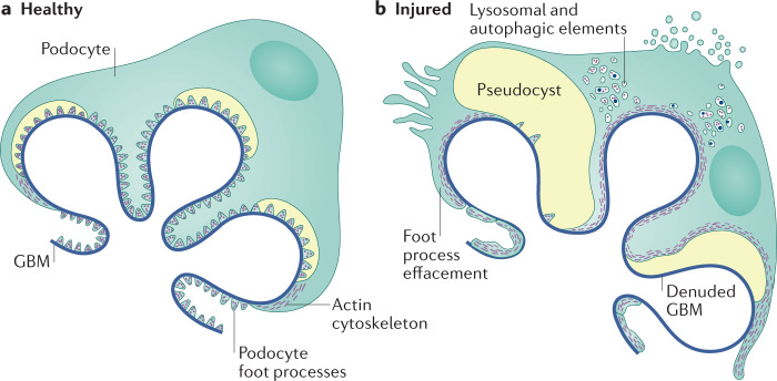
Focal segmental glomerulosclerosis (FSGS) is not a specific disease entity but a lesion that primarily targets the podocyte. In a broad sense, the causes of the lesion can be divided into those triggered by a presumed circulating permeability factor, those that occur secondary to a process that might originate outside the kidneys, those caused by a genetic mutation in a podocyte or glomerular basement membrane protein, and those that arise through an as yet unidentifiable process, seemingly unrelated to a circulating permeability factor. A careful attempt to correctly stratify patients with FSGS based on their clinical presentation and pathological findings on kidney biopsy is essential for sound treatment decisions in individual patients. However, it is also essential for the rational design of therapeutic trials in FSGS. Greater recognition of the pathophysiology underlying podocyte stress and damage in FSGS will increase the likelihood that the cause of an FSGS lesion is properly identified and enable stratification of patients in future interventional trials. Such efforts will facilitate the identification of effective therapeutic agents.


Conflict of interest statement

The authors declare no competing interests.

Figures

Fig. 1
Fig. 1. Podocyte structural changes in FSGS.
a | Healthy podocytes are characterized by the presence of interdigitating foot processes and are attached to the underlying glomerular basement membrane (GBM) by attachment molecules. b | Characteristic structural changes in the injured podocyte include foot process effacement and detachment resulting in denuded areas of glomerular basement membrane. Image courtesy of Mayo Clinic.
Fig. 2
Fig. 2. Foot process effacement in FSGS.
Visualization of foot process effacement in podocytes using focused-ion beam/scanning electron microscopy tomography. Individual podocytes are shown in different colours. a | Foot processes of healthy rat podocytes exhibit a uniform width. b | In diseased podocytes (from rats with puromycin aminonucleoside-induced nephrosis), this uniformity in the foot processes width is lost. c | With disease progression, the podocytes form a large adhesive surface. The yellow masses represent cytoplasmic fragments without connection to neighbouring podocytes. Adapted with permission from ref., American Society of Nephrology.
Fig. 3
Fig. 3. Ultrastructural visualization of foot process effacement.
a,b | Minimal change disease showing diffuse foot process effacement (arrows). c,d | Maladaptive focal segmental glomerulosclerosis (FSGS) showing both preserved (arrowhead) and effaced (arrow) foot processes. e,f | Presumed permeability factor FSGS (ppfFSGS) showing diffuse foot process effacement (arrows).
Fig. 4
Fig. 4. FSGS findings by light microscopy.
A 41-year-old man with a history of sickle cell disease presented with shortness of breath, pleural effusions and lower extremity oedema. His weight was 120.7 kg, serum creatinine 177 µmol/l (2 mg/dl), serum albumin 34 g/l (3.4 g/dl), proteinuria 12 g/24 h. Serology for hepatitis and HIV were negative. Light microscopy of kidney biopsy showed perihilar (a), tip (b), not otherwise specified (NOS) (c) and (d) collapsing lesions. a,b: silver methenamine stain, c,d: periodic acid Schiff stain; magnification ×40. Black arrows show areas of segmental sclerosis; green arrows show a collapsing lesion with epithelial cell hypertrophy and protein reabsorption granules within the epithelial cells. The presence of different Columbia classification subtypes in a single biopsy underlines the inability of light microscopy alone to classify a focal segmental glomerulosclerosis lesion aetiologically. Electron microscopy showed diffuse foot process effacement. The diagnosis of presumed permeability factor focal segmental glomerulosclerosis was made.
Fig. 5
Fig. 5. Electron microscopy evaluation of FSGS.
A 47-year-old man presented with serum creatinine 186 µmol/l (2.1 mg/dl), serum albumin 44 g/l (4.4 g/dl) and proteinuria 3.4 g/24 h. a | Light microscopy showed perihilar segmental sclerosis. Periodic acid Schiff stain; magnification ×40. b | Electron microscopy demonstrated randomly arranged fibrils 15 nm in diameter (that stained Congo red negative, not shown), consistent with a diagnosis of fibrillary glomerulonephritis; magnification ×11,000. c | These fibrils were even more noticeable at higher magnification (×23,000). d | Segmental foot process effacement (arrow); magnification ×6,800. Electron microscopy enabled a diagnosis of fibrillary glomerulonephritis to be made and avoided an erroneous diagnosis of focal segmental glomerulosclerosis (FSGS) based on light microscopy alone.

References

    1. D’Agati VD, Fogo AB, Bruijn JA, Jennette JC. Pathologic classification of focal segmental glomerulosclerosis: a working proposal. Am. J. Kidney Dis. 2004;43:368–382. doi: 10.1053/j.ajkd.2003.10.024. - DOI - PubMed
    1. Velosa JA, Donadio JV, Jr, Holley KE. Focal sclerosing glomerulonephropathy: a clinicopathologic study. Mayo Clin. Proc. 1975;50:121–133. - PubMed
    1. Fogo AB. Causes and pathogenesis of focal segmental glomerulosclerosis. Nat. Rev. Nephrol. 2015;11:76–87. doi: 10.1038/nrneph.2014.216. - DOI - PMC - PubMed
    1. De Vriese AS, Sethi S, Nath KA, Glassock RJ, Fervenza FC. Differentiating primary, genetic, and secondary FSGS in adults: a clinicopathologic approach. J. Am. Soc. Nephrol. 2018;29:759–774. doi: 10.1681/ASN.2017090958. - DOI - PMC - PubMed
    1. Kopp JB, et al. Podocytopathies. Nat. Rev. Dis. Prim. 2020;13:68. doi: 10.1038/s41572-020-0196-7. - DOI - PMC - PubMed

MeSH terms

LinkOut - more resources