

Regional Brain Glucose Metabolism and Its Prognostic Value in Pretreatment Extranodal Natural Killer/T-Cell Lymphoma Patients
- PMID: 34017183
- PMCID: PMC8131093
- DOI: 10.2147/OTT.S308872
Regional Brain Glucose Metabolism and Its Prognostic Value in Pretreatment Extranodal Natural Killer/T-Cell Lymphoma Patients
Abstract
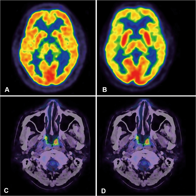
Objective: To explore regional brain glucose metabolic abnormalities of pretreatment stage I/II extranodal natural killer/T-cell lymphoma (ENKTL) patients using positron emission tomography with 2-deoxy-2-[fluorine-18]fluoro-D-glucose integrated with computed tomography (18F-FDG PET/CT) and assess its prognostic value.
Methods: Sixty pretreatment stage I/II ENKTL patients were enrolled in this retrospective study and divided into survival (n = 45) and death (n = 15) groups according to their status at the end of follow-up. A control group consisted of 60 healthy subjects. Regional cerebral glucose metabolism was evaluated on a voxel-by-voxel basis using statistical parametric mapping (SPM8) under a certain significance level (P < 0. 001) and voxel threshold (K = 100 voxels).
Results: Decreased metabolism was noted in patients, involving the bilateral prefrontal and orbitofrontal cortex, partial parietal and occipital cortex, cingulate gyrus and cerebellum; the sensorimotor cortex was largely spared. Increased metabolism was observed in the bilateral putamen, amygdala, and parahippocampal gyrus. Compared with the survival group, the death group had higher metabolism in the bilateral amygdala, putamen, left thalamus, uncus, and parahippocampal gyrus. Only B symptoms were associated with the increased metabolism of basal ganglia and thalamus (BGT). Patients with high metabolic tumor volume, total lesion glycolysis (TLG) and BGT metabolism had a poor prognosis. TLG and maximum standardized uptake value (SUVmax) LBGT/SUVmaxRight cerebellum were associated with Eastern Cooperative Oncology Group (ECOG) and prognostic index of natural killer lymphoma and Epstein-Barr virus-DNA (PINKE) scores. In multivariate analysis, only ECOG was an independent prognostic factor of both progression-free survival (PFS) and overall survival (OS). PINKE was an independent prognostic factor of OS.
Conclusion: Pretreatment stage I/II ENKTL patients exhibited abnormal regional cerebral glucose metabolism. Higher pretreatment glucose metabolism in BGT could predict a relatively poor prognosis but did not surpass the predictive values of ECOG and PINKE in stage I/II ENKTL patients.
Keywords: 18F-FDG PET/CT; extranodal natural killer/T- cell lymphoma; prognostic value; regional cerebral glucose metabolism; statistical parametric mapping.
© 2021 Zhou et al.
Conflict of interest statement
The authors report no conflicts of interest in this work.
Figures


Similar articles
-
[Prognostic value of pretreatment (18)F-FDG PET-CT metabolic parameters in patients with advanced extranodal NK/T cell lymphoma].Zhonghua Zhong Liu Za Zhi. 2019 Nov 23;41(11):831-836. doi: 10.3760/cma.j.issn.0253-3766.2019.11.006. Zhonghua Zhong Liu Za Zhi. 2019. PMID: 31770850 Chinese.
-
Assessment of the prognostic value of interim fluorodeoxyglucose positron emission tomography/computed tomography in nasal-type extranodal natural killer/T-cell lymphoma.Quant Imaging Med Surg. 2021 Apr;11(4):1220-1233. doi: 10.21037/qims-20-620. Quant Imaging Med Surg. 2021. PMID: 33816162 Free PMC article.
-
Assessment of the prognostic capacity of pretreatment, interim, and post-therapy (18)F-FDG PET/CT in extranodal natural killer/T-cell lymphoma, nasal type.Ann Nucl Med. 2015 Jun;29(5):442-51. doi: 10.1007/s12149-015-0964-8. Epub 2015 Mar 24. Ann Nucl Med. 2015. PMID: 25801633
-
Dynamic evaluation of the prognostic value of 18F-FDG PET/CT in extranodal NK/T-cell lymphoma, nasal type.Ann Hematol. 2021 Apr;100(4):1039-1047. doi: 10.1007/s00277-021-04466-3. Epub 2021 Feb 26. Ann Hematol. 2021. PMID: 33634350 Clinical Trial.
-
18F-FDG PET/CT in extranodal natural killer/T-cell lymphoma: a comprehensive evaluation method.Am J Nucl Med Mol Imaging. 2023 Dec 25;13(6):245-258. eCollection 2023. Am J Nucl Med Mol Imaging. 2023. PMID: 38204603 Free PMC article. Review.
Cited by
-
The remodeling of metabolic brain pattern in patients with extracranial diffuse large B-cell lymphoma.EJNMMI Res. 2023 Oct 30;13(1):94. doi: 10.1186/s13550-023-01046-6. EJNMMI Res. 2023. PMID: 37902852 Free PMC article.
-
Construction of an individualized brain metabolic network in patients with advanced non-small cell lung cancer by the Kullback-Leibler divergence-based similarity method: A study based on 18F-fluorodeoxyglucose positron emission tomography.Front Oncol. 2023 Mar 10;13:1098748. doi: 10.3389/fonc.2023.1098748. eCollection 2023. Front Oncol. 2023. PMID: 36969017 Free PMC article.
References
-
- Lin M, Huang G, Sun X, Ni J. The preliminary study of brain metabolic changes in lung cancer patients of different stages. Chin J Clin Med. 2009;16(4):516–518.
-
- Tashiro M, Kubota K, Itoh M, et al. Regional cerebral glucose metabolism of patients with malignant diseases in different clinical phases. Med Sci Monit. 2001;7(2):226–232. - PubMed
-
- Ni J, Lin M, Liu J, Huang G. Regional brain metabolism changes in the body malignant tumor patients without brain metastasis. Chin J Med Imaging Technol. 2010;26(11):2175–2178.
LinkOut - more resources
Full Text Sources
Other Literature Sources
