

Pneumomediastinum in COVID-19: A series of three cases and review of literature
- PMID: 34017591
- PMCID: PMC8114250
- DOI: 10.1177/2050313X211011807
Pneumomediastinum in COVID-19: A series of three cases and review of literature
Abstract
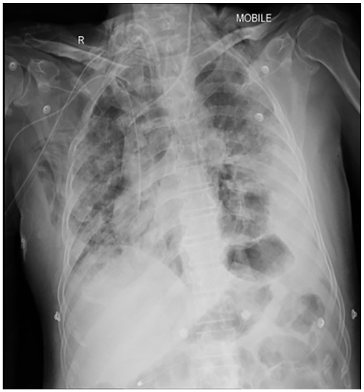
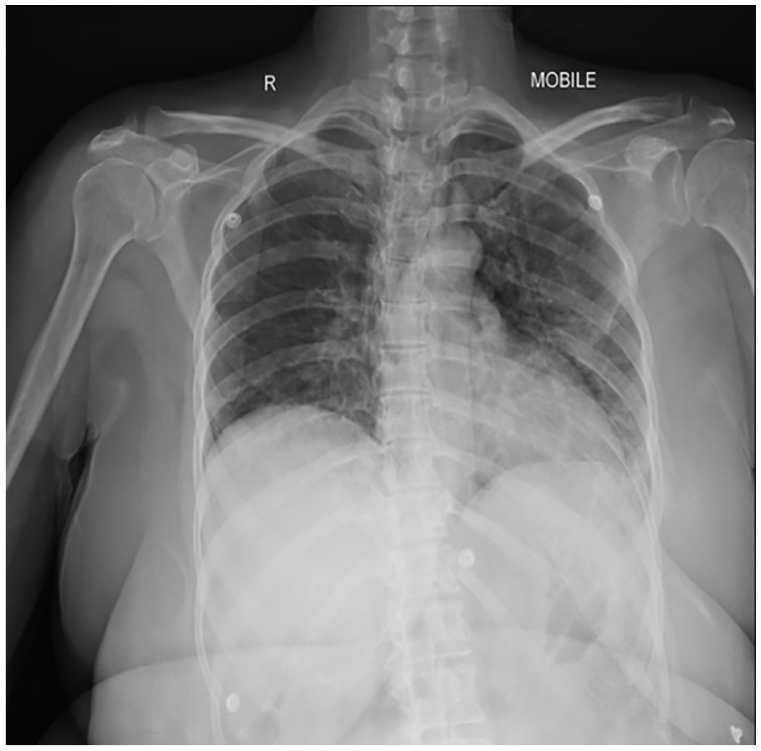
Coronavirus disease-19 caused by severe acute respiratory syndrome Corona virus-2 is characterised by wide heterogeneity in clinical presentation. The typical radiographic findings in COVID-19 include bilateral ground-glass opacities and/or consolidations predominantly affecting the lower lobes and posterior segments of lungs. Other rare abnormal radiographic findings include pneumothorax, pneumomediastinum and pneumopericardium. There has been an increased incidence of pneumomediastinum, a rare but potentially life-threatening complication during this pandemic. It may be spontaneous or secondary. Pneumomediastinum may be due to barotrauma, cytokine storm induced diffuse alveolar injury or direct viral infection of type I and type II pneumocytes. The presence of pneumomediastinum in COVID-19 patients may indicate extensive alveolar membrane destruction and those patients need close monitoring. There are no consensus guidelines in managing COVID-19 patients with pneumomediastinum. Higher mortality rates (70.58%) are reported in intubated COVID-19 patients with pneumomediastinum. The development of pneumomediastinum in COVID-19 should be considered as a poor prognostic factor.
Keywords: COVID-19; Pneumomediastinum; SARS CoV-2; pneumopericardium.
© The Author(s) 2021.
Conflict of interest statement
Declaration of conflicting interests: The author(s) declared no potential conflicts of interest with respect to the research, authorship, and/or publication of this article.
Figures


References
-
- Hamman L. Spontaneous mediastinal emphysema. Bull Johns Hopkins Hospital 1939; 64: 1–21.
-
- Bodey GP. Medical mediastinal emphysema. Ann Intern Med 1961; 54: 46–56.
Publication types
LinkOut - more resources
Full Text Sources
Other Literature Sources
Miscellaneous
