

Clinical value of radiomics and machine learning in breast ultrasound: a multicenter study for differential diagnosis of benign and malignant lesions
- PMID: 34018057
- PMCID: PMC8589755
- DOI: 10.1007/s00330-021-08009-2
Clinical value of radiomics and machine learning in breast ultrasound: a multicenter study for differential diagnosis of benign and malignant lesions
Abstract
Objectives: We aimed to assess the performance of radiomics and machine learning (ML) for classification of non-cystic benign and malignant breast lesions on ultrasound images, compare ML's accuracy with that of a breast radiologist, and verify if the radiologist's performance is improved by using ML.
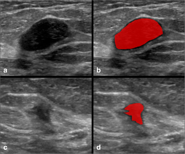
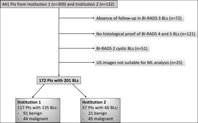
Methods: Our retrospective study included patients from two institutions. A total of 135 lesions from Institution 1 were used to train and test the ML model with cross-validation. Radiomic features were extracted from manually annotated images and underwent a multistep feature selection process. Not reproducible, low variance, and highly intercorrelated features were removed from the dataset. Then, 66 lesions from Institution 2 were used as an external test set for ML and to assess the performance of a radiologist without and with the aid of ML, using McNemar's test.
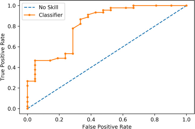
Results: After feature selection, 10 of the 520 features extracted were employed to train a random forest algorithm. Its accuracy in the training set was 82% (standard deviation, SD, ± 6%), with an AUC of 0.90 (SD ± 0.06), while the performance on the test set was 82% (95% confidence intervals (CI) = 70-90%) with an AUC of 0.82 (95% CI = 0.70-0.93). It resulted in being significantly better than the baseline reference (p = 0.0098), but not different from the radiologist (79.4%, p = 0.815). The radiologist's performance improved when using ML (80.2%), but not significantly (p = 0.508).
Conclusions: A radiomic analysis combined with ML showed promising results to differentiate benign from malignant breast lesions on ultrasound images.
Key points: • Machine learning showed good accuracy in discriminating benign from malignant breast lesions • The machine learning classifier's performance was comparable to that of a breast radiologist • The radiologist's accuracy improved with machine learning, but not significantly.
Keywords: Breast cancer; Machine learning; Ultrasound.
© 2021. The Author(s).
Conflict of interest statement
The authors of this manuscript declare no relationships with any companies whose products or services may be related to the subject matter of the article.
Figures


References
-
- Mendelson EB, Böhm-Vélez M, Berg WA et al (2013) ACR BI-RADS® ultrasound. In: American College of Radiology (ed) ACR BI-RADS® Atlas, Breast Imaging Reporting and Data System
Publication types
MeSH terms
LinkOut - more resources
Full Text Sources
Other Literature Sources
