

Review
doi: 10.1007/s10930-021-10000-1.
Epub 2021 May 21.
Update and Supplementary Articles Proteins of SARS CoV-2, Which Causes COVID-19, and the Interacting Proteins
Affiliations
- PMID: 34018094
- PMCID: PMC8137430
- DOI: 10.1007/s10930-021-10000-1
Item in Clipboard
Review
Update and Supplementary Articles Proteins of SARS CoV-2, Which Causes COVID-19, and the Interacting Proteins
Protein J.
2021 Jun.
Abstract
Coronavirus disease 2019 (COVID-19), which is the pandemic caused by the virus, severe acute respiratory syndrome coronavirus-2 (SARS CoV-2), first appearing in December 2019, continues to confound the world. In this update we provide insights into how some of the new mutant variant strains of SARS CoV-2 have evolved to be more infective. We also introduce our supplement of the special issue on the topic of the proteins of SARS CoV-2 in the Protein Journal, which follows this introduction.
Keywords: ACE2 protein; Enzymes; Mutants; Proteins; SARS CoV-2; Spike protein.
Figures
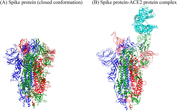
A SARS CoV-2 spike protein (PDB ID: 6VXX). B SARS CoV-2 spike protein bound to the human ACE2-receptor protein (PDB ID: 7A96). The green protomer has its receptor binding domain (RBD) pointing up and is bound to the ACE2 protein (cyan). The red protomer has its RBD pointing up in position to bind to a second ACE2 protein. The blue protomer has its RBD pointing in the closed conformation (Color figure online)
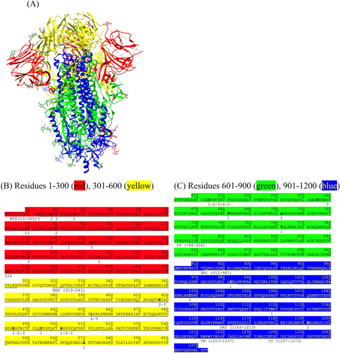
A Spike protein (closed conformation, PDB: 6VXX) colored where position 1–300 is red, position 301–600 is yellow, position 601–900 is green, and position 901–1200 is blue. B Sequence of the spike protein from 100 to 600 (colored as described in (A) of this figure). C Sequence of the spike protein from 601 to 1200 (colored as described in (A) of this figure). The numbers below the bold residues in the sequences refer to the changes in residues for each mutant strain presented in Tables 1 and 2. For mutant strains that have the same mutations, the numbers of the entries are separated by a hyphen—for example, the N501Y mutation is in entries 1, 2, and 3 from Table 1; therefore, position 501 is labeled with “1-2-3”. NTD N-terminal domain, RBD receptor binding domain, HR1 heptad repeat sequence 1, HR2 heptad repeat sequence 2, TM transmembrane domain, CT cytoplasm domain (Color figure online)
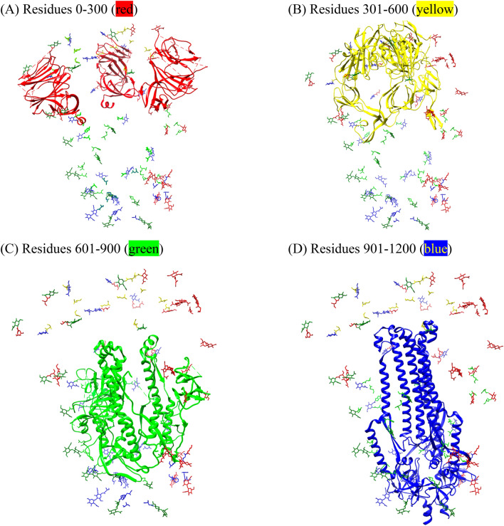
The spike protein with residues colored in the four different ranges: A 0–300 (red), B 301–600 (yellow), C 601–900 (green), and D 901–1200 (blue). Glycan moieties shown for reference (Color figure online)
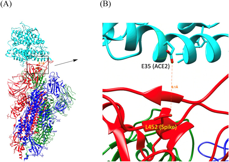
A Spike protein from SARS CoV-2 (tri-colored: red, blue, green) bound to human ACE2 protein (cyan)—PDB ID: 7KNE. Boxed is residue L452 on the spike protein and residue E35 on the ACE2 protein. B The zoomed in picture of L452 (spike) and E35 (ACE2) (Color figure online)
Similar articles
-
A Biochemical Perspective of the Nonstructural Proteins (NSPs) and the Spike Protein of SARS CoV-2.Protein J. 2021 Jun;40(3):260-295. doi: 10.1007/s10930-021-09967-8. Epub 2021 Feb 24. Protein J. 2021. PMID: 33629236 Free PMC article. Review.
-
Susceptibilities of Human ACE2 Genetic Variants in Coronavirus Infection.J Virol. 2022 Jan 12;96(1):e0149221. doi: 10.1128/JVI.01492-21. Epub 2021 Oct 20. J Virol. 2022. PMID: 34668773 Free PMC article.
-
The British variant of the new coronavirus-19 (Sars-Cov-2) should not create a vaccine problem.J Biol Regul Homeost Agents. 2021 Jan-Feb;35(1):1-4. doi: 10.23812/21-3-E. J Biol Regul Homeost Agents. 2021. PMID: 33377359
-
Impact of Genetic Variability in ACE2 Expression on the Evolutionary Dynamics of SARS-CoV-2 Spike D614G Mutation.Genes (Basel). 2020 Dec 24;12(1):16. doi: 10.3390/genes12010016. Genes (Basel). 2020. PMID: 33374416 Free PMC article.
-
Structural basis of severe acute respiratory syndrome coronavirus 2 infection.Curr Opin HIV AIDS. 2021 Jan;16(1):74-81. doi: 10.1097/COH.0000000000000658. Curr Opin HIV AIDS. 2021. PMID: 33186231 Review.
Cited by
-
Efficient inactivation of the contamination with pathogenic microorganisms by a combination of water spray and plasma-activated air.J Hazard Mater. 2023 Mar 15;446:130686. doi: 10.1016/j.jhazmat.2022.130686. Epub 2022 Dec 28. J Hazard Mater. 2023. PMID: 36610342 Free PMC article.
References
-
- https://www.cdc.gov/coronavirus/2019-ncov/science/science-briefs/scienti.... Accessed 18 May 2021
Publication types
MeSH terms
LinkOut - more resources
Full Text Sources
Other Literature Sources
Medical
Miscellaneous
