. 2021 Sep;239(3):669-681.
doi: 10.1111/joa.13440. Epub 2021 May 20.

Myological variation in the forearm anatomy of Callitrichidae and Lemuridae

Affiliations

Myological variation in the forearm anatomy of Callitrichidae and Lemuridae

Edwin Dickinson et al. J Anat. 2021 Sep.

Abstract

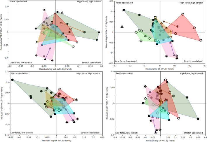
The anatomy of the primate forearm is frequently investigated in terms of locomotor mode, substrate use, and manual dexterity. Such studies typically rely upon broad, interspecific samples for which one or two representative taxa are used to characterize the anatomy of their genus or family. To interpret variation between distantly related taxa, however, it is necessary to contextualize these differences by quantifying variation at lower hierarchical levels, that is, more fine-grained representation within specific genera or families. In this study, we present a focused evaluation of the variation in muscle organization, integration, and architecture within two speciose primate families: the Callitrichidae and Lemuridae. We demonstrate that, within each lineage, several muscle functional groups exhibit substantial variation in muscle organization. Most notably, the digital extensors appear highly variable (particularly among callitrichids), with many unique configurations represented. In terms of architectural variables, both families are more conservative, with the exception of the genus Callimico-for which an increase is observed in forearm muscle mass and strength. We suggest this reflects the increased use of vertical climbing and trunk-to-trunk leaping within this genus relative to the more typically fine-branch substrate use of the other callitrichids. Overall, these data emphasize the underappreciated variation in forearm myology and suggest that overly generalized typification of a taxon's anatomy may conceal significant intraspecific and intrageneric variation therein. Thus, considerations of adaptation within the forearm musculature should endeavor to consider the full range of anatomical variation when making comparisons between multiple taxa within an evolutionary context.

Keywords: PCSA; dexterity; fascicle length; muscle; primate.


Figures

FIGURE 1
FIGURE 1
Photographs taking during the dissection of a specimen of Varecia rubra. (a) and (b) show superficial images of the anterior (flexor) and posterior (extensor) compartments; (c) and (d) show deeper images of the same compartments. Abbreviations as follows: PT, pronator teres; FCR, flexor carpi radialis; PL, palmaris longus; FCU, flexor carpi ulnaris; FDS, flexor digitorum superficialis; FDP, flexor digitorum profundus; PQ, pronator quadratus; ED, extensor digitorum; ECRB, extensor carpi radialis brevis; ECRL, extensor carpi radialis longus; BR, brachioradialis; EDM, extensor digiti minimi; ECU, extensor carpi ulnaris; APL, abductor pollicis longus; EPL, extensor pollicis longus; ED III, extensor digitorum III; EI, extensor indicis; Sup, supinator. Colors of functional groups as follows: yellow = digital flexors, green = wrist flexors, red = digital extensors, blue = wrist extensors, pink = pronators/supinators
FIGURE 2
FIGURE 2
Schematic illustration of digital extensor variations observed within callitrichids and lemurids. Percentages refer to prevalence within callitrichids, followed by lemurids. In all cases, digit numbers in parentheses refer to the digits receiving a tendon from the named muscle body. Muscles present in multiple parts (e.g., [a] and [b] or [a–c]) are organized by frequency of character state. Abbreviations as follows: extensor indicis (Ext. Ind.), extensor digitorum (ED), extensor digiti minimi (EDM), extensor pollicis longus (EPL)
FIGURE 3
FIGURE 3
RMA regressions of each architectural variable (muscle mass, PCSA, and fascicle length) against body mass in each functional muscle group. All variables linearized and log‐transformed prior to analysis. Summary statistics presented in Table 3. In all cases, black line depicts callitrichids, and gray line depicts lemurids. Shapes indicate genus as follows: solid circles = Callithrix, asterisk = Callimico, solid triangles = Leontopithecus, solid squares = Mico, solid diamonds = Saguinus, open circles = Eulemur, open triangles = Hapalemur, open diamonds = Varecia, open squares = Lemur
FIGURE 4
FIGURE 4
Functional space plots showing relationship between relative muscle strength (residuals of PCSA vs body mass—PCSA) and relative muscle excursion potential (residuals of fascicle lengths vs body mass—WFL) in the four major functional compartments of the forearm. Residuals calculated independently within each family following RMA regressions of key variables against body mass. Abbreviations as follows: WE, wrist extensors; DE, digital extensors; WF, wrist flexors; DF, digital flexors. Shapes indicates genus following Figure 3. Colors as follows: dark green = Callithrix, brown = Callimico, purple = Leontopithecus, blue = Mico, pink = Saguinus, red = Eulemur, light green = Varecia, yellow = Lemur. Hapalemur is uncolored as the genus is represented by one data point (open triangle) in each plot
FIGURE 5
FIGURE 5
Weighted fascicle lengths (FL) within each functional group of the forearm within five callitrichid genera. Abbreviations as follows: WF, wrist flexors; WE, wrist extensors; DF, digital flexors; DE, digital extensors; PS, pronators/supinators. Within each plot, central line indicates group mean; intermediate lines (overlap marks) indicate statistical significance between groups if overlap is not observed, or insignificance if two sets overlap; diamond tips indicates 95% confidence intervals for each group, assuming equal variance. All shapes indicate single data points
FIGURE 6
FIGURE 6
Residuals of digital extensor muscle mass (DE MM), wrist flexor muscle mass (WF MM), and pronator/supinator fascicle lengths (PS WFL) across five callitrichid genera, following reduced major axis regressions against body mass. Abbreviations as follows: WF, wrist flexors; DE, digital extensors. Within each plot, central line indicates group mean; intermediate lines (overlap marks) indicate statistical significance between groups if overlap is not observed, or insignificance if two sets overlap; diamond tips indicates 95% confidence intervals for each group, assuming equal variance. All circles indicate single data points

References

    1. Anapol, F. & Barry, K. (1996) Fiber architecture of the extensors of the hindlimb in semiterrestrial and arboreal guenons. American Journal of Physical Anthropology, 99, 429–447. - PubMed
    1. Ashton, E. & Oxnard, C. (1964) Functional adaptations in the primate shoulder girdle. In: Proceedings of the Zoological Society of London. Wiley Online Library, pp. 49–66.
    1. Aversi‐Ferreira, T.A. , Diogo, R. , Potau, J.M. , Bello, G. , Pastor, J.F. & Aziz, M.A. (2010) Comparative Anatomical Study of the Forearm Extensor Muscles of Cebus libidinosus (Rylands et al., 2000; Primates, Cebidae), Modern Humans, and Other Primates, With Comments on Primate Evolution, Phylogeny, and Manipulatory Behavior. The Anatomical Record, 293, 2056–2070. - PubMed
    1. Aziz, M.A. & Dunlap, S.S. (1986) The human extensor digitorum profundus muscle with comments on the evolution of the primate hand. Primates, 27, 293–319.
    1. Barnard, W. (1875) Observations on the membral musculation of Simia satyrus (Orang) and the comparative myology of man and the apes. Proceedings of the American Association for the Advancement of Science, 24, 112–144.

Publication types

MeSH terms

LinkOut - more resources