. 2021 Oct 20;27(11):1804-1812.
doi: 10.1093/ibd/izab093.

Unique Regulation of Coupled NaCl Absorption by Inducible Nitric Oxide in a Spontaneous SAMP1/YitFc Mouse Model of Chronic Intestinal Inflammation

Affiliations

Unique Regulation of Coupled NaCl Absorption by Inducible Nitric Oxide in a Spontaneous SAMP1/YitFc Mouse Model of Chronic Intestinal Inflammation

Subha Arthur et al. Inflamm Bowel Dis. .

Abstract

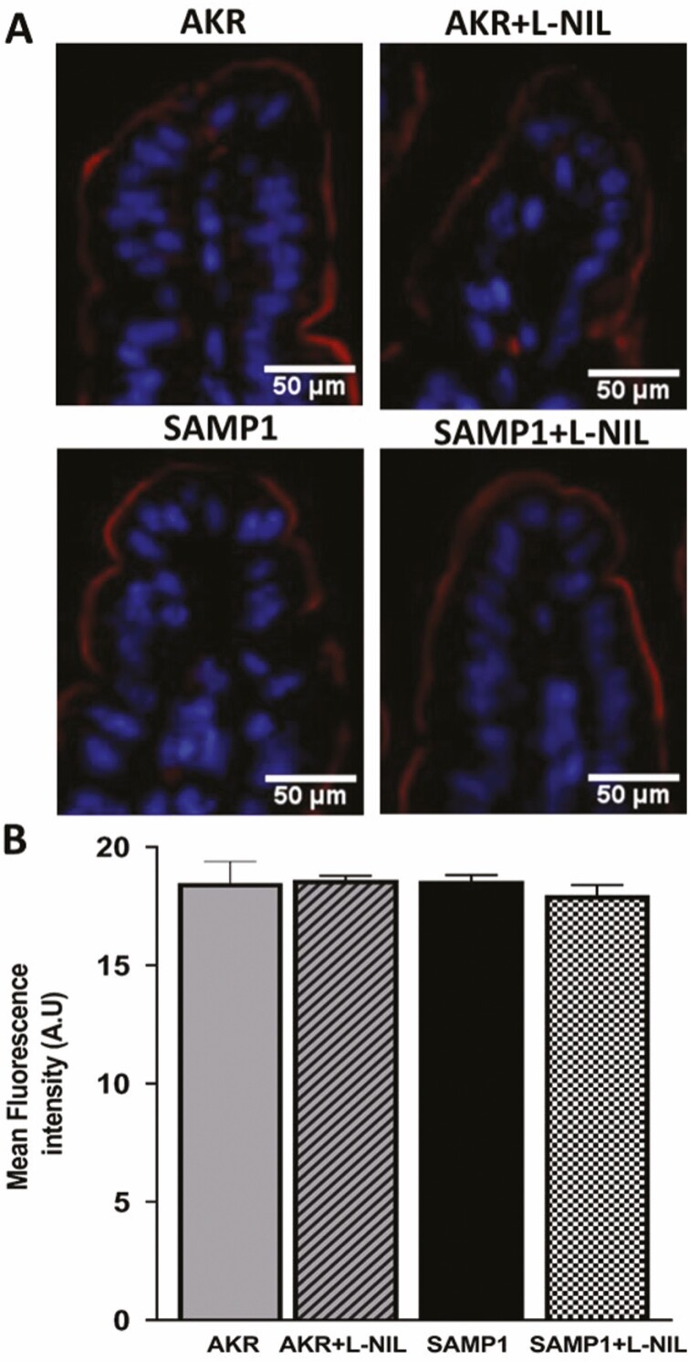
In the small intestine, Na:H (NHE3) and Cl:HCO3 (DRA or PAT1) exchangers present in the brush border membrane (BBM) of absorptive villus cells are primarily responsible for the coupled absorption of NaCl, the malabsorption of which causes diarrhea, a common symptom of inflammatory bowel disease (IBD). Inducible nitric oxide (iNO), a known mediator of inflammation, is increased in the mucosa of the chronically inflamed IBD intestine. An SAMP1/YitFc (SAMP1) mouse, a spontaneous model of chronic ileitis very similar to human IBD, was used to study alterations in NaCl absorption. The SAMP1 and control AKR mice were treated with I-N(6)-(1-Iminoethyl)-lysine (L-NIL) to inhibit iNO production, and DRA/PAT1 and NHE3 activities and protein expression were studied. Though Na:H exchange activity was unaffected, Cl:HCO3 activity was significantly decreased in SAMP1 mice due to a reduction in its affinity for Cl, which was reversed by L-NIL treatment. Though DRA and PAT1 expressions were unchanged in all experimental conditions, phosphorylation studies indicated that DRA, not PAT1, is affected in SAMP1. Moreover, the altered phosphorylation levels of DRA was restored by L-NIL treatment. Inducible NO mediates the inhibition of coupled NaCl absorption by decreasing Cl:HCO3 but not Na:H exchange. Specifically, Cl:HCO3 exchanger DRA but not PAT1 is regulated at the level of its phosphorylation by iNO in the chronically inflamed intestine.

Keywords: Cl:HCO3 exchange; DRA; NHE3; Na:H exchange; PAT1; SAMP1/YitFc; inducible nitric oxide; inflammatory bowel disease.


Figures

Figure 1.
Figure 1.
Ileal cross-sections of the AKR and SAMP1 mice. Hematoxylin and eosin stained (A) AKR and (B) SAMP1 mouse intestine. The SAMP1 mouse intestine shows crypt hypertrophy, villus blunting, and increased intra-epithelial lymphocytes characteristic of IBD.
Figure 2.
Figure 2.
Effect of chronic enteritis and L-NIL treatment on Cl:HCO3 exchange in villus cell BBMV. The Cl:HCO3 exchange, defined as HCO3-dependent and DIDS-sensitive uptake of 36Cl-, was significantly decreased during spontaneous ileitis in SAMP1 mice (n = 3, *P < 0.05). This inhibition was reversed back to normal by in vivo L-NIL treatment. L-NIL had no effect on Cl:HCO3 exchange activity in AKR mice (n = 3).
Figure 3.
Figure 3.
Effect of L-NIL treatment on Na:H exchange activity in BBMV. Na:H exchange, defined as pH-dependent and EIPA-sensitive Na uptake, was present comparably in both AKR and SAMP1 mice villus cell BBMV (n = 3). This indicated that Na:H exchange was unaffected by chronic intestinal inflammation in SAMP1 mice. Further, L-NIL had no effect on Na:H exchange in SAMP1 or AKR mice.
Figure 4.
Figure 4.
Cl:HCO3 exchange kinetic studies. 36Cl- uptake is shown as a function of increasing concentrations of extracellular Cl- at 30 seconds. In all the experimental conditions, as the concentration of extracellular chloride was increased, the uptake of 36Cl- was also stimulated and subsequently became saturated (n = 4). Table 1 shows the kinetic parameters derived from the kinetic experiments. As seen in Table 1, the affinity (1/Km) of Cl:HCO3 exchange for Cl- was significantly decreased in the villus cells from SAMP1 mice compared with the control, but this decreased affinity was completely restored to normal in the villus cells obtained from L-NIL treated SAMP1 mice.
Figure 5.
Figure 5.
Effect of L-NIL on villus cell Cl:HCO3 exchanger DRA protein expression in SAMP1 mouse intestine. Western blot studies revealed that BBM DRA protein level was not affected in SAMP1 mice villus cells nor was it affected by L-NIL treatment in AKR or SAMP1 mice (n = 3), which was further confirmed by the densitometry analyses.
Figure 6.
Figure 6.
Effect of L-NIL on villus cell Cl:HCO3 exchanger PAT1 protein expression in SAMP1 mouse intestine. Western blot studies revealed that BBM PAT1 protein level was not affected in SAMP1 mice villus cells nor was it affected by L-NIL treatment in AKR or SAMP1 mice. Western blot densitometry analyses confirmed these findings (n = 3).
Figure 7.
Figure 7.
Immunofluorescence studies of DRA in SAMP1 mouse intestine. The levels of DRA protein in the BBM of villus cells was similar in AKR and SAMP1 mice, and L-NIL treatment had no effect on DRA in either AKR or SAMP1 mice. B, Densitometric quantitation of DRA fluorescence levels confirmed these findings (n = 4). Images were captured at 10x magnification.
Figure 8.
Figure 8.
Immunofluorescence studies of PAT1 in SAMP1 mouse intestine. Levels of PAT1 protein in the BBM of villus cells was similar in AKR and SAMP1 mice, and L-NIL treatment had no effect on PAT1 levels in either AKR or SAMP1 mice. B, The quantitation of PAT1 fluorescence levels confirmed these findings (n = 4). Images were captured at 10x magnification.
Figure 9.
Figure 9.
Phosphorylation studies of DRA in SAMP1 mice intestine. A, Phosphorylation levels of serine residues in DRA was increased in villus cells from SAMP1 mice. This increased phosphorylation was reversed back to normal by in vivo treatment with L-NIL (n = 3, *P < 0.01, **P < 0.01). B, the phosphorylation of threonine residues in DRA was also increased in villus cells from SAMP1 mice. This increased phosphorylation was also reversed back to normal by in vivo treatment with L-NIL (n = 3, *P < 0.01, **P < 0.01).
Figure 10.
Figure 10.
Phosphorylation studies of PAT1 in SAMP1 mouse intestine. The phosphorylation levels of serine, threonine, and tyrosine residues in PAT1 were barely detectable in AKR mice and were unaffected in villus cells from SAMP1 mouse. Treatment with L-NIL had no effect on the phosphorylation of PAT1 in AKR or SAMP1 mice villus cells (n = 3).
Figure 11.
Figure 11.
Novel model of coupled NaCl absorption. In normal mammalian small intestine, traditional coupled NaCl absorption occurs via the dual operation of Na:H and Cl:HCO3 exchange on the BBM of absorptive villus cells. In the chronically inflamed intestine, this traditional NaCl absorption is likely not altered; however, a novel coupling of Cl:HCO3 exchange with Na-glucose cotransport and the inhibition of both seems to mediate the inhibition of the novel coupled NaCl absorption, leading to diarrhea of IBD. Also, malabsorption of chloride in SAMP1 mice is mediated by altered phosphorylation of DRA, likely through intracellular signaling pathway(s) activated by iNO.

References

    1. Knickelbein R, Aronson PS, Schron CM, et al. . Sodium and chloride transport across rabbit ileal brush border. II. Evidence for Cl-HCO3 exchange and mechanism of coupling. Am J Physiol. 1985;249:G236–G245. - PubMed
    1. Knickelbein RG, Aronson PS, Dobbins JW. Membrane distribution of sodium-hydrogen and chloride-bicarbonate exchangers in crypt and villus cell membranes from rabbit ileum. J Clin Invest. 1988;82:2158–2163. - PMC - PubMed
    1. Sundaram U, Knickelbein RG, Dobbins JW. pH regulation in ileum: Na(+)-H+ and Cl(-)-HCO3- exchange in isolated crypt and villus cells. Am J Physiol. 1991;260:G440–G449. - PubMed
    1. Binder HJ, Singh SK, Geibel JP, et al. . Novel transport properties of colonic crypt cells: fluid absorption and Cl-dependent Na-H exchange. Comp Biochem Physiol A Physiol. 1997;118:265–269. - PubMed
    1. Binder HJ, Ptak T. Jejunal absorption of water and electrolytes in inflammatory bowel disease. J Lab Clin Med. 1970;76:915–924. - PubMed

Publication types