Review
. 2021 May;78(10):4521-4544.
doi: 10.1007/s00018-021-03798-7. Epub 2021 Mar 26.

Adamantinomatous craniopharyngioma as a model to understand paracrine and senescence-induced tumourigenesis

Affiliations
Review

Adamantinomatous craniopharyngioma as a model to understand paracrine and senescence-induced tumourigenesis

Jose Mario Gonzalez-Meljem et al. Cell Mol Life Sci. 2021 May.

Abstract

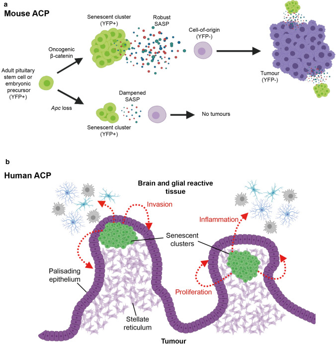
Cellular senescence is a process that can prevent tumour development in a cell autonomous manner by imposing a stable cell cycle arrest after oncogene activation. Paradoxically, senescence can also promote tumour growth cell non-autonomously by creating a permissive tumour microenvironment that fuels tumour initiation, progression to malignancy and metastasis. In a pituitary tumour known as adamantinomatous craniopharyngioma (ACP), cells that carry oncogenic β-catenin mutations and overactivate the WNT signalling pathway form cell clusters that become senescent and activate a senescence-associated secretory phenotype (SASP). Research in mouse models of ACP has provided insights into the function of the senescent cell clusters and revealed a critical role for SASP-mediated activities in paracrine tumour initiation. In this review, we first discuss this research on ACP and subsequently explore the theme of paracrine tumourigenesis in other tumour models available in the literature. Evidence is accumulating supporting the notion that paracrine signalling brought about by senescent cells may underlie tumourigenesis across different tumours and cancer models.

Keywords: Cancer stem cells; Oncogene-induced senescence; Pituitary tumour; SOX2; Senolytics; Therapy-induced senescence; WNT/β-catenin.


Conflict of interest statement

The authors declare that they have no conflict of interest.

Figures

Fig. 1
Fig. 1
Histopathology of human adamantinomatous craniopharyngioma (ACP). TE tumour epithelium, GRT glial reactive tissue, PE palisading epithelium, SR stellate reticulum, WL whorl-like epithelial cell groups. Immunostaining for β-catenin showing nucleo-cytoplasmic accumulation in cells of the WL, whilst the rest of the tumour cells show normal membranous staining. Scale bar 200 μm. The figure is adapted from Martinez-Barbera JP, Andoniadou CL (2020) Biological Behaviour of Craniopharyngiomas. Neuroendocrinology 1–8, with permission of S. Karger AG, Basel
Fig. 2
Fig. 2
Schematic of main components of the canonical WNT signalling pathway. In the absence of WNT ligands, β-catenin, (encoded by the CTNNB1 gene) is normally recruited in a destruction complex containing several proteins including APC (adenomatous polyposis coli), AXIN, CKIα (casein kinase 1 alpha) and GSK3β (glycogen synthase kinase 3β). This results in β-catenin phosphorylation of specific amino acids encoded by CTNNB1 exon 3 and protein degradation by the ubiquitin–proteasome pathway. Consequently, levels of β-catenin protein concentration are low in the cytoplasm and nucleus, hence keeping the target genes in a repressed state. At the same time, two surface E3 ubiquitin ligases, RNF43 and ZNFR3, regulate levels of the WNT ligand–receptor Frizzled through its ubiquitination which leads to its endosomal internalization and degradation. Binding of WNT ligands to their receptor, Frizzled, leads to the formation of a complex alongside coreceptors LRP and Dishevelled (DVL), which captures and disassembles the β-catenin destruction complex and thus prevents β-catenin phosphorylation and degradation. This causes protein stabilization, nucleo-cytoplasmic accumulation of β-catenin and the activation of target genes. Examples of WNT target genes encode for LGR receptors (LGR4-6), which upon binding of R-spondins (Rspo), recruit the RNF43/ZNFR3 complex and, therefore, allows the accumulation of Frizzled in the membrane. This leads to positive feedback and amplification of the WNT signalling pathway. Mutations in exon 3 of CTNNB1, containing the regulatory amino acids of β-catenin responsible for its degradation, prevent β-catenin phosphorylation by the destruction complex. This leads to its nucleo-cytoplasmic accumulation and constitutive overactivation of the WNT/β-catenin pathway even in the absence of WNT ligands and R-spondins. Created with BioRender.com
Fig. 3
Fig. 3
Human adamantinomatous craniopharyngioma (ACP) and ACP murine models contain nucleo-cytoplasmic β-catenin-accumulating cell clusters. a Immunofluorescent staining in human ACP showing the nucleo-cytoplasmic accumulation of β-catenin in cell groups known as “clusters” (arrows), a defining characteristic of these tumours. b Expression of oncogenic β-catenin in Rathke’s Pouch progenitors leads to the formation of clusters in a Hesx1 Cre/+; Ctnnb1 lox(ex3)/+ pre-tumoural pituitary. c Clusters also form upon inducible expression of oncogenic β-catenin in adult pituitary stem cells in Sox2 CreERT2/+; Ctnnb1 lox(ex3)/+ mice. A 16-week post-tamoxifen induction pituitary is shown. Scale bars 100 μm. AL anterior lobe, IL intermediate lobe. The figure is reproduced with permission from Carreno G, Gonzalez-Meljem JM, Haston S, Martinez-Barbera JP (2016) Stem cells and their role in pituitary tumorigenesis. Mol Cell Endocrinol 445:27–34
Fig. 4
Fig. 4
Lineage tracing in mouse ACP models shows the cell non-autonomous origin of tumours. Mouse ACP models were crossed with R26 YFP/+ lineage reporter mice which allows labelling of cells and their descendants upon expression of Cre recombinase. a Double immunofluorescent staining in Sox2 CreERT2/+; Ctnnb1 lox(ex3)/+; R26 YFP/+ pituitaries showing cell clusters (arrowheads) and a large tumoural lesion (asterisk) that accumulate β-catenin. The tumour cells (asterisk) do not express the lineage reporter YFP, indicating they are not descendants of SOX2 + stem cells. Note that the clusters (arrowheads) co-express nucleocytoplasmic β-catenin and YFP, demonstrating that they derive from SOX2 + stem cells. b In Hesx1 Cre/+; Ctnnb1 lox(ex3)/+; R26 YFP/+ mice, most cells of the anterior lobe of the pituitary descend from HESX1 + Rathke’s pouch precursor cells, as shown by YFP expression in a 5-week-old pituitary. After a period of latency, pituitary tissue is displaced by developing tumour tissue that does not express YFP. Scale bar 5 mm. c The absence of Cre-mediated recombination in the tumours is further demonstrated using the mT/mG dual reporter mouse line, in which unrecombined cells express membrane TdTomato protein (red) while pituitary-lineage cells express GFP (green). Note that in the Hesx1 Cre/+; R26 mTmG/+ control pituitary, the anterior lobe (al) tissue is green (recombined) and the posterior lobe (pl) is red (unrecombined since the pl is not derived from the Hesx1 lineage). In an ACP Hesx1 Cre/+; Ctnnb1 lox(ex3)/+; R26 mTmG/+ mouse tumour, most of the tumour cells express TdTomato. Scale bar 1 mm. d Double immunofluorescent staining against the proliferation marker Ki67 and YFP revealing that although most of the cells in the pituitary anterior lobe (al) of a 5-week-old ACP embryonic model are YFP + ve, the tumours in a 20-week-old mouse develop from YFP-ve cells that show a high proliferative activity (asterisk). In very advanced tumours (35 weeks) most of the YFP + ve cells are missing and only sporadic cells are detected in the periphery. Panel a is adapted with permission from Andoniadou CL, Matsushima D, Mousavy-gharavy SN, et al. (2013) The Sox2 + population of the adult murine pituitary includes progenitor/stem cells with tumour-inducing potential. Cell Stem Cell 13:433–445. bd are adapted from Gonzalez-Meljem JM, Haston S, Carreno G, et al. (2017) Stem cell senescence drives age-attenuated induction of pituitary tumours in mouse models of paediatric craniopharyngioma. Nat Commun 8:1819, which is an open-access article licensed under a Creative Commons Attribution 4.0 International License
Fig. 5
Fig. 5
The β-catenin accumulating senescent cell clusters modify the tumour microenvironment (TME) in the pre-tumoural pituitary of the ACP embryonic model. a Immunostaining against fibronectin, laminin and endomucin (EMCN) showing TME alterations prior to tumour initiation in the ACP mouse model. Scale bar 100 μm. b Double immunostaining for the lineage tracing reporter YFP showing an expanded population of EMCN-expressing cells that is not derived from the Hesx1 cell lineage targeted with oncogenic β-catenin. Note that clusters of YFP + ve cells are often surrounded by EMCN + ve cells (arrows). Scale bar 100 μm. c Triple immunostaining showing that in the context of oncogenic β-catenin (Hesx1 Cre/+; Ctnnb1 lox(ex3)/+ pituitary, top panel), large numbers of EMCN + ve cells also co-express SOX9 and interact closely with the senescent clusters (arrows). However, in the context of wild type β-catenin (loss of tumour suppressor Apc in Hesx1 Cre/+; Apc fl/fl pituitaries), senescent clusters are smaller and show an attenuated SASP that fail to induce changes in the TME. Of note, Hesx1 Cre/+; Apc fl/fl do not develop tumours (see text). Scale bar 50 μm. The figure is adapted from Gonzalez-Meljem JM, Haston S, Carreno G, et al. (2017) Stem cell senescence drives age-attenuated induction of pituitary tumours in mouse models of paediatric craniopharyngioma. Nat Commun 8:1819, which is an open-access article licensed under a Creative Commons Attribution 4.0 International License
Fig. 6
Fig. 6
Models depicting the role of senescent cells in mouse and human ACP. a Oncogenic β-catenin expression in either in SOX2 + adult pituitary stem cells or Hesx1 pituitary embryonic progenitors leads to the formation of nucleocytoplasmic β-catenin-accumulating clusters and SASP activation with expression of several cytokines, chemokines and growth factors. Persistent and robust SASP promotes cell transformation of a non-targeted cell (i.e. not expressing oncogenic β-catenin or YFP) in a paracrine manner. In this model SASP-mediated activities of the clusters are required for either tumour initiation, progression or both. b Senescent clusters in human ACP (green cells) are usually found at the base of finger-like tumour projections that invade the brain. The factors secreted by the clusters are proposed to promote tumour cell proliferation of the palisaded epithelium and epithelial bending resulting in tumour invasion. Signals may also promote inflammation in the glial reactive tissue. Created with BioRender.com

References

    1. Hayflick L, Moorhead PS. The serial cultivation of human diploid cell strains. Exp Cell Res. 1961;25:585–621. doi: 10.1016/0014-4827(61)90192-6. - DOI - PubMed
    1. Hayflick L. The limited in vitro lifetime of human diploid cell strains. Exp Cell Res. 1965;37:614–636. doi: 10.1016/0014-4827(65)90211-9. - DOI - PubMed
    1. Orgel LE. The maintenance of the accuracy of protein synthesis and its relevance to ageing. Proc Natl Acad Sci. 1963;49:517–521. doi: 10.1073/PNAS.49.4.517. - DOI - PMC - PubMed
    1. Hernandez-Segura A, Nehme J, Demaria M. Hallmarks of cellular senescence. Trends Cell Biol. 2018;28:436–453. doi: 10.1016/j.tcb.2018.02.001. - DOI - PubMed
    1. Gorgoulis V, Adams PD, Alimonti A, et al. Cellular senescence: defining a path forward. Cell. 2019;179:813–827. doi: 10.1016/j.cell.2019.10.005. - DOI - PubMed

MeSH terms

LinkOut - more resources