

Perivascular spaces and brain waste clearance systems: relevance for neurodegenerative and cerebrovascular pathology
- PMID: 34019111
- PMCID: PMC8460534
- DOI: 10.1007/s00234-021-02718-7
Perivascular spaces and brain waste clearance systems: relevance for neurodegenerative and cerebrovascular pathology
Abstract
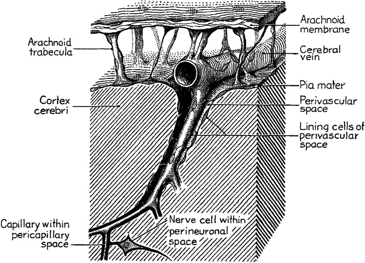
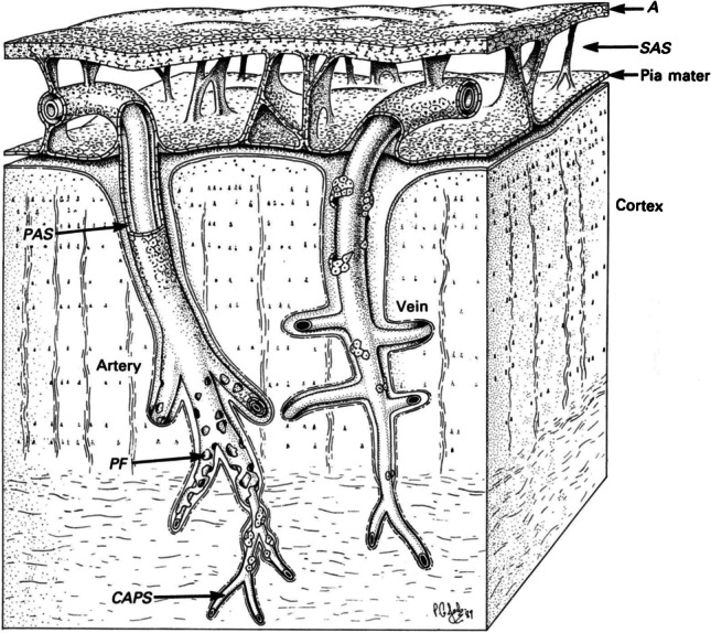
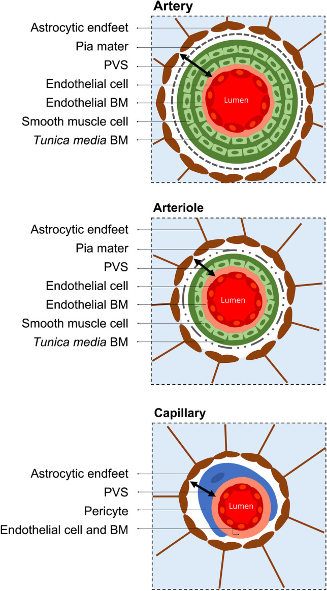
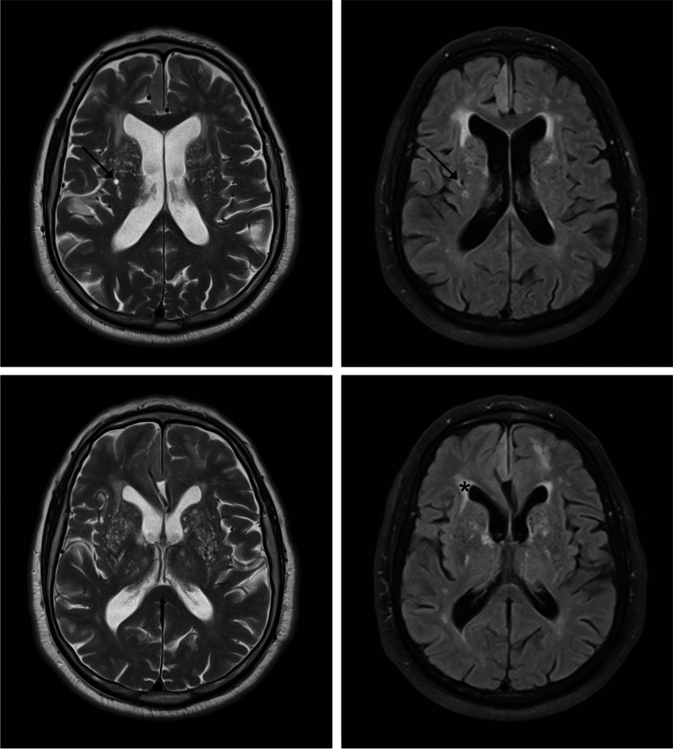
Perivascular spaces (PVS) of the brain, often called Virchow-Robin spaces, comprise fluid, cells and connective tissue, and are externally limited by astrocytic endfeet. PVS are involved in clearing brain waste and belong to the "glymphatic" system and/or the "intramural periarterial drainage" pathway through the basement membranes of the arteries. Related brain waste clearance systems include the blood-brain barrier, scavenger cells, cerebrospinal fluid, perineural lymphatic drainage pathways and the newly characterised meningeal lymphatic vessels. Any functional abnormality of PVS or related clearance systems might lead to accumulation of brain waste. It has been postulated that PVS enlargement can be secondary to accumulation of β-amyloid. Lack of integrity of the vascular wall, microbleeds, cerebral amyloid angiopathy (CAA) and enlarged PVS often occur in the preclinical stages of Alzheimer's disease, preceding substantial brain atrophy. PVS enlargement in the form of état criblé at the basal ganglia has also been considered to reflect focal atrophy, most probably secondary to ischaemic injury, based upon both pathological and imaging arguments. In addition, distinct topographic patterns of enlarged PVS are related to different types of microangiopathy: CAA is linked to enlarged juxtacortical PVS, whereas subjects with vascular risk factors tend to have enlarged PVS in the basal ganglia. Therefore, enlarged PVS are progressively being regarded as a marker of neurodegenerative and cerebrovascular pathology. The present review addresses the evolving concept of PVS and brain waste clearance systems, the potential relevance of their dysfunction to neurodegenerative and cerebrovascular pathology, and potential therapeutic approaches of interest.
Keywords: Brain; Cognitive impairment; Lymphatic; Perivascular spaces; Virchow-Robin spaces; “Glymphatic”.
© 2021. The Author(s).
Conflict of interest statement
None.
Figures
References
-
- Barkhof F, Fox NC, Bastos-Leite AJ, Scheltens P (2011) Normal ageing. In: Neuroimaging in dementia. Springer, pp 43–57. 10.1007/978-3-642-00818-4_4
-
- Rennels ML, Gregory TF, Blaumanis OR, Fujimoto K, Grady PA (1985) Evidence for a ‘paravascular’ fluid circulation in the mammalian central nervous system, provided by the rapid distribution of tracer protein throughout the brain from the subarachnoid space. Brain Res 326(1):47–63. 10.1016/0006-8993(85)91383-6 - PubMed
-
- Iliff JJ, Wang M, Liao Y, Plogg BA, Peng W, Gundersen GA, Benveniste H, Vates GE, Deane R, Goldman SA, Nagelhus EA, Nedergaard M (2012) A paravascular pathway facilitates CSF flow through the brain parenchyma and the clearance of interstitial solutes, including amyloid beta. Sci Transl Med 4 (147):147ra111. 10.1126/scitranslmed.3003748 - PMC - PubMed
Publication types
MeSH terms
LinkOut - more resources
Full Text Sources
Other Literature Sources
Medical
Research Materials
