. 2021 Sep 14;16(9):2169-2181.
doi: 10.1016/j.stemcr.2021.04.018. Epub 2021 May 20.

Increased tissue stiffness triggers contractile dysfunction and telomere shortening in dystrophic cardiomyocytes

Affiliations

Increased tissue stiffness triggers contractile dysfunction and telomere shortening in dystrophic cardiomyocytes

Alex C Y Chang et al. Stem Cell Reports. .

Abstract

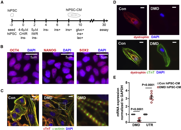
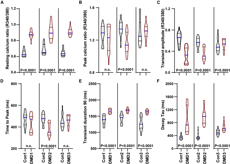
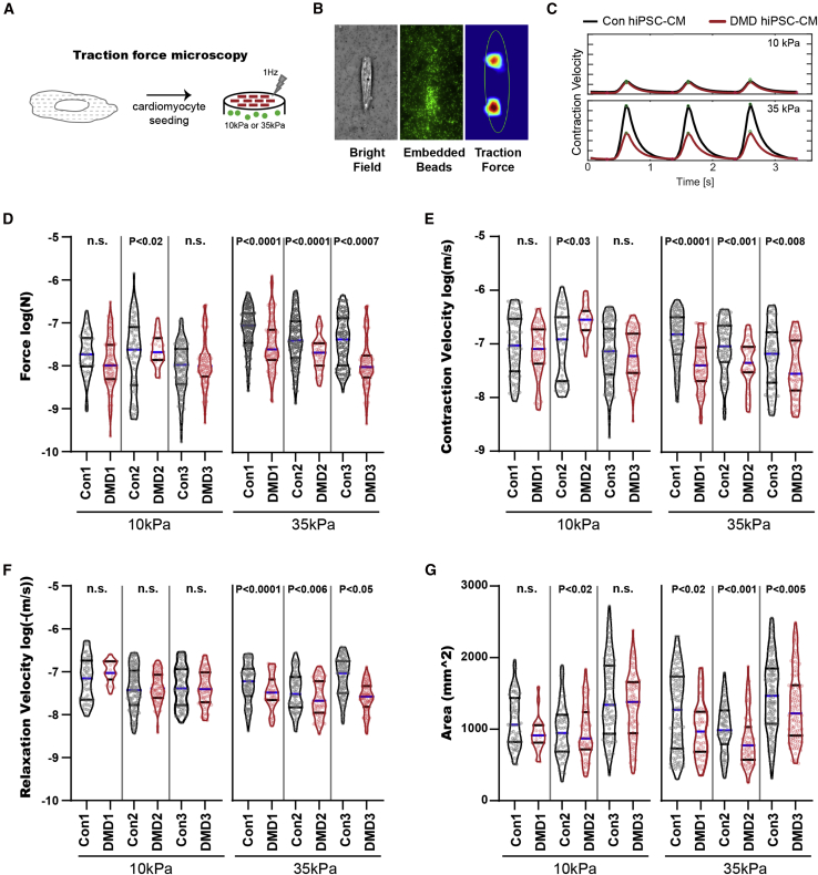
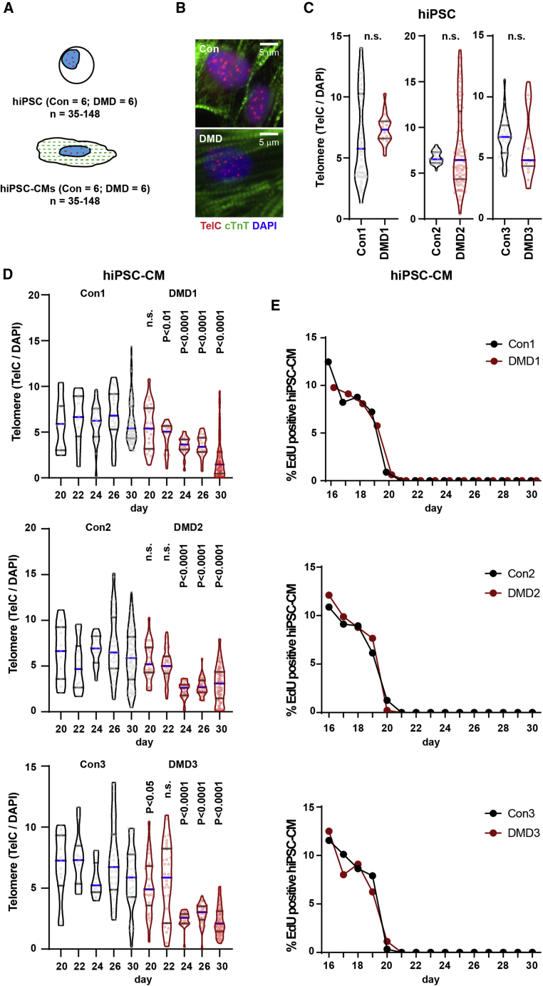
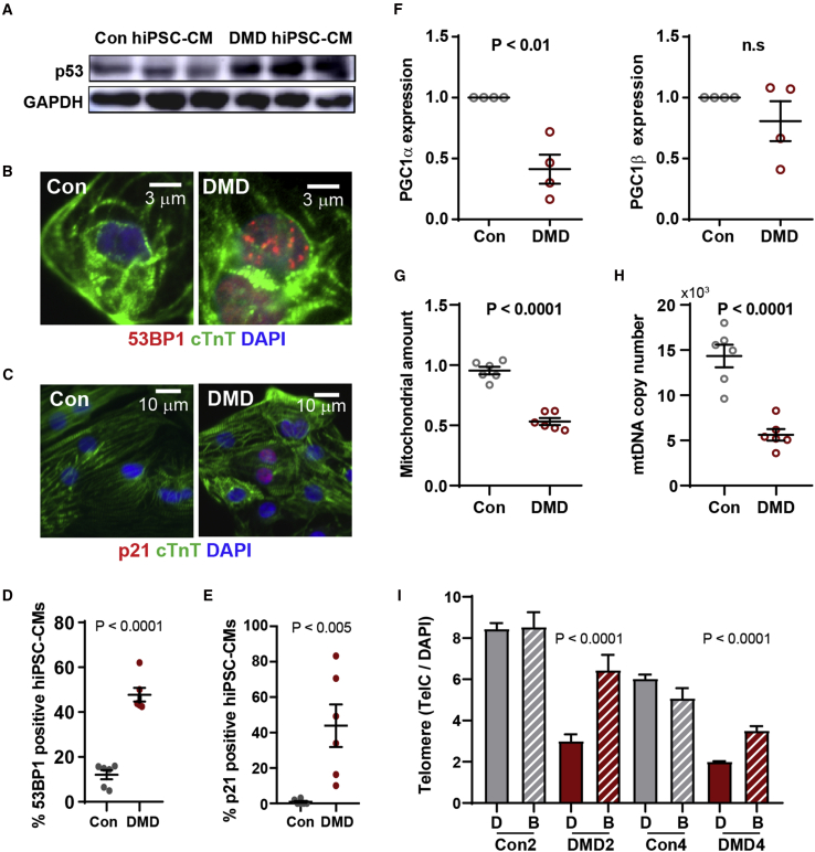
Duchenne muscular dystrophy (DMD) is a rare X-linked recessive disease that is associated with severe progressive muscle degeneration culminating in death due to cardiorespiratory failure. We previously observed an unexpected proliferation-independent telomere shortening in cardiomyocytes of a DMD mouse model. Here, we provide mechanistic insights using human induced pluripotent stem cell-derived cardiomyocytes (hiPSC-CMs). Using traction force microscopy, we show that DMD hiPSC-CMs exhibit deficits in force generation on fibrotic-like bioengineered hydrogels, aberrant calcium handling, and increased reactive oxygen species levels. Furthermore, we observed a progressive post-mitotic telomere shortening in DMD hiPSC-CMs coincident with downregulation of shelterin complex, telomere capping proteins, and activation of the p53 DNA damage response. This telomere shortening is blocked by blebbistatin, which inhibits contraction in DMD cardiomyocytes. Our studies underscore the role of fibrotic stiffening in the etiology of DMD cardiomyopathy. In addition, our data indicate that telomere shortening is progressive, contraction dependent, and mechanosensitive, and suggest points of therapeutic intervention.

Keywords: DMD; dilated cardiomyopathy; fibrosis; hiPSC-CM; telomere.


Conflict of interest statement

The authors declare no competing interests.

Figures

Figure 1
Figure 1
Generation of DMD hiPSC-CMs (A) hiPSC differentiation protocol for the generation of hiPSC-CMs. CHIR, CHIR-99021; IWR, IWR-1; ins, insulin; gluc, glucose; lac, lactate. hiPSCs were differentiated in medium supplemented with B27 minus insulin (ins). On day 6, the medium was supplemented with B27, including insulin. On day 10, hiPSC-CMs were cultured in medium without glucose supplemented with B27 and lactate. (B) Representative micrograph in which hiPSCs were stained with pluripotent stem cell markers OCT4, NANOG, and SOX2. (C) Representative micrograph in which hiPSC-CMs were stained with cardiac troponin T, α-actinin, and DAPI. (D) Representative micrograph in which healthy and DMD hiPSC-CMs were stained with dystrophin, cardiac troponin T, and DAPI. (E) Endogenous dystrophin (DMD) and utrophin (UTR) expression levels were determined by qRT-PCR in hiPSC-CMs (n = 6 independent experiments). Data shown as the mean ± SEM. Student's t test was used to calculate significance.
Figure 2
Figure 2
DMD hiPSC-CMs exhibit aberrant calcium-handling properties Using ratiometric-based Fura2 calcium imaging, (A) resting calcium ratio, (B) peak calcium ratio, (C) transient amplitude, (D) time to peak, (E) transient duration 90, and (F) decay tau per isogenic hiPSC pairs were plotted (isogenic pairs only; n = 3 independent experiments, 19–20 cells analyzed). Data are shown as violin plots where blue median and gray quartiles are shown. Student's t test was used to calculate significance.
Figure 3
Figure 3
DMD hiPSC-CMs exhibit contractile dysfunction under fibrotic microenvironment challenge (A) Contractile assessment of DMD hiPSC-CMs using traction force microscopy where hiPSC-CMs were seeded onto micropatterned tunable hydrogel devices. (B and C) (B) Representative micrographs of contraction force generated by a single hiPSC-CM on 10 or 35 kPa hydrogel (bright field, GFP fluorescent beads, Fourier traction force cytometry) and (C) representative contraction cycles are shown. (D–G) (D) Force, (E) contraction velocity, (F) relaxation velocity, and (G) cell area of single Con and DMD hiPSC-CMs subjected to 10 and 35 kPa hydrogels were measured (n = 3 independent experiments, 19–143 cells analyzed). Data are shown as violin plots where blue median and gray quartiles are shown. Student's t test was used to calculate significance.
Figure 4
Figure 4
DMD hiPSC-CMs exhibit telomere shortening and DNA damage response (A) Telomere length (TelC) was quantified by immunofluorescence staining (Q-FISH) relative to nuclear DAPI staining for hiPSC and hiPSC-CMs. (B) Representative micrographs of hiPSC-CMs stained with cardiac troponin T, TelC and DAPI are shown. (C) Quantification of hiPSC telomere using Q-FISH (n = 3 independent experiments, 17–86 cells analyzed). (D) Telomere Q-FISH reveals progressive telomere loss in DMD hiPSC-CMs between days 20 and 30 (n = 3 independent experiments, 11–179 cells analyzed). (E) hiPSC-CMs were devoid of EdU between days 20 and 30. Data are shown as violin plots where blue median and gray quartiles are shown. Student's t test was used to calculate significance.
Figure 5
Figure 5
DMD hiPSC-CMs exhibit p53 upregulation (A–E) Representative micrographs of (A) p53 activation by immunoblotting (n = 3 independent experiments), (B) DNA damage 53BP1 foci by immunofluorescence (n = 6 independent experiments), and (C) p21 (n = 6 independent experiments) in cardiac troponin T+ hiPSC-CMs are shown and (D and E) quantified, respectively. (F) Reduced expression levels of PGC-1α, master regulator of mitochondrial biogenesis, determined by qRT-PCR (n = 6 independent experiments). (G and H) (G) Mitochondria amount (n = 6 independent experiments) and (H) mitochondrial copy number (n = 6 independent experiments) in hiPSC-CMs were assessed by MitoTracker Green and qRT-PCR using mitochondrial gene (Nd2) to nuclear DNA (Nrf1) primers, respectively. Data represent mean ± SEM. Student's t was test used for statistical analysis. (I) Telomere loss prevented when contraction of DMD hiPSC-CMs was inhibited between days 20 and 30 using blebbistatin was quantified (n = 3 independent experiments, 35–1,834 cells analyzed). Data represent mean ± SEM. Student's t test was used for statistical analysis.

References

    1. Aguado J., Sola-Carvajal A., Cancila V., Revêchon G., Ong P.F., Jones-Weinert C.W., Wallén Arzt E., Lattanzi G., Dreesen O., Tripodo C., et al. Inhibition of DNA damage response at telomeres improves the detrimental phenotypes of Hutchinson-Gilford progeria syndrome. Nat. Commun. 2019;10:4990–5011. doi: 10.1038/s41467-019-13018-3. - DOI - PMC - PubMed
    1. Ain Q., Schmeer C., Penndorf D., Fischer M., Bondeva T., Förster M., Haenold R., Witte O.W., Kretz A. Cell cycle-dependent and -independent telomere shortening accompanies murine brain aging. Aging (Albany NY) 2018;10:3397–3420. doi: 10.18632/aging.101655. - DOI - PMC - PubMed
    1. Amoasii L., Hildyard J.C.W., Li H., Sanchez-Ortiz E., Mireault A., Caballero D., Harron R., Stathopoulou T.-R., Massey C., Shelton J.M., et al. Gene editing restores dystrophin expression in a canine model of Duchenne muscular dystrophy. Science. 2018;8:eaau1549. doi: 10.1126/science.aau1549. - DOI - PMC - PubMed
    1. Arnoult N., Karlseder J. Complex interactions between the DNA-damage response and mammalian telomeres. Nat. Struct. Mol. Biol. 2015;22:859–866. doi: 10.1038/nsmb.3092. - DOI - PMC - PubMed
    1. Bergmann O., Zdunek S., Felker A., Salehpour M., Alkass K., Bernard S., Sjostrom S.L., Szewczykowska M., Jackowska T., Dos Remedios C., Malm T., et al. Dynamics of cell generation and turnover in the human heart. Cell. 2015;161:1566–1575. doi: 10.1016/j.cell.2015.05.026. - DOI - PubMed

Publication types

MeSH terms