

IBS-GEC ESTRO-ABS recommendations for CT based contouring in image guided adaptive brachytherapy for cervical cancer
- PMID: 34019918
- PMCID: PMC8675891
- DOI: 10.1016/j.radonc.2021.05.010
IBS-GEC ESTRO-ABS recommendations for CT based contouring in image guided adaptive brachytherapy for cervical cancer
Abstract
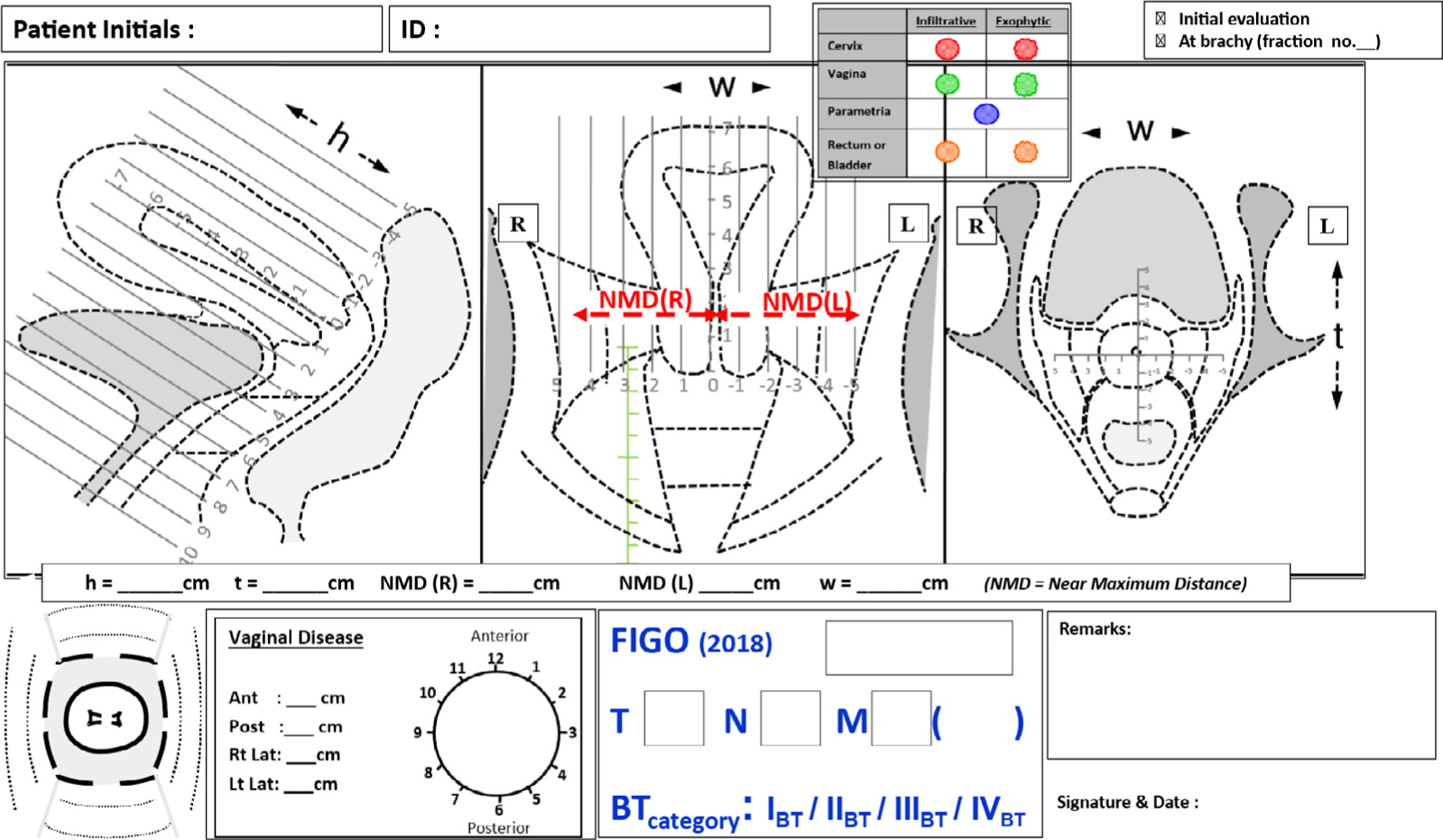
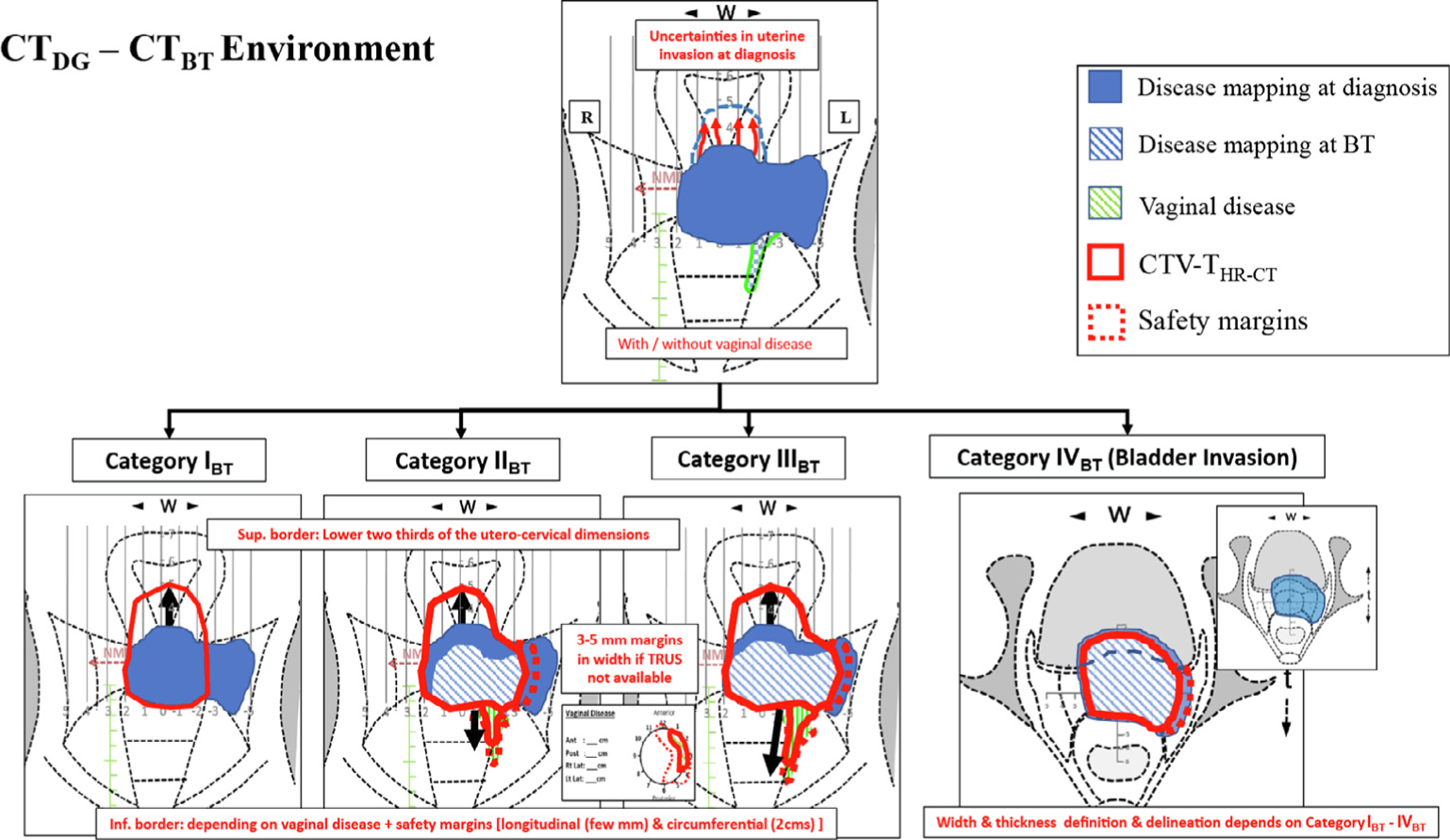
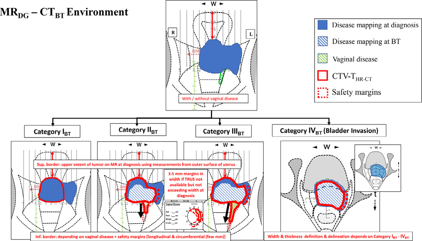
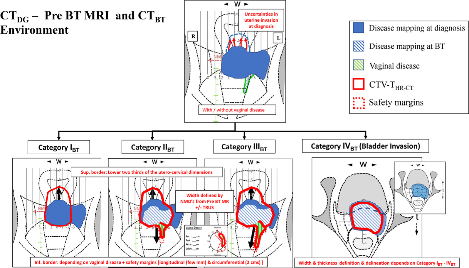
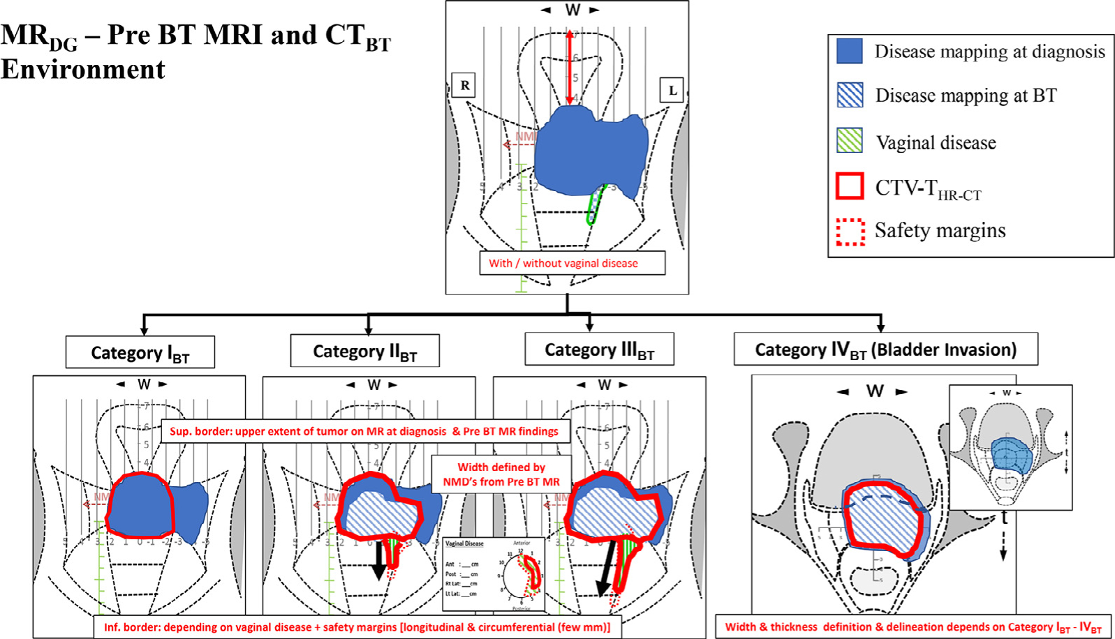
MR Imaging is regarded asthe gold standardfor Image Gudied Adaptive Brachytherapy (IGABT) for cervical cancer. However, its wide applicability is limited by its availability, logistics and financial implications. Use of alternative imaging like CTand Ultrasound (US) for IGABT has been attempted. In order to arrive at a systematic, uniform and international approach for CT based definition and contouring of target structures, GEC ESTRO, IBS and ABS agreed to jointly develop such recommendations based on the concepts and terms as published in the ICRU Report 89. The minimum requirements are clinical examination & documentation, CT or MR imaging at diagnosis and at a minimum, CT imaging with the applicator in place. The recommendations are based on (i) assessment of the GTV at diagnosis and at brachytherapy, (ii) categorizing the response to external radiation into different clinical remission patterns, (iii) defining various clinico-radiological environments and (iv) definition & delineation of a target on CT imaging at the time of brachytherapy with the applicator in situ. CT based target contouring recommendations based on 4 remission categories within 8 defined environments, aim at improving the contouring accuracy for IGABT using CT, US and MRI as available. For each clinico-radiological environment, there is an attempt to minimize the specific uncertainties in order to arrive at the best possible contouring accuracy. Evaluating feasibility & reproducibility, to achieve a benchmark towards a gold standard MR IGABT and further clinical research including outcomes with CT Based IGABT will become the next steps.
Keywords: CT based contouring; CT environments; Cervical cancer; Cervical cancer brachytherapy recommendations; IGABT.
Copyright © 2021 The Authors. Published by Elsevier B.V. All rights reserved.
Conflict of interest statement
Conflict of interest
None.
Figures
References
-
- IARC/WHO. GLOBOCAN 2018: Estimated cancer incidence, mortality and prevalence worldwide in 2018. Cervical Cancer Fact Sheet. Available at: https://gco.iarc.fr/today/data/factsheets/cancers/23-Cervix-Uteri-fact-s....
-
- Dimopoulos JC, Petrow P, Tanderup K, Petric P, Berger D, Kirisits C, et al. Recommendations from Gynaecological (GYN) GEC-ESTRO Working Group (IV): Basic principles and parameters for MR imaging within the frame of image based adaptive cervix cancer brachytherapy. Radiother Oncol 2012;103:113–22. - PMC - PubMed
-
- Haie-Meder C, Chargari C, Rey A, Dumas I, Morice P, Magne N. MRI-based low dose-rate brachytherapy experience in locally advanced cervical cancer patients initially treated by concomitant chemoradiotherapy. Radiother Oncol 2010;96:161–5. - PubMed
-
- Pötter R, Georg P, Dimopoulos JC, Grimm M, Berger D, Nesvacil N, et al. Clinical outcome of protocol based image (MRI) guided adaptive brachytherapy combined with 3D conformal radiotherapy with or without chemotherapy in patients with locally advanced cervical cancer. Radiother Oncol 2011;100:116–23. - PMC - PubMed
MeSH terms
Grants and funding
LinkOut - more resources
Full Text Sources
Other Literature Sources
Medical
