Case Reports
. 2021 May 21;21(1):205.
doi: 10.1186/s12883-021-02228-2.

Neurological manifestations of polyarteritis nodosa: a tour of the neuroaxis by case series

Affiliations
Case Reports

Neurological manifestations of polyarteritis nodosa: a tour of the neuroaxis by case series

Cathra Halabi et al. BMC Neurol. .

Abstract

Background: Heterogenous central nervous system (CNS) neurologic manifestations of polyarteritis nodosa (PAN) are underrecognized. We review three cases of patients with PAN that illustrate a range of nervous system pathology, including the classical mononeuritis multiplex as well as uncommon brain and spinal cord vascular manifestations.

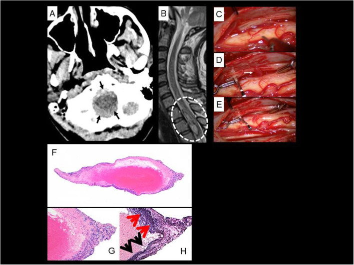
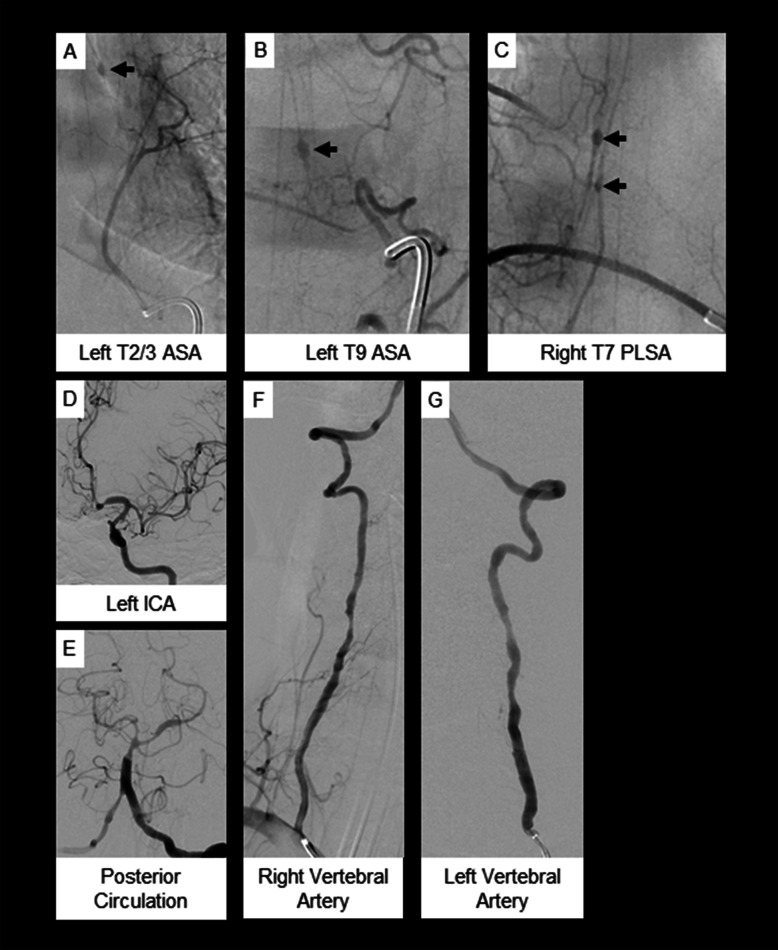
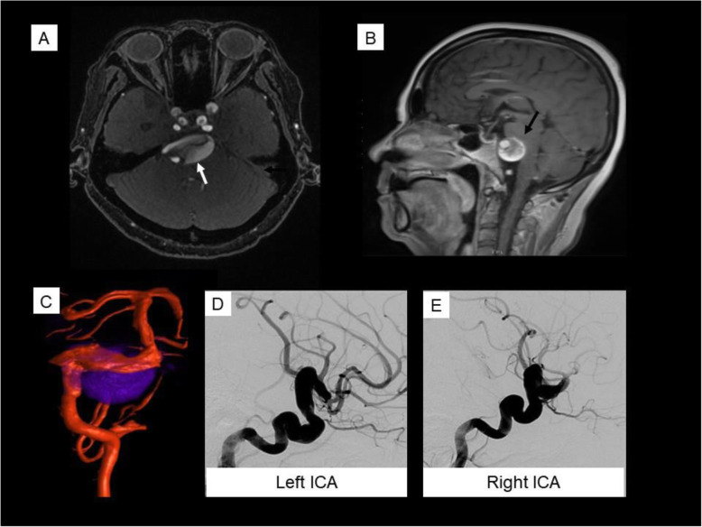
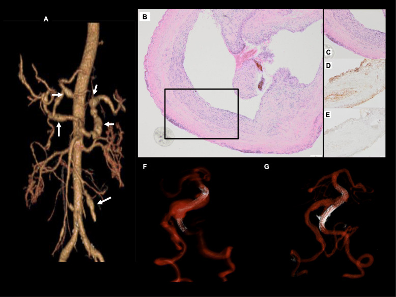
Case presentation: Case 1 presented with mononeuritis multiplex and characteristic skin findings. Case 2 presented with thunderclap headache and myelopathy due to spinal artery aneurysm rupture. Both patients experienced disease remission upon treatment. Case 3 presented with headache and bulbar symptoms due to partially thrombosed intracranial aneurysms, followed by systemic manifestations related to visceral aneurysms. She demonstrated clinical improvement with treatment, was lost to follow-up, then clinically deteriorated and entered hospice care.

Conclusions: Although the peripheral manifestations of PAN are well-known, PAN association with CNS neurovascular disease is relatively underappreciated. Clinician awareness of the spectrum of neurologic disease is required to reduce diagnostic delay and promote prompt diagnosis and treatment with immunosuppressants.

Keywords: Case series; Intracranial aneurysm; Multidisciplinary; Polyarteritis nodosa; Spinal artery aneurysm.


Conflict of interest statement

CFD reports funding from Microvention-Terumo, Inc., outside the submitted work.

CA reports grants from NIH NIAMS 5 T32 AR007304–40 during the conduct of the study.

There are no additional financial or non-financial competing interests to declare.

Figures

Fig. 1
Fig. 1
Case 2. a Axial non-contrast CT image at the level of the foramen magnum shows diffuse subarachnoid hemorrhage (solid arrows). b Sagittal T2W MRI image of the spine shows intradural hemorrhage in the upper thoracic spine (dashed oval) compressing and posteriorly displacing the adjacent thoracic cord. c-e Intraoperative images of fusiform thoracic spinal artery aneurysm resection. d A large fusiform aneurysm was identified (arrow) and isolated with 2 clips placed on either side along the parent vessel. e The fusiform aneurysm and small segment of posterior spinal artery was resected (arrow) using microsurgical technique and separated from the arachnoid of the cord. f Low power magnification view of the biopsy from the posterior spinal artery branch showing disruption of the vessel wall (original X40). g High power view of the vessel wall with lymphocytic infiltration and destruction of vessel wall, consistent with vasculitis (original × 100). h Elastic Van-Gieson stain demonstrating presence of elastic lamina in the lower half of the vessel wall (black arrows) and its destruction on the upper half of the vessel wall (red arrows, original × 100)
Fig. 2
Fig. 2
Case 2. a-c Spinal angiogram and (d-g) cerebral angiogram. a & b Multiple fusiform aneurysms (solid arrows) arising from the anterior spinal artery and (c) posterior lateral spinal artery. d-g Findings consistent with diffuse intra- and extracranial vasculopathy with multifocal narrowing and irregularity
Fig. 3
Fig. 3
Case 3. Giant vertebrobasilar aneurysm. Gadolinium-enhanced MR angiogram (a) and Sagittal T1 sequence (b) demonstrate a partially thrombosed vertebrobasilar aneurysm (white arrow in A). Note the significant mass effect on the pons (black arrow). Fused conventional angiography 3D reconstruction and MR of the vertebrobasilar system with dolichoectatic vessels (red) and outline of partially thrombosed aneurysm (blue) (c). Conventional angiography with left (d) and right (e) lateral carotid injections demonstrating arteriomegaly of the anterior circulation
Fig. 4
Fig. 4
Case 3. a CT angiography 3D reconstruction demonstrating multiple fusiform aneurysms of the hepatic, splenic, and superior mesenteric arteries (arrows). b H&E staining shows the cross-section of the aneurysm tip (original × 40). c Medium power microscopic images showing inflammation involving the aneurysm wall (original × 100). Immunohistochemical staining with the antibodies against p65 subunit of NFkB (d, original × 100) and TNFa (e, original × 100) shows focal and strong positive staining within the inflammatory infiltrates as expected. f, g 3D reconstruction of vertebrobasilar system after conventional angiography and PED deployment into the left vertebral artery (f), and repeat procedure 3 months later with second PED demonstrating overlapping stents and smaller aneurysm (g). Note reduction of flowing portion of the aneurysm sac as contemporaneously performed MR demonstrated continued enlargement of the thrombosed aneurysm sac with mass effect.

Similar articles

Cited by

References

    1. De Virgilio A, Greco A, Magliulo G, Gallo A, Ruoppolo G, Conte M, et al. Polyarteritis nodosa: a contemporary overview. Autoimmun Rev. 2016;15(6):564–570. doi: 10.1016/j.autrev.2016.02.015. - DOI - PubMed
    1. Pagnoux C, Seror R, Henegar C, Mahr A, Cohen P, Le Guern V, et al. Clinical features and outcomes in 348 patients with polyarteritis nodosa: a systematic retrospective study of patients diagnosed between 1963 and 2005 and entered into the French Vasculitis study group database. Arthritis Rheum. 2010;62(2):616–626. doi: 10.1002/art.27240. - DOI - PubMed
    1. Moore PM, Cupps TR. Neurological complications of vasculitis. Ann Neurol. 1983;14(2):155–167. doi: 10.1002/ana.410140202. - DOI - PubMed
    1. Jennette JC, Falk RJ, Andrassy K, Bacon PA, Churg J, Gross WL, et al. Nomenclature of systemic vasculitides. Proposal of an international consensus conference. Arthritis Rheum. 1994;37(2):187–192. doi: 10.1002/art.1780370206. - DOI - PubMed
    1. Ford RG, Siekert RG. Central nervous system manifestations of periarteritis nodosa. Neurology. 1965;15:114–122. doi: 10.1212/WNL.15.2.114. - DOI - PubMed

Publication types

Substances