Review
. 2021 Jun;41(2):247-266.
doi: 10.1016/j.cll.2021.03.005. Epub 2021 Apr 24.

Glycan Imaging Mass Spectrometry: Progress in Developing Clinical Diagnostic Assays for Tissues, Biofluids, and Cells

Affiliations
Review

Glycan Imaging Mass Spectrometry: Progress in Developing Clinical Diagnostic Assays for Tissues, Biofluids, and Cells

Calvin R K Blaschke et al. Clin Lab Med. 2021 Jun.

Abstract

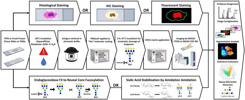
N-glycan imaging mass spectrometry (IMS) can rapidly and reproducibly identify changes in disease-associated N-linked glycosylation that are linked with histopathology features in standard formalin-fixed paraffin-embedded tissue samples. It can detect multiple N-glycans simultaneously and has been used to identify specific N-glycans and carbohydrate structural motifs as possible cancer biomarkers. Recent advancements in instrumentation and sample preparation are also discussed. The tissue N-glycan IMS workflow has been adapted to new glass slide-based assays for effective and rapid analysis of clinical biofluids, cultured cells, and immunoarray-captured glycoproteins for detection of changes in glycosylation associated with disease.

Keywords: Biomarkers; Clinical diagnostics; Glycosylation; Imaging mass spectrometry.


Conflict of interest statement

Disclosure R.R. Drake, P.M. Angel, and A.S. Mehta disclose partial ownership interests in 2 companies: GlycoPath, LLC, Mt Pleasant, South Carolina, and N-Zyme Scientifics, Doylestown, Pennsylvania.

Figures

Figure 1.
Figure 1.
Overview of MALDI-IMS workflow and orthogonal analyses. Middle row: A standardized N-glycan IMS workflow including dewaxing and rehydration, antigen retrieval, release of N-glycans by PNGase F, matrix application and imaging by MALDI-MS. Bottom row: Endoglycosidase F3 and sialic acid-amidation adaptations to the N-glycan IMS workflow for detection of fucosylation and sialylation isomers, respectively. Top row: Orthogonal imaging techniques including histological, immunohistochemistry and immunofluorescence staining. Panel, right side: Integration of data from traditional, adapted, and orthogonal analyses for comprehensive characterization of the tissue N-glycome.
Figure 2.
Figure 2.
Example N-glycan imaging mass spectrometry of a colorectal adenocarcinoma tissue. A) H&E staining showing different tissue structures. B) Overlay image of six N-glycan structures which colocalize with distinct tissue regions. C-H) Individual N-glycan heatmaps from overlay in panel B Hex8HexNAc2 (red), Hex4HexNAc4Fuc2 (yellow), Hex4HexNAc5Fuc1 (purple), Hex5HexNAc4α2,6NeuAc1 (blue), Hex5HexNAc4Fuc1α2,3NeuAc1 (green) and Hex7HexNAc6Fuc1α2,6NeuAc1 (orange). Monosaccharide unit symbols: mannose (green circle), galactose (yellow circle), N-acetylglucosamine (blue square), fucose (red triangle), α2,3-linked sialic acid (purple diamond, right shifted) α2,6-linked sialic acid (purple diamond, left shifted).
Figure 3.
Figure 3.
Profiling of 6 N-glycans from the serum of patients with either liver cirrhosis or hepatocellular carcinoma (HCC) using the method described by Blaschke and colleagues. Monosaccharide unit symbols are as indicated in Figure 2.
Figure 4.
Figure 4.
N-glycan turnover in cell culture by imaging MS strategies. Human aortic endothelial cells are grown in 15N glutamine for 48 hours, adding an 15N at every N-acetylglucosamine, N-acetylgalactose, and N-acetylneuraminic acid. Fully labeled N-glycans with complete incorporation of 15N indicate a complete turnover of that N-glycan. A) Human aortic endothelial cells are grown with 15N glutamine on a conventional cell chamber slide. This image shows cells that have been stained after IMS analysis with Coomassie Blue to allow normalization to cell numbers & protein content. B) Example image data of the same region of cultured cells. Image is of high mannose N-glycan Man9. Cells were scanned at a step size of 25 μm. Blue indicates no turnover, yellow indicates full turnover, white is partial incorporation of the label, indicating a slower turnover rate for some cells (red arrows). C) Example N-glycan profile showing the high mannose peak Man9. Monosaccharide unit symbols are as indicated in Figure 2.
Figure 5.
Figure 5.
Antibody panel based imaging of 3 N-glycans for 7 glycoproteins captured from a pool of serum collected from patients with liver cirrhosis using the method described by Black and colleagues. Monosaccharide unit symbols are as indicated in Figure 2. A1AT = alpha-1-antitrypsin, Hapto = haptoglobin, Hemo = hemoglobin, IgG = Immunoglobulin G, LMWK = low-molecular-weight kininogen, Trans = transferrin, AGP = alpha-1-acid glycoprotein.

References

    1. Nairn AV, York WS, Harris K, Hall EM, Pierce JM, Moremen KW. Regulation of glycan structures in animal tissues: Transcript profiling of glycan-related genes. J Biol Chem. 2008;283(25):17298–17313. doi:10.1074/jbc.M801964200 - DOI - PMC - PubMed
    1. Apweiler R, Hermjakob H, Sharon N. On the frequency of protein glycosylation, as deduced from analysis of the SWISS-PROT database. Biochim Biophys Acta - Gen Subj. 1999;1473(1):4–8. doi:10.1016/S0304-4165(99)00165-8 - DOI - PubMed
    1. Moremen KW, Tiemeyer M, Nairn A V. Vertebrate protein glycosylation: Diversity, synthesis and function. Nat Rev Mol Cell Biol. 2012;13(7):448–462. doi:10.1038/nrm3383 - DOI - PMC - PubMed
    1. Tanaka T, Yoneyama T, Noro D, et al. Aberrant N-glycosylation profile of serum immunoglobulins is a diagnostic biomarker of urothelial carcinomas. Int J Mol Sci. 2017;18(12):1–14. doi:10.3390/ijms18122632 - DOI - PMC - PubMed
    1. Gu J, Isaji T, Xu Q, et al. Potential roles of N-glycosylation in cell adhesion. Glycoconj J. 2012;29(8-9):599–607. doi:10.1007/s10719-012-9386-1 - DOI - PubMed

Publication types

MeSH terms