. 2021 May;8(1):e000622.
doi: 10.1136/bmjgast-2021-000622.

Peritoneal or mesenteric tumours revealing histiocytosis

Affiliations

Peritoneal or mesenteric tumours revealing histiocytosis

Fleur Cohen-Aubart et al. BMJ Open Gastroenterol. 2021 May.

Abstract

Objective: Peritoneal or mesenteric tumours may correspond to several tumour types or tumour-like conditions, some of them being represented by histiocytosis. This rare condition often poses diagnostic difficulties that can lead to important time delay in targeted therapies. Our aim was to describe main features of histiocytoses with mesenteric localisation that can improve the diagnostic process.

Design: We performed a retrospective study on 22 patients, whose peritoneal/mesenteric biopsies were infiltrated by histiocytes.

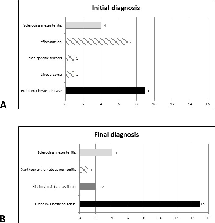
Results: Abdominal pain was the revealing symptom in 10 cases, and 19 patients underwent surgical biopsies. The diagnosis of histiocytosis was proposed by initial pathologists in 41% of patients. The other initial diagnoses were inflammation (n=7), sclerosing mesenteritis (n=4) and liposarcoma (n=1). The CD163/CD68+CD1a- histiocytes infiltrated subserosa and/or deeper adipose tissues in 16 and 14 cases, respectively. A BRAFV600E mutation was detected within the biopsies in 11 cases, and two others were MAP2K1 mutated. The final diagnosis was histiocytosis in 18 patients, 15 of whom had Erdheim-Chester disease. The median diagnostic delay of histiocytosis was 9 months. Patients treated with BRAF or MEK inhibitors showed a partial response or a stable disease. One patient died soon after surgery, and five died by the progression of the disease.

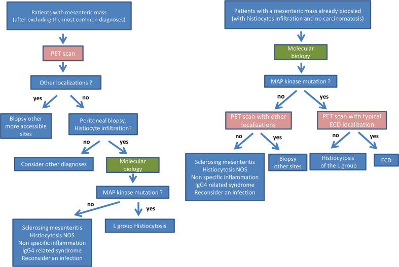
Conclusion: Diagnosis of masses arising in the mesentery should be carefully explored as one of the possibilities in histiocytosis. This diagnosis is frequently missed on mesenteric biopsies. Molecular biology for detecting the mutations in BRAF or in genes of the MAP kinase pathway is a critical diagnostic tool.

Keywords: abdominal pain; cancer; molecular biology.


Conflict of interest statement

Competing interests: None declared.

Figures

Figure 1
Figure 1
Initial and final diagnoses of patients within this cohort. Diagnosis of histiocytosis was proposed in 9/22 patients in initial pathology reports, and in 17 patients after central review.
Figure 2
Figure 2
CT scan of patients with mesenteric involvement by histiocytosis. Patient #11 (A) with mesenteric tumour (long arrows) surrounding mesenteric vessels, initially diagnosed as sclerosing mesenteritis. This patient had typical Erdheim-Chester lesions consisting in ‘coated aorta’ (short arrows) and ‘hairy kidneys’ (arrow heads). This patient also had intraperitoneal effusion in the parieto-colic area (star) and around the liver. Axial CT scan, contrast injection in portal phase. Patient #2 with mesenteric infiltration (arrows) before (B), and with partial response after 4.5 months of treatment with trametinib (C). Axial CT scan, without contrast injection. Patient #15 with mesenteric infiltration (arrows) in the pelvis, before (D) and after 7 months (E) and 39 months (F) of treatment with vemurafenib. Small lymph nodes were also present in this area. Axial CT scan, contrast injection.
Figure 3
Figure 3
Histology of the patients with mesenteric involvement by histiocytosis. Low magnification showing diffuse, mainly superficial, involvement of epiploon by histiocytosis (patient #13, H&E ×1 (A) and CD163 ×1 (B)). Patchy superficial involvement of epiploon by histiocytosis (patient #22, H&E ×12 (C), H&E ×40 (D) and CD163 ×40 (E)). Diffuse superficial involvement of serosa by eosinophilic histiocytes (patient #9, H&E ×200 (F), phosphoERK ×200 (G)). Infiltration by eosinophilic (H) or foamy (I) histiocytes (patient #2, H&E ×200 (H), patient #10, H&E ×40 (I)).
Figure 4
Figure 4
Recommendations for diagnostic process in patients with mesenteric mass of unknown aetiology.

References

    1. Emory TS, Monihan JM, Carr NJ, et al. . Sclerosing mesenteritis, mesenteric panniculitis and mesenteric lipodystrophy: a single entity? Am J Surg Pathol 1997;21:392–8. 10.1097/00000478-199704000-00004 - DOI - PubMed
    1. Moore FO, Berne JD, Fox AD. Mesenteric panniculitis and Erdheim-Chester disease: xanthogranulomatous diseases confused with malignancy. J Am Coll Surg 2007;204:326–7. 10.1016/j.jamcollsurg.2006.06.032 - DOI - PubMed
    1. Alexiou J, Klastersky J. Erdheim-Chester disease: a case report. Am J Case Rep 2015;16:361–6. 10.12659/AJCR.892750 - DOI - PMC - PubMed
    1. Balasubramanian G, Modiri A, Affi M, et al. . A fatal case of Erdheim-Chester disease with hepatic involvement. ACG Case Rep J 2017;4:e95. 10.14309/crj.2017.95 - DOI - PMC - PubMed
    1. Wang F, Cao X, Niu N, et al. . Multisystemic imaging findings in Chinese patients with Erdheim-Chester disease. AJR Am J Roentgenol 2019;213:1179–86. 10.2214/AJR.19.21523 - DOI - PubMed

Publication types

Substances