Comparative Study
. 2021 Aug;378(2):146-156.
doi: 10.1124/jpet.121.000675. Epub 2021 May 21.

A Comparison of Breathing Stimulants for Reversal of Synthetic Opioid-Induced Respiratory Depression in Conscious Rats

Affiliations
Comparative Study

A Comparison of Breathing Stimulants for Reversal of Synthetic Opioid-Induced Respiratory Depression in Conscious Rats

Kaye E Dandrea et al. J Pharmacol Exp Ther. 2021 Aug.

Abstract

Potent synthetic opioids are an important cause of death in the United States' opioid epidemic, and a breathing stimulant may have utility in treating opioid overdose. We hypothesized that sufentanil-induced respiratory depression may be reversed by breathing stimulant administration. Using nose-only plethysmography and arterial blood analysis, we compared effects of several breathing stimulants in reversing sufentanil-induced respiratory depression in conscious rats. We studied taltirelin (1 mg/kg i.v.), PKTHPP (5 mg/kg i.v.), CX717 (30 mg/kg i.v.), BIMU8 (1 mg/kg i.v.), A85380 (30 μg/kg i.v.), and 8-hydroxy-2-(di-n-propylamino)tetralin (8-OH-DPAT) (150 μg/kg i.v./i.m.) and used sufentanil (10 μg/kg i.v.). By plethysmography (in % baseline, mean ± S.E.M.), taltirelin restored ventilation in sufentanil-treated rats (from 50 ± 5% to 102 ± 8%) by increased breathing rates (from 80 ± 4% to 160 ± 12%). By arterial blood analysis, however, taltirelin did not correct hypoxia, decreased hypercarbia only after 45 minutes, and worsened metabolic acidosis (base excess from +0 ± 1 to -7 ± 1 mEq/l). Additionally, taltirelin increased exhaled carbon dioxide, an estimate of oxygen consumption, by up to 64%. PKTHPP, CX717, BIMU8, and A85380 failed to significantly change ventilation or arterial blood values in sufentanil-treated rats. 8-OH-DPAT, however, improved ventilation (from 54 ± 8% to 92 ± 10%), reversed hypercarbia (from 64 ± 6 to 47 ± 2 mmHg), and shortened time to righting from 43 ± 4 to 15 ± 1 minutes in sufentanil-treated rats placed supine. Taltirelin has limited therapeutic potential, as its ventilatory effects are offset by metabolic acidosis, possibly from increased oxygen consumption. At the doses studied, PKTHPP, CX717, BIMU8, and A85380 have limited effects in reversing sufentanil-induced respiratory depression; 8-OH-DPAT, however, warrants further study. SIGNIFICANCE STATEMENT: Respiratory depression is an important cause of death after potent synthetic opioid overdose. 8-Hydroxy-2-(di-n-propylamino)tetralin or related compounds may be useful in treating respiratory depression as caused by potent synthetic opioids.


Figures

Fig. 1.
Fig. 1.
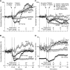
Rat breathing and CO2 production after morphine and taltirelin treatment with and without isoflurane anesthesia. Average respiratory rate (A), minute ventilation (B), tidal volume (C), and exhaled CO2 (D) as measured by nose-only plethysmography vs. time. Animals received 10 mg/kg i.v. morphine (M) over 5 minutes or 1 ml i.v. saline (NS) at 15 minutes followed by 1 mg/kg i.v. taltirelin (T) or 1 ml i.v. saline (NS) at 20 minutes; only one group received anesthesia with continuous 1.5% inhaled isoflurane (w/Iso) throughout. For each animal, data were normalized in (A) through (C) to 15 minutes of baseline data; each data point represents an average of 1 minute of data ± S.E.M.; n = 6–8 animals in (A) through (C) and n = 5–6 in (D). “ns” indicates no significance (P > 0.05); asterisks (∗ and ∗∗∗) indicate statistical significance (P < 0.05, P < 0.001, respectively) by one-way ANOVA test with a Sidak’s multiple comparisons post-test at data points 15, 30, or 45 minutes after first morphine or saline administration. For all conscious experiments, average respiratory rate (rate), minute ventilation (MV), tidal volume (TV), and CO2 production at baseline were 121 ± 4 breaths/min, 53 ± 3 ml/min/100 g, 0.44 ± 0.02 ml/100 g, and 42.6 ± 7.4 ml/kg/min, respectively; n = 21; n = 16 (CO2 production). For isoflurane-anesthetized animals, average respiratory rate, minute ventilation, tidal volume, and CO2 production at baseline were 71 ± 3 breaths/min, 33 ± 1 ml/min/100 g, 0.46 ± 0.02 ml/100 g, and 15.4 ± 0.9 ml/kg/min, respectively; n = 6.
Fig. 2.
Fig. 2.
Conscious rat breathing and CO2 production after sufentanil and taltirelin treatment. Avg. respiratory rate (A), minute ventilation (B), tidal volume (C), and exhaled CO2 (D) as measured by nose-only plethysmography vs. time. Animals received 10 μg/kg i.v. sufentanil (S) by bolus (over ∼5-10 seconds) or 1 ml i.v. saline (NS) at 15 minutes followed by 1 mg/kg i.v. taltirelin (T) or 1 ml i.v. saline (NS) at 20 minutes. Data were averaged and normalized similar to Fig. 1 and were collected from n = 6 animals. “ns” indicates no significance (P > 0.05); asterisks (∗ and ∗∗∗) indicate statistical significance (P < 0.05, P < 0.0001, respectively) by one-way ANOVA test with Sidak’s multiple comparisons post-test at data points 15, 30, or 45 minutes after first sufentanil or saline administration. Baseline avg. respiratory rate, minute ventilation, tidal volume, and CO2 production at baseline were 126 ± 3 breaths/min, 55 ± 2 ml/min/100 g, 0.44 ± 0.02 ml/100 g, and 33.0 ± 1.8 ml/kg/min, respectively; n = 18.
Fig. 3.
Fig. 3.
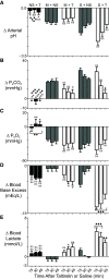
Morphine, sufentanil, and taltirelin effects on arterial blood pH (A), carbon dioxide (B), and oxygen (C) partial pressures and base excess (D) and lactate (E) levels in conscious, air-breathing rats. Data were collected using animals different than those of Figs. 1 and 2. Drug dosing and administration order and times were as in Figs. 1 and 2: 10 mg/kg i.v. morphine (M) (over 5 minutes) or 10 μg/kg i.v. sufentanil (S) by bolus (over ∼5-10 seconds) or 1 ml i.v. saline (NS) at 15 minutes followed by 1 mg/kg i.v. taltirelin (T) or 1 ml i.v. saline (NS) at 20 minutes. Data indicate change (Δ) in measured value from baseline—collected just prior to first opioid/saline administration—and 15, 30, and 45 minutes after taltirelin (or saline) administration. Data are avg. from 6–8 animals (n = 6–8). “ns” indicates no significance (P > 0.05); asterisks and diamonds (∗/⧫, ∗∗/⧫⧫, and ∗∗∗/⧫⧫⧫) indicate statistical significance (P < 0.05, P < 0.01, and P < 0.001) by one-way ANOVA test with a Sidak’s multiple comparisons post-test relative to morphine plus saline (M+NS) or sufentanil plus saline (S+NS) animals, respectively. Baseline arterial blood pH, PaCO2, PaO2, base excess, and lactate were 7.48 ± 0.01, 38.9 ± 0.8 mmHg, 90 ± 2 mmHg, +5.0 ± 0.3 mEq/L, and 0.7 ± 0.1 mmol/L, respectively; n = 21.
Fig. 4.
Fig. 4.
Conscious rat breathing and CO2 production after sufentanil, 8-OH-DPAT, and prazosin treatment. Avg. respiratory rate (A), minute ventilation (B), tidal volume (C), and exhaled CO2 (D) as measured by nose-only plethysmography vs. time. Animals received 10 μg/kg i.v. sufentanil (S) by bolus (over ∼5 to 10 seconds) at 15 minutes followed by 150 μg/kg i.v. 8-OH-DPAT (D) or 150 μg/kg i.v. 8-OH-DPAT plus 250 μg/kg i.v. prazosin (P) at 20 minutes and 1 mg/kg i.v. naloxone at 60 minutes. Data were averaged and normalized similar to Fig. 1 and were collected from n = 6 animals. “ns” indicates no significance (P > 0.05); asterisks (∗ and ∗∗∗) indicate statistical significance (P < 0.05, P < 0.001, respectively) by one-way ANOVA test with Sidak’s multiple comparisons post-test at data points 15, 30, or 45 minutes after sufentanil. Baseline avg. respiratory rate, minute ventilation, tidal volume, and CO2 production for all experiments were 127 ± 4 breaths/min, 59 ± 2 ml/min/100 g, 0.47 ± 0.02 ml/100 g, and 29.2 ± 1.7 ml/kg/min, respectively; n = 18.
Fig. 5.
Fig. 5.
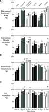
Conscious rat breathing and CO2 production after sufentanil, 8-OH-DPAT, prazosin, CX717, BIMU8, and A85380 treatment. Avg. respiratory rate (A), minute ventilation (B), tidal volume (C), and exhaled CO2 (D) as measured by nose-only plethysmography at three time points (30, 45, and 60 minutes); this includes 8-OH-DPAT data shown in Fig. 4. Rats received 10 μg/kg i.v. sufentanil at 15 minutes followed by 1) 1 ml of saline, 2) 150 μg/kg i.v. 8-OH-DPAT, 3) 150 μg/kg i.v. 8-OH-DPAT plus 250 μg/kg i.v. prazosin, 4) 30 mg/kg i.v. CX717, 5) 30 μg/kg i.v. A85380, or 6) 1 mg/kg i.v. BIMU8 at 20 minutes. Data were averaged and normalized similar to Fig. 1 and were collected from n = 6 animals for each compound. “ns” indicates no significance (P > 0.05); asterisks (∗ and ∗∗∗) indicate statistical significance (P < 0.05, P < 0.001, respectively) by one-way ANOVA test with Sidak’s multiple comparisons post-test at data points. The avg. baseline respiratory rate, minute ventilation, tidal volume, and CO2 production for all experiments were 128 breaths/min, 61 ± 1 ml/min/100 g, 0.47 ± 0.01 ml/100 g, and 29.8 ± 1.0 ml/kg/min, respectively; n = 36.
Fig. 6.
Fig. 6.
Sufentanil, 8-OH-DPAT, prazosin, CX717, BIMU8, and A85380 effects on arterial blood pH (A), carbon dioxide (B), and oxygen (C) partial pressures and base excess (D) and lactate (E) levels in conscious, air-breathing rats. Blood gas data were collected on the same animals simultaneously with the breathing data of Figs. 4 and 5, such that drug dosing and administration order and times are the same. Data indicate change (Δ) in measured value from baseline—collected just prior to first opioid administration—and 15, 30, and 45 minutes after study drug(s) (or saline) administration. Data are avg. from six animals (n = 6). “ns” indicates no significance (P > 0.05); asterisks and diamonds (∗, ∗∗, and ∗∗∗) indicate statistical significance (P < 0.05, P < 0.01, and P < 0.001) by one-way ANOVA test with a Sidak’s multiple comparisons post-test relative to sufentanil plus saline (S+NS) animals, respectively, at the same time point. Baseline arterial blood pH, PaCO2, PaO2, base excess, and lactate were 7.44 ± 0.01, 41.0 ± 0.6 mm Hg, 90 ± 1 mm Hg, +3.0 ± 0.3 mEq/L, and 1.38 ± 0.1 mmol/L, respectively; n = 36.
Fig. 7.
Fig. 7.
Conscious rat breathing and CO2 production after intravenous sufentanil and intramuscular 8-OH-DPAT treatment. Avg. respiratory rate (A), minute ventilation (B), tidal volume (C), and exhaled CO2 (D) as measured by nose-only plethysmography vs. time. Animals received 10 μg/kg i.v. sufentanil (S) by bolus (over ∼5 to 10 seconds) at 15 minutes followed by 150 μg/kg i.m. 8-OH-DPAT (D) or 1 ml/kg i.m. normal saline at 20 minutes and 1 mg/kg i.v. naloxone at 60 minutes. Data were averaged and normalized similar to Fig. 1 and were collected from n = 6 animals. “ns” indicates no significance (P > 0.05); asterisks (∗ and ∗∗) indicate statistical significance (P < 0.05, P < 0.01, respectively) by one-way ANOVA test with Sidak’s multiple comparisons post-test at data points 15, 30, or 45 minutes after sufentanil. Baseline avg. respiratory rate, minute ventilation, tidal volume, and CO2 production for all experiments were 120 ± 3 breaths/min, 58 ± 1 ml/min/100 g, 0.48 ± 0.01 ml/100 g, and 28.9 ± 1.2 ml/kg/min, respectively; n = 24.

Similar articles

Cited by

References

    1. Barnett A, Goldstein J, Fiedler E, Taber R (1975) Etonitazine-induced rigidity and its antagonism by centrally acting muscle relaxants. Eur J Pharmacol 30:23–28. - PubMed
    1. Bennett JA, Abrams JT, Van Riper DF, Horrow JC (1997) Difficult or impossible ventilation after sufentanil-induced anesthesia is caused primarily by vocal cord closure. Anesthesiology 87:1070–1074. - PubMed
    1. Bilbey DL, Salem H, Grossman MH (1960) The anatomical basis of the straub phenomenon. Br J Pharmacol Chemother 15:540–543. - PMC - PubMed
    1. Boghosian JD, Luethy A, Cotten JF (2018) Intravenous and Intratracheal Thyrotropin Releasing Hormone and Its Analog Taltirelin Reverse Opioid-Induced Respiratory Depression in Isoflurane Anesthetized Rats. J Pharmacol Exp Ther 366:105–112. - PMC - PubMed
    1. Cheng JP, Leary JB, Sembhi A, Edwards CM, Bondi CO, Kline AE (2016) 5-hydroxytryptamine1A (5-HT1A) receptor agonists: A decade of empirical evidence supports their use as an efficacious therapeutic strategy for brain trauma. Brain Res 1640 (Pt A):5–14. - PMC - PubMed

Publication types