. 2021 Jun 30;41(26):5579-5594.
doi: 10.1523/JNEUROSCI.1741-20.2021. Epub 2021 May 21.

Protein Phosphatase 2B Dual Function Facilitates Synaptic Integrity and Motor Learning

Affiliations

Protein Phosphatase 2B Dual Function Facilitates Synaptic Integrity and Motor Learning

Zhanmin Lin et al. J Neurosci. .

Abstract

Protein phosphatase 2B (PP2B) is critical for synaptic plasticity and learning, but the molecular mechanisms involved remain unclear. Here we identified different types of proteins that interact with PP2B, including various structural proteins of the postsynaptic densities (PSDs) of Purkinje cells (PCs) in mice. Deleting PP2B reduced expression of PSD proteins and the relative thickness of PSD at the parallel fiber to PC synapses, whereas reexpression of inactive PP2B partly restored the impaired distribution of nanoclusters of PSD proteins, together indicating a structural role of PP2B. In contrast, lateral mobility of surface glutamate receptors solely depended on PP2B phosphatase activity. Finally, the level of motor learning covaried with both the enzymatic and nonenzymatic functions of PP2B. Thus, PP2B controls synaptic function and learning both through its action as a phosphatase and as a structural protein that facilitates synapse integrity.SIGNIFICANCE STATEMENT Phosphatases are generally considered to serve their critical role in learning and memory through their enzymatic operations. Here, we show that protein phosphatase 2B (PP2B) interacts with structural proteins at the synapses of cerebellar Purkinje cells. Differentially manipulating the enzymatic and structural domains of PP2B leads to different phenotypes in cerebellar learning. We propose that PP2B is crucial for cerebellar learning via two complementary actions, an enzymatic and a structural operation.

Keywords: Purkinje cells; cerebellar learning; protein phosphatase 2B.


Figures

Figure 1.
Figure 1.
Selective inhibition of phosphatase activity of PP2B does not fully reproduce the phenotype of the L7-PP2B KO. A, Motor performance during the VOR in the light (VVOR) revealed no aberrations in the FK506-injected group (i.e., group in which phosphatase function of PP2B was specifically inhibited) compared with controls and L7-PP2B KOs (all p values > 0.5). Error bars indicate ± SEM. B, Short-term learning paradigm of gain decrease. L7-PP2B KO mice were unable to learn (***p values < 0.001 with respect to both vehicle-only and FK506 group). Error bars indicate ± SEM. C, Long-term learning paradigm of 5 d phase-reversal eye movement. y axis indicates the value of gain*cos(phase); the lower this value, the better the learning. Phase-reversal learning in the FK506-injected group is impaired compared with controls (p < 0.001) but is better than that of L7-PP2B KO mice (p < 0.001), suggesting that, in addition to its function as a phosphatase, PP2B may also have a structural role. Error bars indicate ± SEM.
Figure 2.
Figure 2.
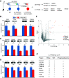
Specific presynaptic and postsynaptic KO of PP2B at PF-PC synapses resulted in downregulation of presynaptic and postsynaptic proteins, respectively. A, Scheme of cerebellar cortex circuity and iTRAQ experimental setup (left) and list of significantly downregulated proteins in α6-PP2B (presynaptic KO) and L7-PP2B (postsynaptic KO) synaptosomes identified by 8-plex iTRAQ (n = 4:4 mice per run, repeated twice) (right). The presynaptic axon terminals of WT and PP2B KO GCs, which are making synaptic contact with the spine heads of PCs, are represented in black and green, respectively. WT PCs and PP2B KO PC (both cell bodies and spine heads) are represented in blue and red, respectively. Arrows leaving the PCs indicate that they form the sole output of the cerebellar cortex, with their axons traversing toward the cerebellar nuclei. B, Immunoblots of L7-PP2B KO and WT littermates synaptosomes (n = 6:6 mice, 10-12 PND). Whereas CNB and CNA confirmed successful KO of PP2B (*p < 0.05), calbindin and PSD95 signals suggest that the number of PCs and synapses are not affected by knocking out PP2B, respectively (p = 0.9009, p = 0.2413, unpaired parametric t test). C, Immunoblots showing that multiple PSD proteins are downregulated in L7-PP2B KO. D, Immunoblots confirming that several glutamate receptor subunits are significantly downregulated in L7-PP2B KO. Error bars indicate ± SEM (*p < 0.05). E, Phospho-proteomic volcano plot reveals changed detection of protein/phosphor sites in L7-PP2B mice (P2, 10-plex TMT, n = 5 × 3 mice:5 × 3 mice, 10-12 PND). The x axis and y axis indicate fold change and log(p value), respectively. Area of upper quadrant on the right shows proteins with a phosphorylation fold change > 2 (unpaired t test, p < 0.01) following comparison of WT and L7-PP2B mice P2 fractions. For example, only 1 site of Cacna1g is less phosphorylated in L7-PP2B mice, whereas others are more phosphorylated. Moreover, a number of other sites of other proteins, such as Shank1, Shank2, and pcp2, are only more phosphorylated. F, Summary table of changes in protein levels as observed following iTRAQ (8-plex), TMT (10-plex) for total proteins, and for phospho sites determined by phospho-proteomics.
Figure 3.
Figure 3.
PP2B stably and directly interacts with multiple PSD proteins. A, Illustration of IP-MS/MS workflow. Top, IP-MS/MS for cerebellar PP2B in WT mice P2 fractions. Bottom, Scheme of IP-BN (blue native)-PAGE-MS/MS. B, IP-MS/MS result. Proteins identified as PP2B interactors. These proteins were identified in IP with 2 PP2B antibodies, and at least twice for each antibody (see Materials and Methods; Extended Data Figure 3-1). Proteins were manually grouped by their known overall functions. The number in the brackets indicates the number of proteins belonging to that group. Only one protein (C1QC) from the IP-MS/MS result was not included in the graph. C, The result of IP/BN-PAGE/MS from WT cerebellar P2 fractions. Detection of different proteins is presented in columns, fractions analyzed by MS are presented in rows, and colors are coded with normalized iBAQ value (in percentage of total). CNA, CNB, and calmodulin are well colocalized. The interactors of CNA, including CaMK2, Homer3, Shank1, Shank2, Grid2, and mGluR1, are enriched in the high-molecular weight fractions; proteins may form multiple complexes. D, HEK293 cell co-IP experiments confirming direct interactions with the candidate proteins from cerebellum IP, Homer3, mGluR1, Shank1, and CaMK2b, as illustrated by the enrichment of the first column compared with the second and third column. This figure is constructed by horizontally combining Western blots from different individual target proteins.
Figure 4.
Figure 4.
Ultrastructure of PF-PC synapses. A, Electron micrograph of a PF terminal contacting two PC spines in WT (left) or a single spine in L7-PP2B KO mice (middle, right). Black and white asterisks indicate PF terminals and PC spines, respectively. Scale bars: left, 0.3 µm; middle, 0.4 µm; right, 0.4. B, Quantification of average area and relative thickness (ratio of thickness per length) of PSDs for WT and L7-PP2B mice (25 synapses per lobule per mouse). p = 0.699 and p = 0.015 for area and relative thickness, respectively (Mann–Whitney test). Error bars indicate ± SEM, *p < 0.05.
Figure 5.
Figure 5.
Cluster size of PP2B-PSD interactors as revealed with dSTORM. A, Example of dSTORM aligned with confocal image of a cultured PC, illustrated with Homer3 dSTORM and calbindin staining. Scale bar: right, 400 nm. B, Example of dSTORM images of cultured PC spines immunostained with PP2B PSD interactors. Scale bars, 100 nm. C, Top and bottom, Cumulative and box plots, respectively. In the box plots, we compared WT (blue), WT with pharmacologically long-term blockage of phosphatase activity of PP2B by FK506 (FK, green), and KO (red). **p < 0.01; ***p < 0.001; Dunn test with Bonferroni correction. D, Same as in C, but for comparisons of KO infected with AAV encoding WT-PP2B (WTR; see Fig. 6A), KO infected with AAV encoding enzyme-dead PP2B (PD; see Fig. 6A), and KO infected with empty AAV (KO). E, A model summarized from the results above. The cluster sizes of Homer3, mGluR1 were smaller in the PP2B KO, whereas Shank2, Shank1, and Grid2 showed bigger cluster sizes in PP2B KO. Moreover, clusters of Homer3, mGluR1, and Shank2 were rescued to WT level following expression of enzyme-dead PP2B in KO cells, indicating their regulation by PP2B via a phosphatase-independent manner. Instead, the cluster size of Shank1 and Grid2 did depend on PP2B phosphatase activity, as the enzyme-dead expression led to the same level as in L7-PP2B KO mice.
Figure 6.
Figure 6.
Single-molecule tracking of GluR2 in PC. A, Left, Example of a cultured PC (DIV 21). Scale bar, 10 μm. Middle, Example of a piece of PC dendrite visualized by transfection of L7-mCherry. Scale bar, 1 μm. Right, Example of surface GluR2 tracks from the inset in the middle panel. Scale bar, 1 μm. B, The surface GluR2 tracking results under different conditions, which are summarized in the histogram on the right (color coding at x axis applies for all panels). WT PCs show a larger percentage of mobile GluR2 than the FK and PP2B KO cells. Shades and error bars indicate SEM. Whereas the expression of WT PP2B in KO cells rescued the mobility (WTR, purple), the expression of enzyme-dead PP2B in KO cells (PD, orange) led to the same level of mobility as PP2B KOs (one-way ANOVA with Tukey's multiple comparisons, WT vs FK p = 0.0218; WT vs KO p < 0.0001; WT vs WTR p = 0.1232; WTR vs KO p = 0.0008; WTR vs PD p < 0.0001). C, The surface GluR2 STORM in WT, FK, and KO cultured PCs. Left, Two examples of surface GluR2 dSTORM on a PC spine: one from the side and one from top. Middle, right, Cumulative and box plots, respectively. Dunn test with Bonferroni correction: WT versus FK p < 0.0001, WT versus KO p < 0.0001. D, E, mEPSCs recorded from acute slices of WT and L7-PP2B KO mice. D, Left, Traces represent example traces of mEPSC recordings. Right, Histogram represents the mEPSC amplitude from all recordings. Bin size = 1 pA. E, The amplitude of the mEPSCs is increased in the FK-treated PCs as well as the L7-PP2B KO PCs (Mann–Whitney test, p = 0.019, n = 19: 8 cells, WT:KO, from 3 pairs of mice), whereas their frequencies appear unaffected (Mann–Whitney test, p = 0.979). *p < 0.05; **p < 0.01; ***p < 0.001.
Figure 7.
Figure 7.
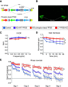
Expression of enzyme-dead PP2B partially rescues motor learning deficits in L7-PP2B KOs. A, Schematic of the Cre-dependent WT-PP2B and enzyme-dead PP2B AAVs. The enzyme-dead PP2B is made by introducing a single amino-acid mutation in CNA at H151A. B, Example of bilateral injections of AAV into the flocculus. Text box below represents the experimental groups. Blue represents control (WT littermates injected with CAG-EGFP, n = 9 mice). Purple represents WTR (L7-PP2B KOs reexpressed with WT-PP2B, n = 11 mice). Orange represents PD (L7-PP2B KOs injected with enzyme-dead PP2B, n = 12 mice). Red represents L7-PP2B KO (n = 7 mice). C, Motor performance of VOR in light (VVOR). D, Short-term learning paradigm for gain-decrease (ANOVA with Bonferroni correction, p < 0.001; PD vs WTR: p = 0.015; PD vs control: p < 0.001; KO vs control: p <0.001; KO vs WTR: p = 0.007; KO vs PD p > 0.5; control vs WTR: p = 0.031) *p < 0.05. E, Long-term learning paradigm following 5 d phase-reversal training. y axis indicates the value of gain*cos(phase); the lower this value, the better the learning. Phase-reversal comparison for the last 2 d showed that L7-PP2B KO mice injected with PD learned better than the L7-PP2B KO mice (unpaired t test, p = 0.038). Control WT mice injected with GFP and L7-PP2B KOs with WT-PP2B learned better than the PD KO group, indicating that both the enzymatic and nonenzymatic functions of PP2B may play a role in VOR learning.

Similar articles

Cited by

References

    1. Albus J (1971) A theory of cerebellar function. Math Biosci 10:25–61. 10.1016/0025-5564(71)90051-4 - DOI
    1. Bienenstock EL, Cooper LN, Munro PW (1982) Theory for the development of neuron selectivity: orientation specificity and binocular interaction in visual cortex. J Neurosci 2:32–48. - PMC - PubMed
    1. Bingol B, Wang CF, Arnott D, Cheng D, Peng J, Sheng M (2010) Autophosphorylated CaMKIIalpha acts as a scaffold to recruit proteasomes to dendritic spines. Cell 140:567–578. 10.1016/j.cell.2010.01.024 - DOI - PubMed
    1. Boele HJ, Peter S, ten Brinke MM, Verdonschot L, IJpelaar ACH, Rizopoulos D, Gao Z, Koekkoek SK, De Zeeuw CI (2018) Impact of parallel fiber to Purkinje cell long-term depression is unmasked in absence of inhibitory input. Sci Adv 4:eaas9426. 10.1126/sciadv.aas9426 - DOI - PMC - PubMed
    1. Böhm G, Prefot P, Jung S, Selzer S, Mitra V, Britton D, Kuhn K, Pike I, Thompson AH (2015) Low-pH solid-phase amino labeling of complex peptide digests with TMTs improves peptide identification rates for multiplexed global phosphopeptide analysis. J Proteome Res 14:2500–2510. 10.1021/acs.jproteome.5b00072 - DOI - PubMed

Publication types

LinkOut - more resources