

Anti-CD20 Depletes Meningeal B Cells but Does Not Halt the Formation of Meningeal Ectopic Lymphoid Tissue
- PMID: 34021057
- PMCID: PMC8143698
- DOI: 10.1212/NXI.0000000000001012
Anti-CD20 Depletes Meningeal B Cells but Does Not Halt the Formation of Meningeal Ectopic Lymphoid Tissue
Abstract
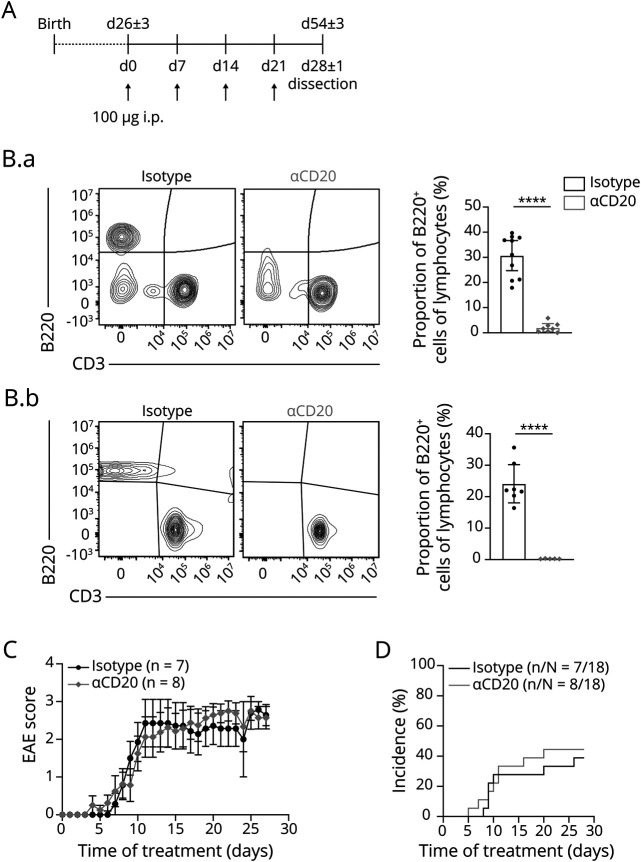
Objective: To investigate whether anti-CD20 B-cell-depleting monoclonal antibodies (ɑCD20 mAbs) inhibit the formation or retention of meningeal ectopic lymphoid tissue (mELT) in a murine model of multiple sclerosis (MS).
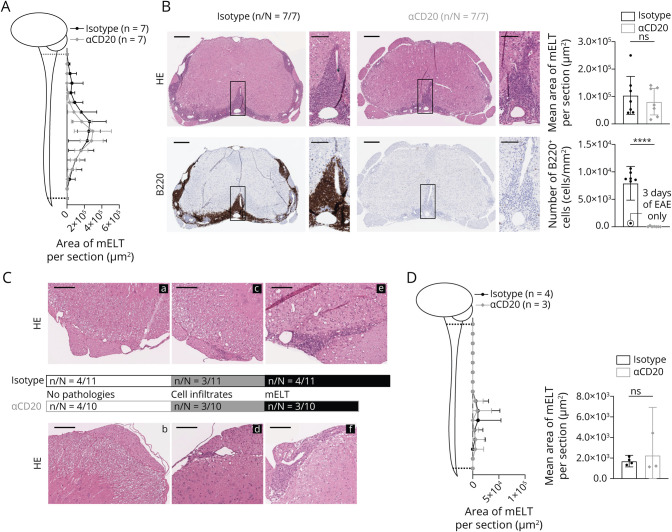
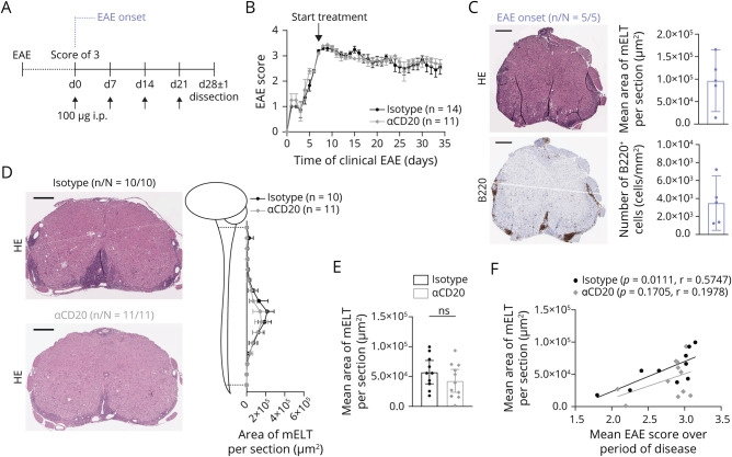
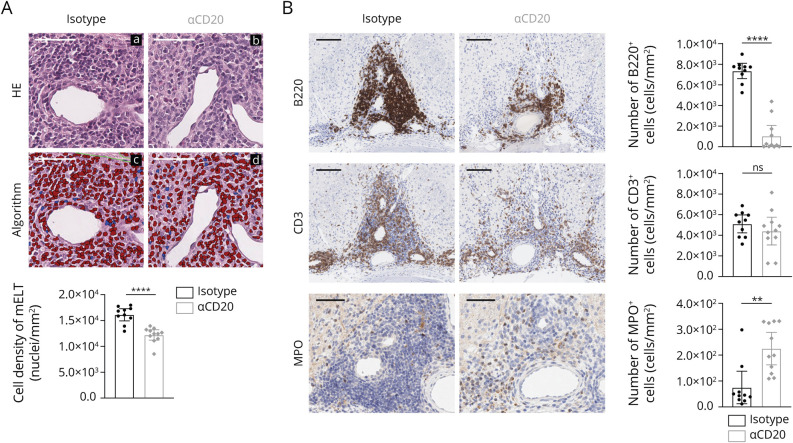
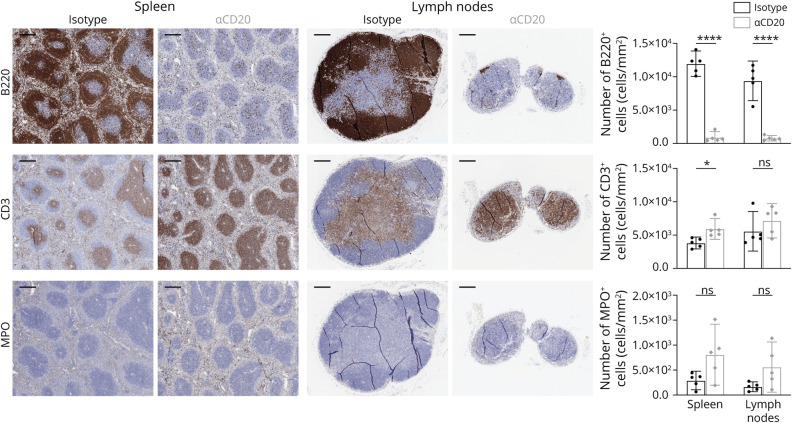
Methods: We used a spontaneous chronic experimental autoimmune encephalomyelitis (EAE) model of mice with mutant T-cell and B-cell receptors specific for myelin oligodendrocyte glycoprotein (MOG), which develop meningeal inflammatory infiltrates resembling those described in MS. ɑCD20 mAbs were administered in either a preventive or a treatment regimen. The extent and cellular composition of mELT was assessed by histology and immunohistochemistry.
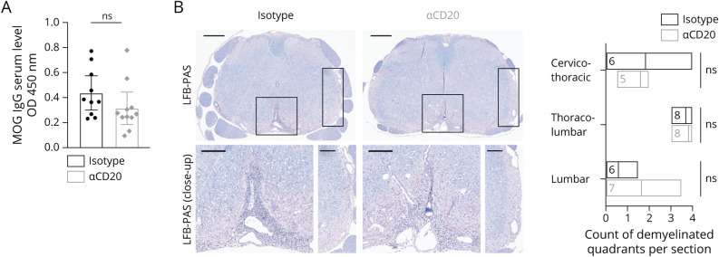
Results: ɑCD20 mAb, applied in a paradigm to either prevent or treat EAE, did not alter the disease course in either condition. However, ɑCD20 mAb depleted virtually all B cells from the meningeal compartment but failed to prevent the formation of mELT altogether. Because of the absence of B cells, mELT was less densely populated with immune cells and the cellular composition was changed, with increased neutrophil granulocytes.
Conclusions: These results demonstrate that, in CNS autoimmune disease, meningeal inflammatory infiltrates may form and persist in the absence of B cells. Together with the finding that ɑCD20 mAb does not ameliorate spontaneous chronic EAE with mELT, our data suggest that mELT may have yet unknown capacities that are independent of B cells and contribute to CNS autoimmunity.
Copyright © 2021 The Author(s). Published by Wolters Kluwer Health, Inc. on behalf of the American Academy of Neurology.
Figures
Comment in
-
Can Systemic Anti-CD20 B Cell-Depleting Antibodies Eliminate Meningeal Follicles in Multiple Sclerosis?Neurol Neuroimmunol Neuroinflamm. 2021 May 17;8(4):e1000. doi: 10.1212/NXI.0000000000001000. Print 2021 Jul. Neurol Neuroimmunol Neuroinflamm. 2021. PMID: 34001659 Free PMC article. No abstract available.
Similar articles
-
Siponimod Inhibits the Formation of Meningeal Ectopic Lymphoid Tissue in Experimental Autoimmune Encephalomyelitis.Neurol Neuroimmunol Neuroinflamm. 2021 Dec 15;9(1):e1117. doi: 10.1212/NXI.0000000000001117. Print 2022 Jan. Neurol Neuroimmunol Neuroinflamm. 2021. PMID: 34911793 Free PMC article.
-
Single-cell profiling indicates a high similarity between immune cells in the cerebrospinal fluid and in meningeal ectopic lymphoid tissue in experimental autoimmune encephalomyelitis.Front Immunol. 2024 Jun 12;15:1400641. doi: 10.3389/fimmu.2024.1400641. eCollection 2024. Front Immunol. 2024. PMID: 38933267 Free PMC article.
-
B-cell activation influences T-cell polarization and outcome of anti-CD20 B-cell depletion in central nervous system autoimmunity.Ann Neurol. 2010 Sep;68(3):369-83. doi: 10.1002/ana.22081. Ann Neurol. 2010. PMID: 20641064 Free PMC article.
-
CD20 therapies in multiple sclerosis and experimental autoimmune encephalomyelitis - Targeting T or B cells?Mult Scler Relat Disord. 2016 Sep;9:110-7. doi: 10.1016/j.msard.2016.07.011. Epub 2016 Jul 20. Mult Scler Relat Disord. 2016. PMID: 27645355 Review.
-
Therapies for multiple sclerosis targeting B cells.Croat Med J. 2019 Apr 30;60(2):87-98. doi: 10.3325/cmj.2019.60.87. Croat Med J. 2019. PMID: 31044580 Free PMC article. Review.
Cited by
-
Effects of a Fully Humanized Type II Anti-CD20 Monoclonal Antibody on Peripheral and CNS B Cells in a Transgenic Mouse Model of Multiple Sclerosis.Int J Mol Sci. 2022 Mar 15;23(6):3172. doi: 10.3390/ijms23063172. Int J Mol Sci. 2022. PMID: 35328594 Free PMC article.
-
Editorial: Community series in trends in neuroimmunology: cross-talk between brain-resident and peripheral immune cells in both health and disease, volume II.Front Immunol. 2025 Jul 3;16:1644278. doi: 10.3389/fimmu.2025.1644278. eCollection 2025. Front Immunol. 2025. PMID: 40677724 Free PMC article. No abstract available.
-
Single-Cell Profiling Indicates a Proinflammatory Role of Meningeal Ectopic Lymphoid Tissue in Experimental Autoimmune Encephalomyelitis.Neurol Neuroimmunol Neuroinflamm. 2024 Jan;11(1):e200185. doi: 10.1212/NXI.0000000000200185. Epub 2023 Dec 15. Neurol Neuroimmunol Neuroinflamm. 2024. PMID: 38100739 Free PMC article.
-
Siponimod Inhibits the Formation of Meningeal Ectopic Lymphoid Tissue in Experimental Autoimmune Encephalomyelitis.Neurol Neuroimmunol Neuroinflamm. 2021 Dec 15;9(1):e1117. doi: 10.1212/NXI.0000000000001117. Print 2022 Jan. Neurol Neuroimmunol Neuroinflamm. 2021. PMID: 34911793 Free PMC article.
-
High efficacy therapy to prevent the formation of meningeal tertiary lymphoid organs after CXCL13 index screening in early multiple sclerosis.Front Neurosci. 2025 Mar 17;19:1558810. doi: 10.3389/fnins.2025.1558810. eCollection 2025. Front Neurosci. 2025. PMID: 40165834 Free PMC article.
References
-
- Hauser SL, Waubant E, Arnold DL, et al. . B-cell depletion with rituximab in relapsing-remitting multiple sclerosis. N Engl J Med. 2008;358(7):676-688. - PubMed
-
- Hauser SL, Bar-Or A, Comi G, et al. . Ocrelizumab versus interferon beta-1a in relapsing multiple sclerosis. N Engl J Med. 2017;376(3):221-234. - PubMed
-
- Montalban X, Hauser SL, Kappos L, et al. . Ocrelizumab versus placebo in primary progressive multiple sclerosis. N Engl J Med. 2017;376(3):209-220. - PubMed
-
- Ancau M, Berthele A, Hemmer B. CD20 monoclonal antibodies for the treatment of multiple sclerosis: up-to-date. Expert Opin Biol Ther. 2019;19(8):829-843. - PubMed
Publication types
MeSH terms
Substances
LinkOut - more resources
Full Text Sources
Other Literature Sources