. 2021 Sep;19(9):1559-1570.
doi: 10.1158/1541-7786.MCR-20-1017. Epub 2021 May 21.

Defining the Energetic Basis for a Conformational Switch Mediating Ligand-Independent Activation of Mutant Estrogen Receptors in Breast Cancer

Affiliations

Defining the Energetic Basis for a Conformational Switch Mediating Ligand-Independent Activation of Mutant Estrogen Receptors in Breast Cancer

Christopher G Mayne et al. Mol Cancer Res. 2021 Sep.

Abstract

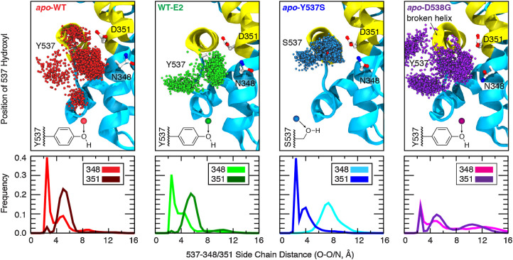
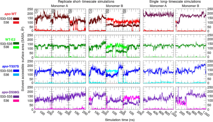
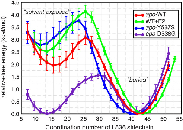
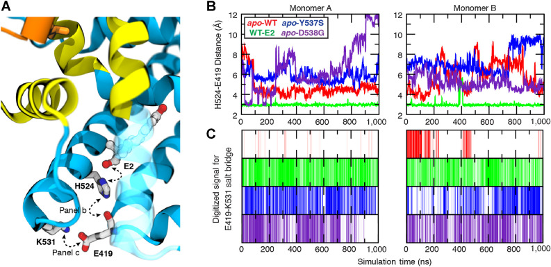
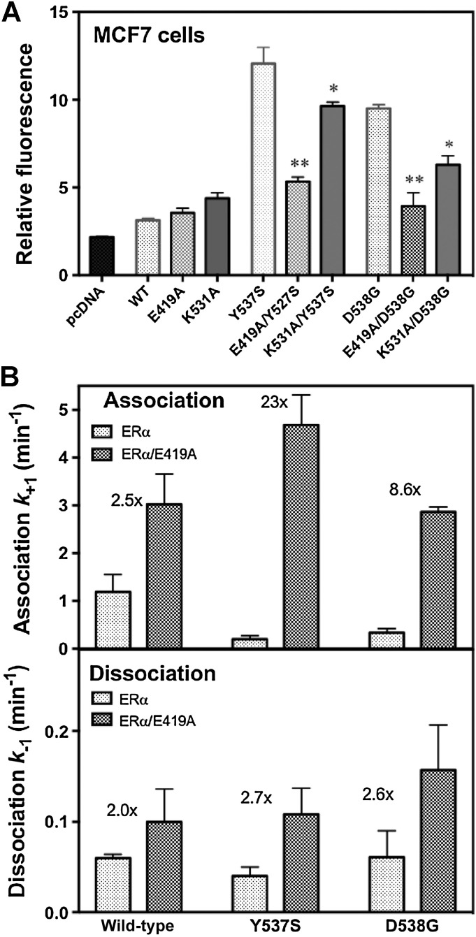
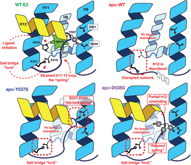
Although most primary estrogen receptor (ER)-positive breast cancers respond well to endocrine therapies, many relapse later as metastatic disease due to endocrine therapy resistance. Over one third of these are associated with mutations in the ligand-binding domain (LBD) that activate the receptor independent of ligand. We have used an array of advanced computational techniques rooted in molecular dynamics simulations, in concert with and validated by experiments, to characterize the molecular mechanisms by which specific acquired somatic point mutations give rise to ER constitutive activation. By comparing structural and energetic features of constitutively active mutants and ligand-bound forms of ER-LBD with unliganded wild-type (WT) ER, we characterize a spring force originating from strain in the Helix 11-12 loop of WT-ER, opposing folding of Helix 12 into the active conformation and keeping WT-ER off and disordered, with the ligand-binding pocket open for rapid ligand binding. We quantify ways in which this spring force is abrogated by activating mutations that latch (Y537S) or relax (D538G) the folded form of the loop, enabling formation of the active conformation without ligand binding. We also identify a new ligand-mediated hydrogen-bonding network that stabilizes the active, ligand-bound conformation of WT-ER LBD, and similarly stabilizes the active conformation of the ER mutants in the hormone-free state. IMPLICATIONS: Our investigations provide deep insight into the energetic basis for the structural mechanisms of receptor activation through mutation, exemplified here with ER in endocrine-resistant metastatic breast cancers, with potential application to other dysregulated receptor signaling due to driver mutations.


Figures

Figure 1. Effect of ligand binding and mutation on hydrogen-bonding contacts. The dynamics of hydrogen-bonding partners Y/S537 (H12) with N348 and D351 (H3) were evaluated from replicate 200-ns simulations of dimeric ERα complexes (1.2 μs aggregate sampling for each system). Top, the position of the Y/S537 side-chain oxygen is shown every 200 ps (3,000 spheres; only monomer A is shown for clarity). Bottom, the shortest distance between side-chain heteroatoms of Y/S537(O) and N348(O/N) or D351(O/O) were binned (width = 0.5 Å) to yield frequency histograms of each interaction.
Figure 1.
Effect of ligand binding and mutation on hydrogen-bonding contacts. The dynamics of hydrogen-bonding partners Y/S537 (H12) with N348 and D351 (H3) were evaluated from replicate 200-ns simulations of dimeric ERα complexes (1.2 μs aggregate sampling for each system). Top, the position of the Y/S537 side-chain oxygen is shown every 200 ps (3,000 spheres; only monomer A is shown for clarity). Bottom, the shortest distance between side-chain heteroatoms of Y/S537(O) and N348(O/N) or D351(O/O) were binned (width = 0.5 Å) to yield frequency histograms of each interaction.
Figure 2. Solvent exposure of consecutive hydrophobic residues in the H11–12 loop. The solvent accessible surface area (SASA) was measured for residues 533–535 (Val, Val, Pro, dark coloring) and for 536 (Leu, light coloring), separately, in replicate 200-ns (left 2) and in single 1-μs (right 2) equilibrium simulations.
Figure 2.
Solvent exposure of consecutive hydrophobic residues in the H11–12 loop. The solvent accessible surface area (SASA) was measured for residues 533–535 (Val, Val, Pro, dark coloring) and for 536 (Leu, light coloring), separately, in replicate 200-ns (left 2) and in single 1-μs (right 2) equilibrium simulations.
Figure 3. Free energy profile of Leu536 conformational change. The average relative free energy and associated error were computed for conformations of Leu536 rotating from a solvent-exposed conformation (low coordination number) to a buried conformation (high coordination number).
Figure 3.
Free energy profile of Leu536 conformational change. The average relative free energy and associated error were computed for conformations of Leu536 rotating from a solvent-exposed conformation (low coordination number) to a buried conformation (high coordination number).
Figure 4. Ligand-mediated hydrogen-bonding network correlates to receptor activity. The stability of key interactions forming a ligand-mediated hydrogen-bond network was quantified from microsecond simulations. A, The network is initiated by the 17β-hydroxyl group of E2 and proceeds through His524:Nε hydrogen bond to the carbonyl of Glu419, terminating with a salt bridge formed between Lys531 and Glu419. B, The His524–Glu419 interaction was monitored by measuring the distance between His524:Nε (donor) and Glu419 carbonyl oxygen (acceptor), whereas (C) the presence of the terminal salt bridge between Glu419 and Lys531 was monitored by digitizing the signal based on geometric constraints.
Figure 4.
Ligand-mediated hydrogen-bonding network correlates to receptor activity. The stability of key interactions forming a ligand-mediated hydrogen-bond network was quantified from microsecond simulations. A, The network is initiated by the 17β-hydroxyl group of E2 and proceeds through His524:Nε hydrogen bond to the carbonyl of Glu419, terminating with a salt bridge formed between Lys531 and Glu419. B, The His524–Glu419 interaction was monitored by measuring the distance between His524:Nε (donor) and Glu419 carbonyl oxygen (acceptor), whereas (C) the presence of the terminal salt bridge between Glu419 and Lys531 was monitored by digitizing the signal based on geometric constraints.
Figure 5. Ligand-binding kinetics and cellular activity of the ER. A, MCF-7 cells were transfected with plasmids for control, WT ERα and the eight indicated mutant ERs, as well as an ER-responsive luciferase plasmid, and constitutive transcriptional activity was monitored in the absence of added estrogen. The * indicates a significance of <0.05 and ** a significance <0.01. B, Ligand association and dissociation rates of the LBDs of WT, Y537S, and D538G ERα were monitored under pseudo first-order conditions using the fluorescent ligand, THC-ketone. Rate constants are shown in the presence of the Glu419–Lys531 salt bridge (gray bars) and in its absence due to the additional E419A (stippled bars). The fold increase in ligand association rate from removal of the salt bridge is indicated by the number above the bars for each receptor.
Figure 5.
Ligand-binding kinetics and cellular activity of the ER. A, MCF-7 cells were transfected with plasmids for control, WT ERα and the eight indicated mutant ERs, as well as an ER-responsive luciferase plasmid, and constitutive transcriptional activity was monitored in the absence of added estrogen. The * indicates a significance of <0.05 and ** a significance <0.01. B, Ligand association and dissociation rates of the LBDs of WT, Y537S, and D538G ERα were monitored under pseudo first-order conditions using the fluorescent ligand, THC-ketone. Rate constants are shown in the presence of the Glu419–Lys531 salt bridge (gray bars) and in its absence due to the additional E419A (stippled bars). The fold increase in ligand association rate from removal of the salt bridge is indicated by the number above the bars for each receptor.
Figure 6. The “Spring-Loading” Model consolidating the effects of ligand binding and the activating mutations in ER. In WT receptor, a ligand-mediated hydrogen-bonding network forms, crisscrossing H7, H8, and H11, and terminating with a salt bridge formed across the base of the ligand-binding pocket. In the absence of ligand, His524 is no longer ordered, and the remainder of the network fails to form. Introduction of either the Y537S or D538G mutations, however, overcomes the strain energy of the H11–12 loop to allow the terminal salt bridge to form in the absence of ligand. Specifically, the Y537S mutation yields an optimal hydrogen bond between H3 and H12, operating as a “latch” holding H12 in the activated conformation. The D538G mutation, by contrast, induces a partial unwinding of H12, which serves to relax the backbone strain energy of the spring-like loop.
Figure 6.
The “Spring-Loading” Model consolidating the effects of ligand binding and the activating mutations in ER. In WT receptor, a ligand-mediated hydrogen-bonding network forms, crisscrossing H7, H8, and H11, and terminating with a salt bridge formed across the base of the ligand-binding pocket. In the absence of ligand, His524 is no longer ordered, and the remainder of the network fails to form. Introduction of either the Y537S or D538G mutations, however, overcomes the strain energy of the H11–12 loop to allow the terminal salt bridge to form in the absence of ligand. Specifically, the Y537S mutation yields an optimal hydrogen bond between H3 and H12, operating as a “latch” holding H12 in the activated conformation. The D538G mutation, by contrast, induces a partial unwinding of H12, which serves to relax the backbone strain energy of the spring-like loop.

Similar articles

Cited by

References

    1. The Cancer Genome Atlas Research Network, Weinstein JN, Collisson EA, Mills GB, Shaw KRM, Ozenberger BA, et al. . The cancer genome atlas pan-cancer analysis project. Nat Genet. 2013;45:1113–20. - PMC - PubMed
    1. Razavi P, Chang MT, Xu G, Bandlamudi C, Ross DS, Vasan N, et al. . The genomic landscape of endocrine-resistant advanced breast cancers. Cancer Cell 2018;34:427–38. - PMC - PubMed
    1. Lefebvre C, Bachelot T, Filleron T, Pedrero M, Campone M, Soria JC, et al. . Mutational profile of metastatic breast cancers: a retrospective analysis. PLoS Med 2016;13:e1002201. - PMC - PubMed
    1. Henzler-Wildman K, Kern D. Dynamic personalities of proteins. Nature 2007;450:964–72. - PubMed
    1. Berendsen H. Collective protein dynamics in relation to function. Curr Opin Struct Biol 2000;10:165–9. - PubMed

Publication types