

Characteristics of mental health implications and plasma metabolomics in patients recently recovered from COVID-19
- PMID: 34021116
- PMCID: PMC8138845
- DOI: 10.1038/s41398-021-01426-3
Characteristics of mental health implications and plasma metabolomics in patients recently recovered from COVID-19
Abstract
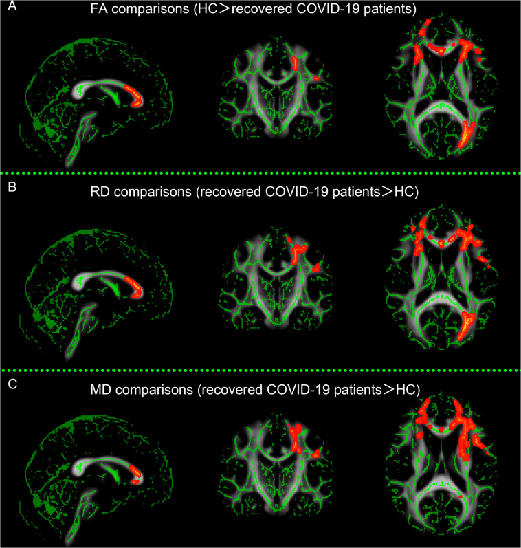
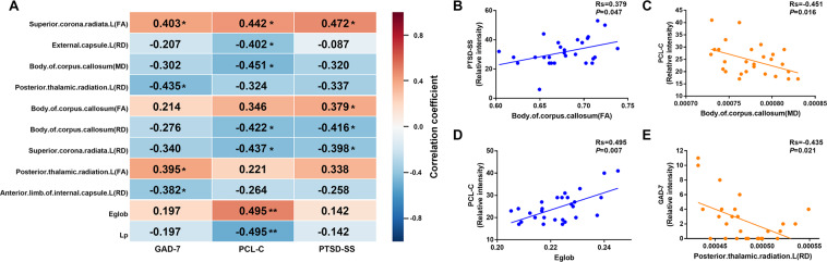
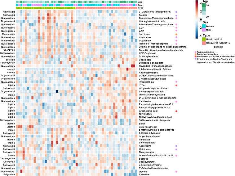
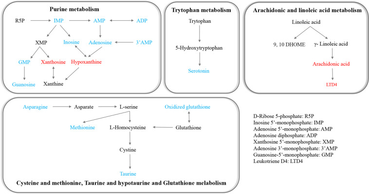
This study aimed to explore the associations between cerebral white matter (WM) alterations, mental health status, and metabolism in recovered COVID-19 patients. We included 28 recovered COVID-19 patients and 27 healthy controls between April 2020 and June 2020. Demographic data, the mental health scores, diffusion-tensor imaging (DTI) data, and plasma metabolomics were collected and compared between the two groups. Tract-based spatial statistics and graph theory approaches were used for DTI data analysis. Untargeted metabolomics analysis of the plasma was performed. Correlation analyses were performed between these characteristics. Recovered COVID-19 patients showed decreased fractional anisotropy, increased mean diffusivity and radial diffusivity values in widespread brain regions, and significantly lower global efficiency, longer shortest path length, and less nodal local efficiency in superior occipital gyrus (all, P < 0.05, Bonferroni corrected). Our results also demonstrated significantly different plasma metabolic profiling in recovered COVID-19 patients even at 3 months after their hospital discharge, which was mainly related to purine pathways, amino acids, lipids, and amine metabolism. Certain regions with cerebral WM alterations in the recovered patients showed significant correlations with different metabolites and the mental health scores. We observed multiple alterations in both WM integrity and plasma metabolomics that may explain the deteriorated mental health of recovered COVID-19 patients. These findings may provide potential biomarkers for the mental health evaluation for the recovered COVID-19 patients and potential targets for novel therapeutics.
Conflict of interest statement
The authors declare no competing interests.
Figures

Similar articles
-
A Diffusion Tensor Imaging Study on White Matter Abnormalities in Patients with Type 2 Diabetes Using Tract-Based Spatial Statistics.AJNR Am J Neuroradiol. 2016 Aug;37(8):1462-9. doi: 10.3174/ajnr.A4740. Epub 2016 Mar 17. AJNR Am J Neuroradiol. 2016. PMID: 26988810 Free PMC article.
-
Diffusion tensor imaging study of pediatric patients with congenital hydrocephalus: 1-year postsurgical outcomes.J Neurosurg Pediatr. 2016 Sep;18(3):306-19. doi: 10.3171/2016.2.PEDS15628. Epub 2016 May 20. J Neurosurg Pediatr. 2016. PMID: 27203134 Free PMC article.
-
Cerebral Reorganization after Hemispherectomy: A DTI Study.AJNR Am J Neuroradiol. 2016 May;37(5):924-31. doi: 10.3174/ajnr.A4647. Epub 2016 Jan 14. AJNR Am J Neuroradiol. 2016. PMID: 26767710 Free PMC article.
-
The role of diffusion tensor imaging and fractional anisotropy in the evaluation of patients with idiopathic normal pressure hydrocephalus: a literature review.Neurosurg Focus. 2016 Sep;41(3):E12. doi: 10.3171/2016.6.FOCUS16192. Neurosurg Focus. 2016. PMID: 27581308 Review.
-
White matter microstructural damage in early treated phenylketonuric patients.Orphanet J Rare Dis. 2018 Oct 26;13(1):188. doi: 10.1186/s13023-018-0912-5. Orphanet J Rare Dis. 2018. PMID: 30367646 Free PMC article.
Cited by
-
Identifying cerebral microstructural changes in patients with COVID-19 using MRI: A systematic review.Brain Circ. 2023 Mar 24;9(1):6-15. doi: 10.4103/bc.bc_77_22. eCollection 2023 Jan-Mar. Brain Circ. 2023. PMID: 37151797 Free PMC article. Review.
-
Abnormal brain diffusivity in participants with persistent neuropsychiatric symptoms after COVID-19.NeuroImmune Pharm Ther. 2023 Mar 25;2(1):37-48. doi: 10.1515/nipt-2022-0016. Epub 2023 Jan 5. NeuroImmune Pharm Ther. 2023. PMID: 37067870 Free PMC article.
-
[COVID-19: neurological manifestations-update : What we know so far].Radiologe. 2021 Oct;61(10):902-908. doi: 10.1007/s00117-021-00907-2. Epub 2021 Sep 9. Radiologe. 2021. PMID: 34499188 Free PMC article. Review. German.
-
Longitudinal alterations in morphological brain networks and cognitive function in common-type COVID-19: a 3-month follow-up study.Front Neurol. 2025 Apr 15;16:1549195. doi: 10.3389/fneur.2025.1549195. eCollection 2025. Front Neurol. 2025. PMID: 40303891 Free PMC article.
-
On the merits and potential of advanced neuroimaging techniques in COVID-19: A scoping review.Neuroimage Clin. 2024;42:103589. doi: 10.1016/j.nicl.2024.103589. Epub 2024 Mar 6. Neuroimage Clin. 2024. PMID: 38461701 Free PMC article.
References
-
- WHO. Coronavirus Disease (COVID-2019) Situation Reports-71 (World Health Organization, 2020). .
Publication types
MeSH terms
Grants and funding
LinkOut - more resources
Full Text Sources
Other Literature Sources
Medical
Miscellaneous
