

Development of fully automated anterior chamber cell analysis based on image software
- PMID: 34021183
- PMCID: PMC8140074
- DOI: 10.1038/s41598-021-89794-0
Development of fully automated anterior chamber cell analysis based on image software
Abstract
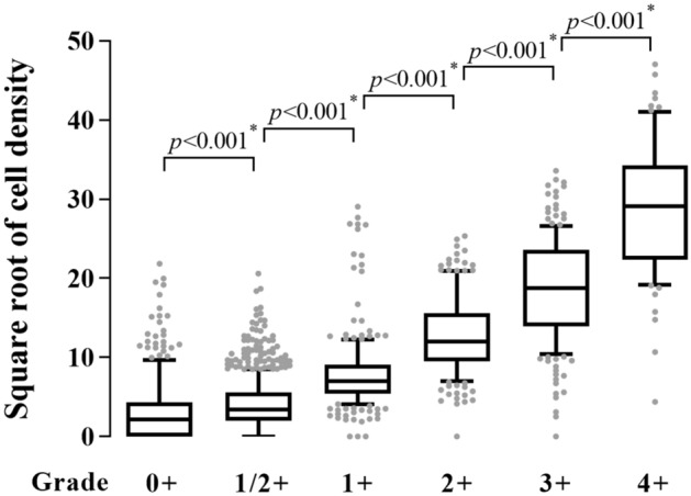
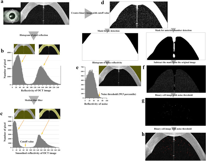
Optical coherence tomography (OCT) is a noninvasive method that can quickly and accurately examine the eye at the cellular level. Several studies have used OCT for analysis of anterior chamber cells. However, these studies have several limitations. This study was performed to supplement existing reports of automated analysis of anterior chamber cell images using spectral domain OCT (SD-OCT) and to compare this method with the Standardization of Uveitis Nomenclature (SUN) grading system. We analyzed 2398 anterior segment SD-OCT images from 34 patients using code written in Python. Cell density, size, and eccentricity were measured automatically. Increases in SUN grade were associated with significant cell density increases at all stages (p < 0.001). Significant differences were observed in eccentricity in uveitis, post-surgical inflammation, and vitreous hemorrhage (p < 0.001). Anterior segment SD-OCT is reliable, fast, and accurate means of anterior chamber cell analysis. This method showed a strong correlation with the SUN grade system. Also, eccentricity could be helpful as a supplementary evaluation tool.
Conflict of interest statement
The authors declare no competing interests.
Figures


References
-
- Kempen JH, Ganesh SK, Sangwan VS, Rathinam SR. Interobserver agreement in grading activity and site of inflammation in eyes of patients with uveitis. Am. J. Ophthalmol. 2008;146:813e811–818e811. - PubMed
-
- Wong IG, Nugent AK, Vargas-Martín F. The effect of biomicroscope illumination system on grading anterior chamber inflammation. Am. J. Ophthalmol. 2009;148:516e512–520e512. - PubMed
Publication types
MeSH terms
LinkOut - more resources
Full Text Sources
Other Literature Sources
