. 2021 May 21;11(1):10694.
doi: 10.1038/s41598-021-89889-8.

Validation of the relationship between coagulopathy and localization of hydroxyethyl starch on the vascular endothelium in a rat hemodilution model

Affiliations

Validation of the relationship between coagulopathy and localization of hydroxyethyl starch on the vascular endothelium in a rat hemodilution model

Ryu Azumaguchi et al. Sci Rep. .

Erratum in

Abstract

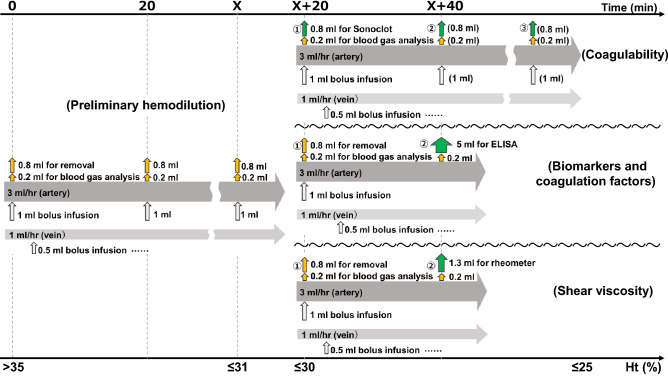
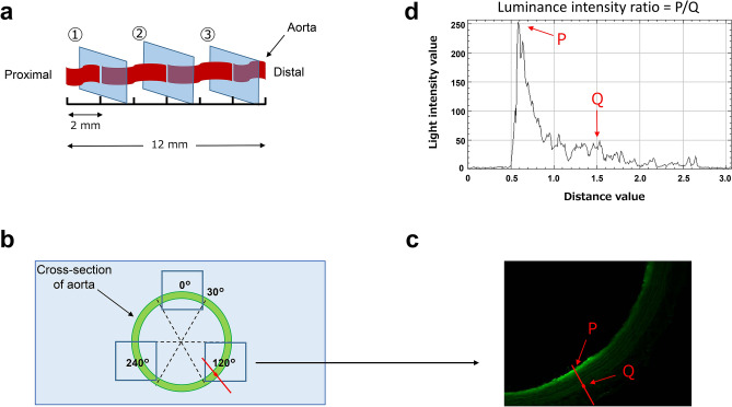
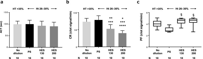
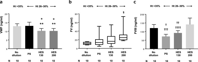
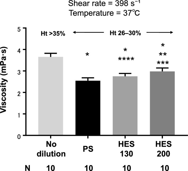
Various anticoagulant properties have been associated with hydroxyethyl starch (HES). However, the mechanism remains unclear and it has not been fully considered whether these properties are beyond the dilutional effect itself. The aim of this study was to reproduce the coagulopathy induced by HES and to test the hypothesis that the coagulopathy is caused by endothelial or glycocalyx damage due to localization of HES on the endothelium, which is caused by the high shear viscosity of dilutional blood. Using a rat model, we compared blood coagulability measured by Sonoclot, levels of endothelial and glycocalyx damage markers and coagulation factors, and blood shear viscosity when hemodilution was performed with physiological saline (PS), 6% HES 130/0.4 in PS, and 10% HES 200/0.5 in PS. We also evaluated the localization rates of fluorescently labeled HES on endothelium in the isolated aorta. HES decreased the fibrin gel formation rate more than did PS. HES was shown to cover the endothelium, possibly due to its high shear viscosity, and this mechanism potentially acted to protect, rather than damage, the endothelium and glycocalyx. However, this covering effect may be the cause of coagulopathy due to inhibition of von Willebrand factor secretion from the endothelium.


Conflict of interest statement

The authors declare no competing interests.

Figures

Figure 1
Figure 1
Hemodilution protocol. All hemodilution procedures were performed consecutively, first by the preliminary hemodilution method, followed by secondary hemodilution in different ways depending on what was measured. Ht hematocrit, ELISA enzyme-linked immunosorbent assay. X time at which the Ht level reached ≤ 31%. Yellow arrow, external; green arrow, external (sampling); white arrow, internal.
Figure 2
Figure 2
Evaluation of fluorescently labeled HES localized in the isolated aorta. (a) Collection of cross-sections from the isolated aorta. (b) Three images (squares) were taken in one cross-section divided into 6 parts. (c) A normal line was drawn from the point of highest luminance intensity on the endothelium (P) to the outer membrane (Q). (d) Endothelial luminance intensity ratio (P/Q).
Figure 3
Figure 3
Coagulability measurement using Sonoclot. ACT activated clotting time, CR clot rate, PF platelet function. PS, physiological saline; HES 130, 6% hydroxyethyl starch 130/0.4 in PS (6% Voluven); HES 200, 10% hydroxyethyl starch 200/0.5 in PS (10% Pentaspan); Ht, hematocrit. *P < 0.0001 vs no-dilution, **P = 0.0007 vs no-dilution, ***P < 0.0001 vs PS, ****P = 0.047 vs HES130, §P = 0.002 vs PS.
Figure 4
Figure 4
Quantification of Quantification of ET and GCX damage markers by ELISA. ET, endothelium; GCX, glycocalyx; ELISA, enzyme-linked immunosorbent assay; TM, thrombomodulin;aPC, activated protein C; SDC-1, syndecan-1; HSPG, heparan sulfate proteoglycan. *P = 0.008 vs no-dilution, **P = 0.003 vs PS, ***P = 0.016 vs no-dilution, ****P =0.005 vs PS, §P= 0.021 vs no-dilution, P = 0.0009 vs no-dilution, ††P = 0.018 vs PS,†††P = 0.003 vs no-dilution, ††††P = 0.045 vs PS.
Figure 5
Figure 5
Quantification of coagulation factors by ELISA. ELISA enzyme-linked immunosorbent assay, VWF von Willebrand factor, FV coagulation factor V, FVIII coagulation factor VIII. *P = 0.003 vs no-dilution, **P = 0.001 vs PS, §P = 0.03 vs no-dilution, P < 0.0001 vs no-dilution, ††P = 0.004 vs no-dilution, †††P < 0.0001 vs HES200, ††††P = 0.0002 vs HES200.
Figure 6
Figure 6
Shear viscosity measurement by rheometer. PS physiological saline, HES130 6% hydroxyethyl starch 130/0.4 in PS (6% Voluven), HES200 10% hydroxyethyl starch 200/0.5 in PS (10% Pentaspan). *P < 0.0001 vs no-dilution, **P < 0.0001 vs PS, ***P = 0.01 vs HES130, ****P = 0.032 vs PS.
Figure 7
Figure 7
Left: Representative cross-sectional images of the isolated aorta after infusion of fluorescently labeled HES. (a) Fluorescein sodium salt in HES130 (control), (b) FITC-HES130, (c) fluorescein sodium salt in HES200 (control), (d) FITC-HES200. Right: Luminance intensity ratio of endothelium to outer membrane after infusion of fluorescently labeled HES in the isolated aorta. HES130 6% hydroxyethyl starch 130/0.4 in PS (6% Voluven), HES200 10% hydroxyethyl starch 200/0.5 in PS (10% Pentaspan), FITC fluorescein isothiocyanate. *P < 0.0001 vs fluorescein sodium salt in HES130 (control), **P < 0.0001 vs fluorescein sodium salt in HES200 (control).

References

    1. Myburgh JA, Mythen MG. Resuscitation fluids. N. Engl. J. Med. 2013;369:1243–1251. doi: 10.1056/NEJMra1208627. - DOI - PubMed
    1. Navickis RJ, Haynes GR, Wilkes MM. Effect of hydroxyethyl starch on bleeding after cardiopulmonary bypass: A meta-analysis of randomized trials. J. Thorac. Cardiovasc. Surg. 2012;144:223–230. doi: 10.1016/j.jtcvs.2012.04.009. - DOI - PubMed
    1. Hartog CS, Reuter D, Loesche W, Hofmann M, Reinhart K. Influence of hydroxyethyl starch (HES) 130/0.4 on hemostasis as measured by viscoelastic device analysis: A systematic review. Intensive Care Med. 2011;37:1725–1737. doi: 10.1007/s00134-011-2385-z. - DOI - PubMed
    1. Levi M, Jonge E. Clinical relevance of the effects of plasma expanders on coagulation. Semin. Thromb. Hemost. 2007;33:810–815. doi: 10.1055/s-2007-1000370. - DOI - PubMed
    1. Deusch E, Gamsjäger T, Kress HG, Kozek-Langenecker SA. Binding of hydroxyethyl starch molecules to the platelet surface. Anesth. Analg. 2003;97:680–683. doi: 10.1213/01.ANE.0000073353.31894.BE. - DOI - PubMed

Publication types