Review
. 2021 Nov 10;62(8):1231-1238.
doi: 10.1093/pcp/pcab066.

Tools for Assessing Cell-Cycle Progression in Plants

Affiliations
Review

Tools for Assessing Cell-Cycle Progression in Plants

Clara Echevarría et al. Plant Cell Physiol. .

Abstract

Estimation of cell-cycle parameters is crucial for understanding the developmental programs established during the formation of an organism. A number of complementary approaches have been developed and adapted to plants to assess the cell-cycle status in different proliferative tissues. The most classical methods relying on metabolic labeling are still very much employed and give valuable information on cell-cycle progression in fixed tissues. However, the growing knowledge of plant cell-cycle regulators with defined expression pattern together with the development of fluorescent proteins technology enabled the generation of fusion proteins that function individually or in conjunction as cell-cycle reporters. Together with the improvement of imaging techniques, in vivo live imaging to monitor plant cell-cycle progression in normal growth conditions or in response to different stimuli has been possible. Here, we review these tools and their specific outputs for plant cell-cycle analysis.

Keywords: Arabidopsis; Cell cycle; Live-imaging; Metabolic labeling; Reporter genes; plants.


Figures

Fig. 1
Fig. 1
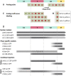
Summary of the available tools to  assess cell-cycle status. (A–C) Following cell-cycle progression using thymidine analogs. (A) Measure of G2 length: a cell population is treated by a short pulse of EdU. Cells undergoing S-phase are labeled and cell cycle is led to progress during different chase time periods until labeled mitoses appear. (B) Different patterns observed for early, mid and late S-phase nuclei. (C) Cells are continuously incorporating EdU as they progress through S-phase. The proportion of labeled cells during a time period is an estimation of the cell-cycle duration. (D) Distribution (bar position), fluorescence intensity (gray saturation) and expression pattern inside the nucleus (homogenous or speckled) throughout the different cell-cycle phases for the translational reporters described in this report. The arrowhead in the bar indicates that the marker remains in the next mitotic cycle, a blunt end indicates the degradation of the protein. The tag name position is indicative of a N-terminal (left) or C-terminal (right) fusion. Promoter used in the construct is only specified in case the protein is not expressed under its native regulator.
Fig. 2
Fig. 2
Expression of a selection of cell-cycle reporters in Arabidopsis. (A) H4::DB-VENUS expression in the shoot meristem labels cells in S-G2-M (image provided by A. Jones and J. Murray). (B) Cytrap expression in the root meristem: pHTR2::CDT1a (C3)-RFP (red) and pCYCB1;1::NCYCB1;1-GFP (green) label cells in S-G2 and G2-M, respectively (image provided by M. Umeda). (C) PlaCCI expression in the root meristem: pCDT1a::CDT1a-CFP (cyan), pHTR13::HTR13-mCherry (red) and pCYCB1;1::NCYCB1;1-YFP (yellow) label cells in G1, G1-S-G2 and G2-M, respectively. Scale bars: 20 µm (A) 25 µm (B–C).

References

    1. Bajar B.T., Lam A.J., Badiee R.K., Oh Y.-H., Chu J., Zhou X.X., et al. (2016) Fluorescent indicators for simultaneous reporting of all four cell cycle phases. Nat. Methods 13: 993–996. - PMC - PubMed
    1. Boehm E.M., Gildenberg M.S. and Washington M.T. (2016) The many roles of PCNA in Eukaryotic DNA replication. Enzymes 39: 231–254. - PMC - PubMed
    1. Boisnard-Lorig C., Colon-Carmona A., Bauch M., Hodge S., Doerner P., Bancharel E., et al. (2001) Dynamic analyses of the expression of the HISTONE: YFPfusion protein in arabidopsis show that syncytial endosperm is divided in mitotic domains. Plant Cell 13: 495–509. - PMC - PubMed
    1. Campilho A., Garcia B., Toorn H.V.D., Wijk H.V., Campilho A. and Scheres B. (2006) Time-lapse analysis of stem-cell divisions in the Arabidopsis thaliana root meristem. Plant J. 48: 619–627. - PubMed
    1. Caro E. and Gutierrez C. (2007) A green GEM: intriguing analogies with animal geminin. Trends Cell Biol. 17: 580–585. - PubMed

MeSH terms

Substances